| SKU | M00297 |
|---|---|
| Application | Immunocytochemistry |
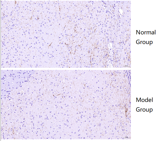
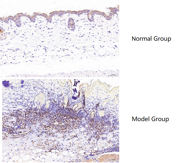
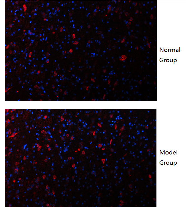
| Sample | mouse brain |
| Sample Processing Description | Normal brain tissue and brain tissue from a cerebral infarction model |
| Other Reagents | Goat serum, DAB Chromogenic Solution, Anti-fade mounting medium |
| Primary Antibody | c-Fos Rabbit Monoclonal Antibody |
| Primary Incubation | 1:200, overnight at 4 ℃ |
| Secondary Antibody | DyLight 594 Goat Anti-Rabbit IgG (H+L) |
| Secondary Incubation | 45 min at 37℃ |
| Detection | Imaging system:Leica DM2500 |
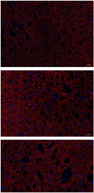
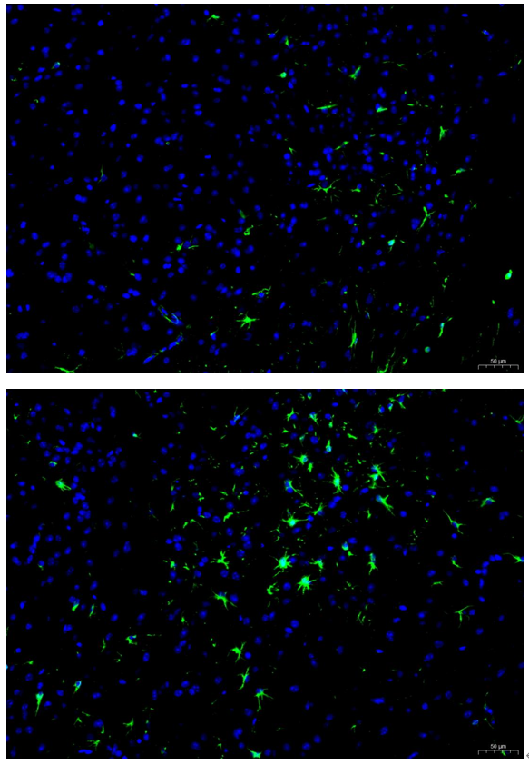
| Results Summary | c-Fos is a marker of activated neural cells, especially neurons. During the acute phase of cerebral infarction, as a strong stress and injury stimulus, it drives c-Fos expression in the brain—particularly in the peri-infarct region—far higher than in normal resting brain. The experimental results show clear staining of positive cells, and the expression levels are consistent with expectations. |