| SKU | DZ41310 |
|---|---|
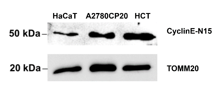
| Application | Western Blot |
| Sample | Human HaCaT cell, Human A2780 cell, Human HCT cell |
| Sample Processing Description | Dissection, Homogenization, Sample boiling in 1X Lamelli, SDS-PAGE, Standard Western Blotting |
| Primary Antibody | Human CCNE1 Antibody |
| Primary Incubation | 1:1000, overnight at 4 ℃ |
| Secondary Antibody | Anti-rabbit/mouse IgG horseradish peroxidase-conjugated |
| Secondary Incubation | 1:10000, 1-2 hours in room temperature |
| Detection | Biorad Chemidoc |
| Results Summary | Using freshly prepared blocking buffer helped reduce background. Results were reproducible and matched the expected patterns. |