| SKU | M00422 |
|---|---|
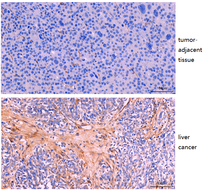
| Application | Immunohistochemistry |
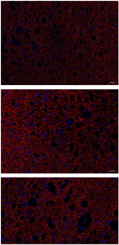
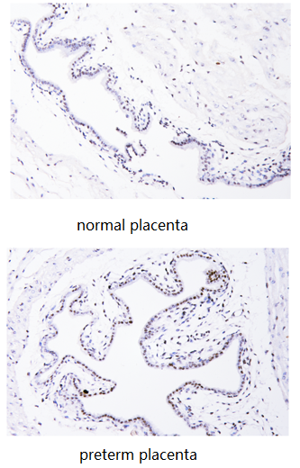
| Sample | human liver cancer |
| Sample Processing Description | Collected clinical surgical human hepatocellular carcinoma and adjacent peritumoral tissues were fixed in formalin and embedded in paraffin. |
| Other Reagents | Goat serum, DAB Chromogenic Solution |
| Primary Antibody | FAP1 Rabbit Monoclonal Antibody |
| Primary Incubation | 1:200, overnight at 4 ℃ |
| Secondary Antibody | Two-step IHC kit (Immunohistochemistry kit |
| Secondary Incubation | 30 min at 37℃ |
| Detection | Imaging system:Leica DM2500 |
| Results Summary | FAP (fibroblast activation protein) expression is mainly associated with stromal cells in the tumor microenvironment and is very low in normal human tissues. Its role in hepatocellular carcinoma (HCC) is complex, but overall it functions as an important pro-tumor factor. High FAP expression is associated with poor prognosis. It can remodel the extracellular matrix, promote tumor invasion and metastasis, and suppress anti-tumor immunity. In this study, FAP showed low expression in peritumoral tissues (close to normal tissue) and high expression in the tumor stroma of HCC. |