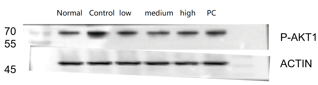
| SKU | P00024-2 |
|---|---|
| Application | Western Blot |
| Sample | rat colon tissue |
| Sample Processing Description | RIPA lysis buffer supplemented with PMSF (100:1) was used to lyse the samples for 10 min. The lysates were centrifuged at 12,000 rpm for 15 min, and the supernatants were collected. Fivefold loading buffer was added, and the samples were denatured at 100 °C for 10 min before loading onto SDS-PAGE. |
| Other Reagents | Blocking buffer |
| Primary Antibody | Phospho-AKT1 (T450) Rabbit Monoclonal Antibody |
| Primary Incubation | 1:1000, overnight at 4 ℃ |
| Secondary Antibody | HRP Conjugated AffiniPure Goat Anti-Rabbit IgG (H+L) |
| Secondary Incubation | 1:2000, 1 hour in room temperature |
| Detection | Substrate: ECL, Imaging system:ChemiDoc MP |
| Results Summary | The figure shows representative Western blot results of AKT1 (Phospho-T450) and the internal control Actin in rat colon tissues from different groups. Among the low, medium, and high doses of the traditional Chinese medicine, the high-dose group exhibits the best therapeutic effect. The target bands are clear and well defined, and the experimental results are satisfactory. |