| SKU | PB9034 |
|---|---|
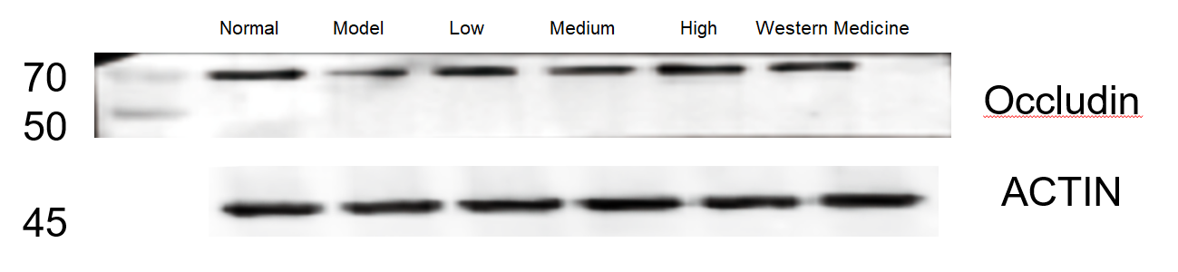
| Application | Western Blot |
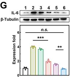
| Sample | mouse PACs cells |
| Sample Processing Description | After centrifugation, the supernatant was collected. A small aliquot of the protein solution was taken for BCA quantification, and the remaining protein solution was mixed with an equal volume of loading buffer, then denatured in a 100 °C water bath for 5 minutes. |
| Other Reagents | Protein-Free Rapid Blocking Buffer |
| Primary Antibody | Anti-IL6 Antibody Picoband® |
| Primary Incubation | 1:1000, overnight at 4 ℃ |
| Secondary Antibody | HRP Conjugated AffiniPure Goat Anti-rabbit IgG (H+L) |
| Secondary Incubation | 1 h in RT |
| Detection | Substrate: ECL substrate, Imaging system:ChemiDoc MP |
| Results Summary | The levels of IL-6 and IL-1β in the pancreas are key indicators for evaluating whether the nanodrug exerts anti-inflammatory effects. Initially, antibodies from two domestic and international suppliers were used for WB experiments, but their specificity was relatively poor. Subsequently, the antibody from Boster was adopted, which demonstrated strong specificity, high titer, and cost-effectiveness. |