| SKU | PB9533 |
|---|---|
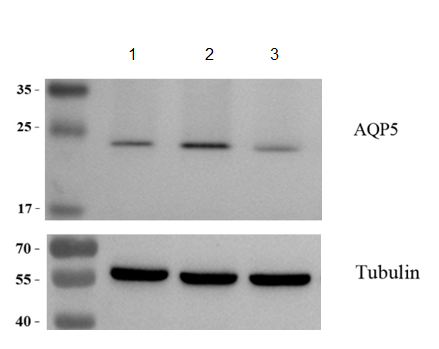
| Application | Western Blot |
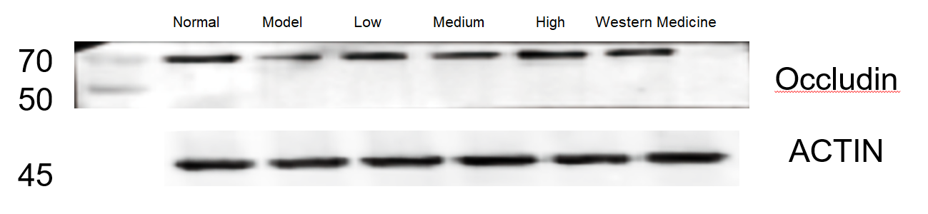
| Sample | rat colon tissue |
| Sample Processing Description | Samples were lysed in RIPA buffer containing PMSF protease inhibitor (100:1) for 10 min, centrifuged at 12,000 rpm for 15 min, and the supernatant was mixed with 5× loading buffer, boiled at 100 °C for 10 min, and loaded onto SDS-PAGE. |
| Other Reagents | 5% Non-fat milk |
| Primary Antibody | CDK1 Antibody Picoband® |
| Primary Incubation | 1:1000, overnight at 4 ℃ |
| Secondary Antibody | HRP Conjugated AffiniPure Goat Anti-rabbit IgG (H+L) |
| Secondary Incubation | 1:5000, 1 h in RT |
| Detection | Substrate: ECL, Imaging system:ChemiDoc MP |
| Results Summary | The image shows WB results of CDK1 and the loading control Actin in rat colon across different groups; CDK1 expression was elevated in the model group and most effectively reduced in the high-dose herbal treatment group, with clear and distinct target bands. |