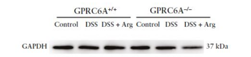
Customer Testimonials

Peer Reviews from Real Labs

Browse peer-submitted results linked to specific products—ratings, images, and practical notes included. Contribute your own result to strengthen the evidence base and claim rewards after verification.

Video Interviews

See real workflows and decision points—what researchers tried, what worked, and why. Share your story in a short interview and claim rewards!

Get Interview Rewards

Filter by category, reactivity, and host to find the most relevant peer results.

The Anti-GAPDH antibody (A00227-1) demonstrated high sensitivity and clear WB bands in mouse intestinal tissue, offering excellent cost-effectiveness and reliability, and is highly recommended for use.

Excellent, submitted by on
SKU A00227-1
Application Western Blot
Sample mouse intesinal tissue
Sample Processing Description Mouse colon tissue was lysed in RIPA buffer containing a protease inhibitor cocktail. Protein concentration was determined using the Pierce™ BCA Protein Assay Kit, and equal amounts of protein were loaded after boiling denaturation.
Other Reagents 5% non-fat milk
Primary Antibody GAPDH Antibody Picoband®
Primary Incubation 1:1000, overnight at 4 ℃
Secondary Antibody goat anti rabbit secondary antibodies
Secondary Incubation 1:5000, 1 h in RT
Detection Substrate: ECL substrate, Image system:ChemiDoc MP
Results Summary This antibody is highly sensitive, produces clear WB bands, is reusable, offers excellent cost-effectiveness, and demonstrates a clear advantage over similar international products, making it highly recommended for use.

Using Boster’s IL-6 antibody (RP1012) in WB on HK-2 cells produced clear, specific bands with high sensitivity and cost-effectiveness, outperforming previously tested antibodies.

Excellent, submitted by on
SKU RP1012
Application Western Blot
Sample human HK-2 cells
Sample Processing Description Cell samples were directly lysed in RIPA buffer, mixed with loading buffer at the appropriate ratio, and denatured at 98 °C. Twenty microliters of each protein sample were loaded per lane onto SDS-PAGE.
Other Reagents 5% non-fat milk
Primary Antibody Interleukin-6 IL6 Antibody Picoband®
Primary Incubation 1:1000, overnight at 4 ℃
Secondary Antibody HRP Conjugated AffiniPure Goat Anti-rabbit IgG (H+L)
Secondary Incubation 1 h in RT
Detection Substrate: ECL substrate
Results Summary Previously, WB experiments were performed using antibodies from two domestic and international suppliers, which showed poor specificity and were expensive. Subsequently, Boster’s IL-6 antibody (Cat. RP1012) was used, demonstrating high specificity, strong titer, cost-effectiveness, and yielding clear bands.

In WB using Anti-CDK1 (Phospho-T450) antibody (Cat# PB9533-50 µL), CDK1 expression in rat colon was increased in the model group and most effectively reduced in the high-dose herbal treatment group, with clear and well-defined target bands.

Excellent, submitted by on
SKU PB9533
Application Western Blot
Sample rat colon tissue
Sample Processing Description Samples were lysed in RIPA buffer containing PMSF protease inhibitor (100:1) for 10 min, centrifuged at 12,000 rpm for 15 min, and the supernatant was mixed with 5× loading buffer, boiled at 100 °C for 10 min, and loaded onto SDS-PAGE.
Other Reagents 5% Non-fat milk
Primary Antibody CDK1 Antibody Picoband®
Primary Incubation 1:1000, overnight at 4 ℃
Secondary Antibody HRP Conjugated AffiniPure Goat Anti-rabbit IgG (H+L)
Secondary Incubation 1:5000, 1 h in RT
Detection Substrate: ECL, Imaging system:ChemiDoc MP
Results Summary The image shows WB results of CDK1 and the loading control Actin in rat colon across different groups; CDK1 expression was elevated in the model group and most effectively reduced in the high-dose herbal treatment group, with clear and distinct target bands.

In WB using Occludin antibody (Cat# A01246), clear bands showed decreased Occludin in the model group and best recovery in the high-dose herbal treatment group.

Excellent, submitted by on
SKU A01246
Application Western Blot
Sample rat colon tissue
Sample Processing Description RIPA lysis buffer containing protease inhibitor PMSF (100:1) was used to lyse samples for 10 min, followed by centrifugation at 12,000 rpm for 15 min; the supernatant was mixed with 5× loading buffer, boiled at 100 °C for 10 min, and loaded onto SDS-PAGE.
Other Reagents 5% Non-fat milk
Primary Antibody Occludin OCLN Antibody
Primary Incubation 1:1000, overnight at 4 ℃
Secondary Antibody HRP Conjugated AffiniPure Goat Anti-rabbit IgG (H+L)
Secondary Incubation 1:5000, 1 h in RT
Detection Substrate: ECL substrate, Imaging system:ChemiDoc MP
Results Summary The image shows WB results of Occludin and the loading control Actin in rat colon across different groups; Occludin expression was reduced in the model group, with the high-dose herbal treatment showing the best recovery and clear, well-defined target bands.

In IHC using Ki67 antibody (Cat# PB9026), strong and specific nuclear staining was observed in nude mouse tumor tissue, confirming high Ki67 expression and active cell proliferation.

Excellent, submitted by on
SKU PB9026
Application Western Blot
Sample Nude mouse tumor tissue
Sample Processing Description Paraffin-embedded tumor tissue sections.
Primary Antibody Ki67/MKI67 Antibody Picoband®
Primary Incubation 1:1000, overnight at 4 ℃
Secondary Antibody HRP-conjugated Anti-Rabbit IgG Secondary Antibody
Secondary Incubation 1 hour in room temperature
Detection Substrate: ECL reagent, Imaging system:ChemiDoc MP
Results Summary This antibody is highly specific and efficient, with a clean background and no nonspecific bands. The target band has sharp and well-defined edges.

In the IF experiment using Anti-IBA-1 antibody (Cat# M01394-4), the antibody clearly and specifically labeled microglial cells in mouse cerebral infarction tissue, showing distinct staining and well-defined cellular morphology.

Excellent, submitted by on
SKU M01394-4
Application Immunofluorescence
Sample mouse brain tissue
Sample Processing Description Mouse cerebral infarction model; brain tissues were collected, fixed in formaldehyde for 48 hours, and then sagittally paraffin-embedded.
Other ReagentsGoat serum, DAPI Staining Solution, Antifade fluorescence mounting medium.
Primary Antibody Iba1 Rabbit Monoclonal Antibody
Primary Incubation 1:100, overnight at 4 ℃
Secondary Antibody Goat Anti-Rabbit IgG (H+L) Secondary Antibody, Fluoro594 Conjugated (BA1142, Boster)
Secondary Incubation 45 min at 37℃
Detection Imaging system:Leica DMi3000
Results Summary IBA-1 is a well-established marker of microglia in the nervous system. Microglia are the primary immune effector cells in the central nervous system and respond rapidly to CNS injury by proliferating, upregulating or re-expressing MHC antigens, migrating, and adopting a phagocyte-like morphology. In this study, IBA-1 was used to label microglia in cerebral infarction samples, showing clear staining and accurate cellular morphology.

In this IHC experiment using Anti-SLC22A3 antibody (Cat# M04914), strong and clear expression was observed in cervical cancer tumor cells, with further comparison planned between tumor and adjacent tissues.

Excellent, submitted by on
SKU M04067-2
Application Immunohistochemistry
Sample human cervical cancer tissue
Sample Processing Description Cervical cancer tissue collected from clinical surgery, fixed in formalin and paraffin-embedded
Other ReagentsGoat serum, DAB substrate solution
Primary Antibody SLC22A3 Rabbit Monoclonal Antibody
Primary Incubation 1:200, overnight at 4 ℃
Secondary Antibody Two-step IHC detection kit
Secondary Incubation 30 min at 37℃
Detection Imaging system:Leica DM2500
Results Summary IHC using SLC22A3 antibody (M04914) showed strong and clear expression in tumor cells of human cervical carcinoma, and the next step will be to compare expression between tumor and adjacent normal tissue.

In this IHC experiment using Anti-SERPINB3 antibody (Cat# M04067-2) on human cervical cancer paraffin sections, SERPINB3 was highly expressed in tumor cells with clear positive staining and low background, demonstrating excellent antibody performance.

Excellent, submitted by on
SKU M04067-2
Application Immunohistochemistry
Sample human cervical cancer tissue
Sample Processing Description Cervical cancer tissue collected from clinical surgery, fixed in formalin and paraffin-embedded
Other ReagentsGoat serum, DAB substrate solution
Primary Antibody SerpinB3 Rabbit Monoclonal Antibody
Primary Incubation 1:400, overnight at 4 ℃
Secondary Antibody Two-step IHC detection kit
Secondary Incubation 30 min at 37℃
Detection Imaging system:Leica DM2500
Results Summary The results demonstrated high expression of SERPINB3 in cervical cancer tumor cells with clear positive staining; further studies will compare its expression levels between tumor and adjacent non-tumor tissues.

In this IHC experiment using Anti-CD4 antibody (Cat# M00344) on human cervical cancer paraffin sections, CD4-positive helper T cells were clearly and specifically stained with minimal background, demonstrating excellent antibody performance.

Excellent, submitted by on
SKU M00344
Application Western blot
Sample human cervical cancer paraffin sections
Sample Processing Description Cervical cancer tissue collected from clinical surgery, fixed in formalin and paraffin-embedded
Other ReagentsGoat serum, DAB substrate solution
Primary Antibody CD4 Rabbit Monoclonal Antibody
Primary Incubation 1:200, overnight at 4 ℃
Secondary Antibody Two-step IHC detection kit
Secondary Incubation 30 min at 37℃
Detection Substrate: ECL substrate, Imaging system:ChemiDoc MP
Results Summary CD4 is a marker of helper T cells; in this IHC experiment, CD4 staining clearly identified helper T cells in human cervical cancer samples, showing excellent specificity and performance of the antibody.

In this Western blot experiment using Anti-AQP1 antibody (Cat# BM5035) on MADB106 rat mammary carcinoma cells, AQP1 protein levels were markedly increased in cells treated with the agonist and significantly decreased in cells treated with the inhibitor.

Excellent, submitted by on
SKU M00865
Application Western blot
Sample MADB106 rat mammary carcinoma cells
Sample Processing Description MADB106 cells under normal culture conditions , MADB106 cells treated with agonist , MADB106 cells treated with inhibitor
Other ReagentsRIPA Lysis Buffer, Protease Inhibitor, Resolving Gel Solution ,Transfer Buffer ,Blocking Buffer
Primary Antibody AQP1 Rabbit Monoclonal Antibody
Primary Incubation 1:3000, overnight at 4 ℃
Secondary Antibody 1:10,000, HRP-conjugated Goat Anti-Rabbit IgG
Secondary Incubation 1h at RT
Detection Substrate: ECL substrate, Imaging system:ChemiDoc MP
Results Summary AQP1 (Aquaporin 1) is the first discovered water channel protein, whose primary function is efficient water transport. Recent studies have revealed that AQP1 plays a critical role in cancer progression, promoting tumor growth in breast cancer by enhancing angiogenesis and cell migration. In this experiment, AQP1 protein levels were markedly increased in cells treated with the agonist and significantly decreased in cells treated with the inhibitor.