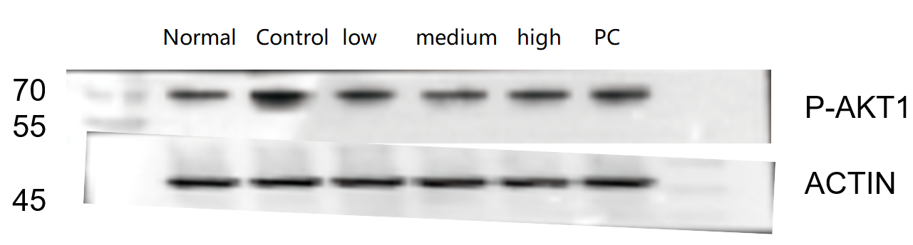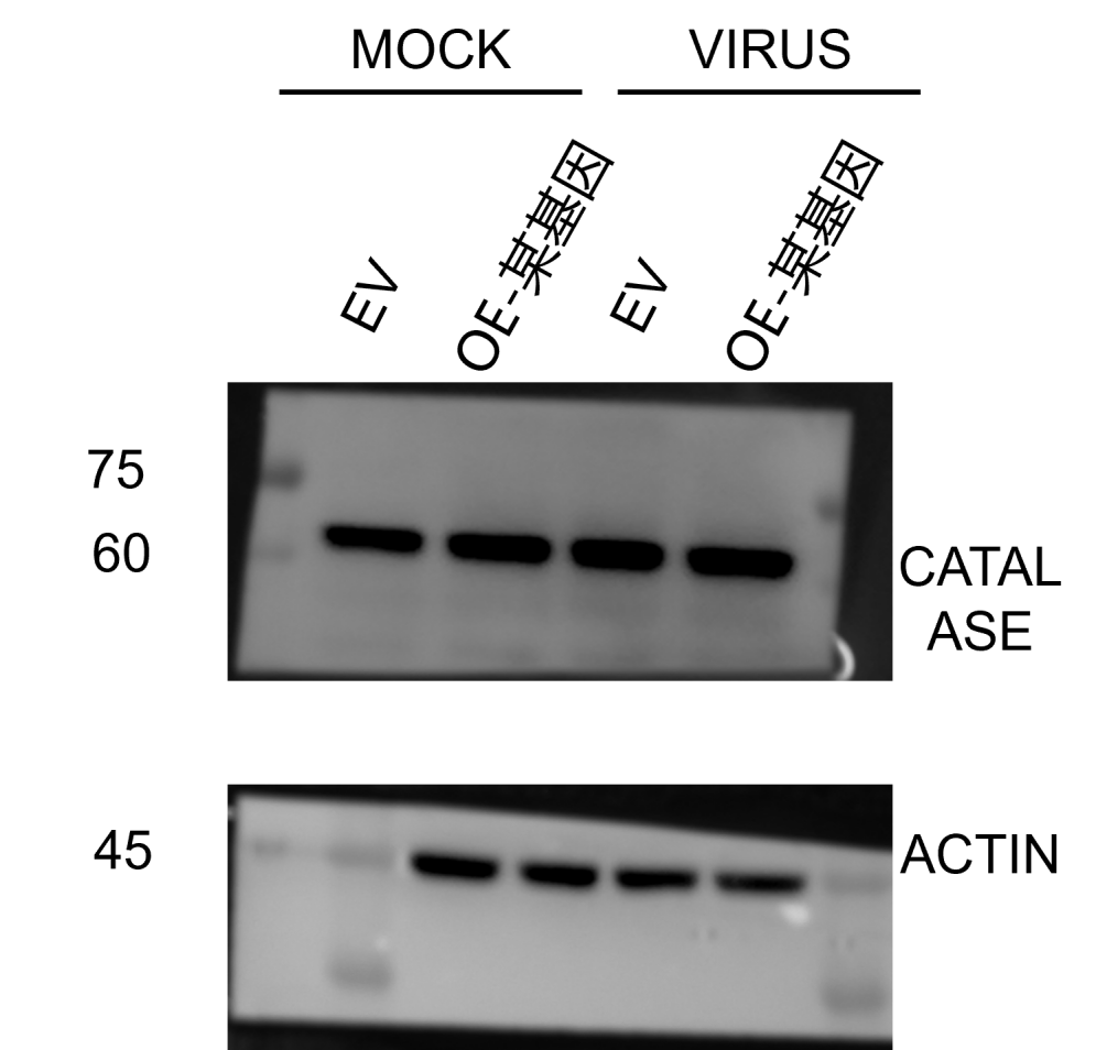
| SKU | M00024-1 |
|---|---|
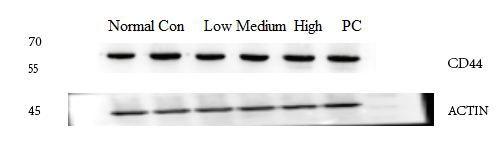
| Application | Western Blot |
| Sample | rat colon tissue |
| Sample Processing Description | RIPA lysis buffer: Add protease inhibitor PMSF (100:1), lyse for 10 min, centrifuge at 12,000 rpm for 15 min, transfer the supernatant and mix with 5× loading buffer, denature at 100 °C for 10 min, and load onto SDS-PAGE. |
| Other Reagents | Blocking buffer |
| Primary Antibody | AKT1 Monoclonal Antibody |
| Primary Incubation | 1:1000, overnight at 4 ℃ |
| Secondary Antibody | HRP Conjugated AffiniPure Goat Anti-Rabbit IgG (H+L) |
| Secondary Incubation | 1:1000, 1 hour in room temperature |
| Detection | Substrate: ECL, Imaging system:ChemiDoc MP |
| Results Summary | The figure shows a schematic of Western blot results for the target protein AKT1 and the internal control Actin in rat colon across different groups. No significant differences were observed between groups; the target bands are clear and distinct, and the experimental results are satisfactory. |