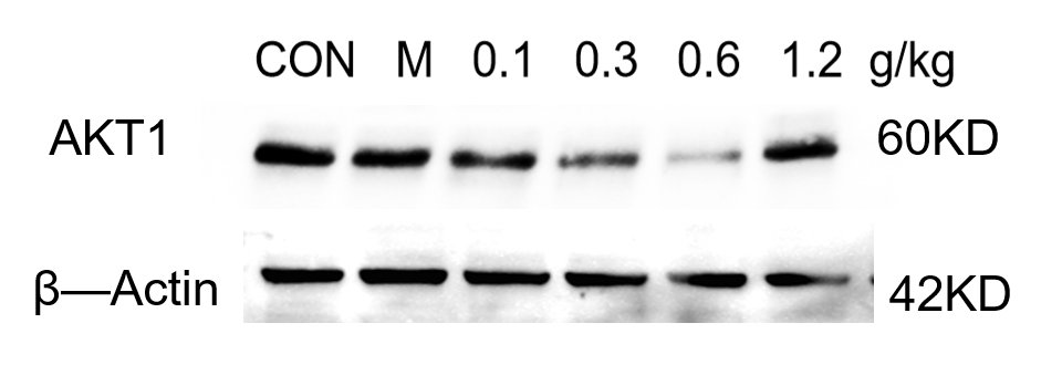
Customer Testimonials

Peer Reviews from Real Labs

Browse peer-submitted results linked to specific products—ratings, images, and practical notes included. Contribute your own result to strengthen the evidence base and claim rewards after verification.

Video Interviews

See real workflows and decision points—what researchers tried, what worked, and why. Share your story in a short interview and claim rewards!

Get Interview Rewards

Filter by category, reactivity, and host to find the most relevant peer results.

The antibody shows a clear target, correct localization, minimal background, and overall good results.

Excellent, submitted by on
SKU P00024-4
Application Western Blot
Sample mouse 4T1 cells
Sample Processing Description normal 4T1 cells, LPS-stimulated 4T1 cells, cells treated with low, medium, or high doses of the drug, and cells treated with a positive control drug.
Other Reagents RIPA lysis buffer, Protease inhibitor, Electrophoresis buffer, Transfer buffer, Blocking buffer
Primary Antibody Anti-AKT1,2,3 Antibody Picoband®
Primary Incubation 1:10000, overnight at 4 ℃
Secondary Antibody HRP Goat Anti-Rabbit IgG
Secondary Incubation 1:10000, 1 hour in room temperature
Detection Substrate: ECL, Imaging system:ChemiDoc MP
Results Summary Under equal loading conditions, the total AKT protein levels were similar across all groups, while the amount of phosphorylated AKT1 showed significant changes. The blank group had the lowest level, the model group had the highest, and in the drug-treated groups, the levels decreased progressively from low to medium to high dose. The positive control drug group was close to the high-dose group.

The antibody shows a clear target, correct localization, minimal background, and overall good results.

Excellent, submitted by on
SKU M00003
Application Western Blot
Sample mouse 4T1 cells
Sample Processing Description normal 4T1 cells, LPS-stimulated 4T1 cells, cells treated with low, medium, or high doses of the drug, and cells treated with a positive control drug.
Other Reagents RIPA lysis buffer, Protease inhibitor, Electrophoresis buffer, Transfer buffer, Blocking buffer
Primary Antibody Anti-ATF4 Rabbit Monoclonal Antibody
Primary Incubation 1:10000, overnight at 4 ℃
Secondary Antibody HRP Goat Anti-Rabbit IgG
Secondary Incubation 1:10000, 1 hour in room temperature
Detection Substrate: ECL, Imaging system:ChemiDoc MP
Results Summary With equal loading amounts, the total MTOR protein levels were similar across all groups. However, the levels of phosphorylated MTOR showed significant changes: the blank group had the lowest, the model group the highest, and in the drug-treated groups, the phosphorylation levels decreased sequentially from low, medium, to high dose. The positive control group was comparable to the high-dose group.

Cell samples were lysed by sonication in RIPA buffer containing protease and phosphatase inhibitors, followed by centrifugation for 10 minutes. The supernatant was mixed with loading buffer at a 4:1 ratio and boiled for 10 minutes. Fifteen microliters of

Excellent, submitted by on
SKU M00003
Application Western Blot
Sample human OCI-LY1 cell
Sample Processing Description Cell samples were lysed by sonication in RIPA buffer containing protease and phosphatase inhibitors, followed by centrifugation for 10 minutes. The supernatant was mixed with loading buffer at a 4:1 ratio and boiled for 10 minutes. Fifteen microliters of each sample were loaded per well.
Other Reagents 5% Non-fat milk
Primary Antibody mTOR/Tor Rabbit Monoclonal Antibody
Primary Incubation 1:3000, overnight at 4 ℃
Secondary Antibody HRP-conjugated Anti-Rabbit IgG Secondary Antibody
Secondary Incubation 1 hour in room temperature
Detection Substrate: ECL reagent, Imaging system:ChemiDoc MP
Results Summary This antibody is highly specific and efficient, with a clean background and no nonspecific bands. The target band has sharp and well-defined edges.

This antibody is highly specific and efficient, with a clean background and no nonspecific bands. The target band has well-defined edges.

Excellent, submitted by on
SKU P00003
Application Western Blot
Sample human OCI-LY1 cell
Sample Processing Description Cell samples were lysed by sonication in RIPA buffer containing protease and phosphatase inhibitors, followed by centrifugation for 10 minutes. The supernatant was mixed with loading buffer at a 4:1 ratio and boiled for 10 minutes. Fifteen microliters of each sample were loaded per well.
Other Reagents 5% Non-fat milk
Primary Antibody Phospho-mTOR (S2448) Rabbit Monoclonal Antibody
Primary Incubation 1:3000, overnight at 4 ℃
Secondary Antibody HRP-conjugated Anti-Rabbit IgG Secondary Antibody
Secondary Incubation 1 hour in room temperature
Detection Substrate: ECL reagent, Imaging system:ChemiDoc MP
Results Summary This antibody is highly specific and efficient, with a clean background and no nonspecific bands. The target band has sharp and well-defined edges.

This antibody is highly specific and efficient, with a clear target band and no nonspecific bands.

Excellent, submitted by on
SKU M00220-1
Application Western Blot
Sample mouse lung cancer tissue
Sample Processing Description Tissue samples were directly lysed in RIPA buffer, mixed with loading buffer at the appropriate ratio, and denatured by heating at 98°C. Twenty microliters of each protein sample were loaded per lane onto an SDS-PAGE gel.
Other Reagents 5% Non-fat milk
Primary Antibody CD86/B7 2 Rabbit Monoclonal Antibody
Primary Incubation 1:2000, overnight at 4 ℃
Secondary Antibody HRP-conjugated Anti-Rabbit IgG Secondary Antibody
Secondary Incubation 1 hour in room temperature
Detection Substrate: ECL reagent, Imaging system:Tanon
Results Summary This antibody is highly specific and efficient, with a clear target band and no nonspecific bands.

The Anti-Cytokeratin antibody (M02416-2) produced clear and specific target bands in WB analysis of untreated and drug-treated HTR8 cells, demonstrating reliable and consistent detection without non-specific signals.

Excellent, submitted by on
SKU M02416-2
Application Western Blot
Sample human HTR8 cells
Sample Processing Description Cells were lysed with RIPA lysis buffer, mixed with β-mercaptoethanol, denatured at 100°C for 10 minutes, and then loaded onto SDS-PAGE.
Other Reagents blocking buffer
Primary Antibody Cytokeratin 7 KRT7 Rabbit Monoclonal Antibody
Primary Incubation 1:1000, overnight at 4 ℃
Secondary Antibody HRP-conjugated Anti-Rabbit IgG Secondary Antibody
Secondary Incubation 1 hour in room temperature
Detection Substrate: ECL reagent, Imaging system:ChemiDoc MP
Results Summary The figure shows WB results of Cytokeratin in untreated and drug-treated HTR8 cells, with clear and distinct bands at the expected position, demonstrating reliable and specific detection.

This antibody is suitable for detecting PTEN protein in mouse hippocampus by Western blot, showing clear, distinct, and highly specific bands.

Excellent, submitted by on
SKU M00006-1
Application Western Blot
Sample Mouse hippocampus tissue
Sample Processing Description The mouse hippocampus was lysed with RIPA buffer containing a protease inhibitor cocktail. After protein quantification, samples were mixed with 5× protein loading buffer and heated for 10 minutes to denature. Load 5 μL of protein per lane and apply to SDS-PAGE.
Other Reagents 5% Non-fat milk
Primary Antibody Anti-PTEN Rabbit Monoclonal Antibody
Primary Incubation overnight at 4 ℃
Secondary Antibody HRP-conjugated Anti-Rabbit IgG Secondary Antibody
Secondary Incubation 1 hour in room temperature
Detection Substrate: Ultra-sensitive ECL luminescent reagent, Imaging system:ChemiDoc MP
Results Summary This antibody is suitable for detecting PTEN protein in mouse hippocampus by Western blot, showing clear, distinct, and highly specific bands.

The HIF1A antibody was used to detect the expression of the target protein in human uterine tissue. The Western blot results showed clear bands, and the antibody maintained good performance after reuse.

Excellent, submitted by on
SKU M00024-1
Application Western Blot
Sample Mouse hippocampus tissue
Sample Processing Description The mouse hippocampus was lysed with RIPA buffer containing a protease inhibitor cocktail. After protein quantification, samples were mixed with 5× protein loading buffer and heated for 10 minutes to denature. Load 5 μL of protein per lane and apply to SDS-PAGE.
Primary Antibody Anti-HIF-1 alpha Rabbit Monoclonal Antibody
Primary Incubation 1:1000, overnight at 4 ℃
Secondary Antibody HRP-conjugated Anti-Rabbit IgG Secondary Antibody
Secondary Incubation 1 hour in room temperature
Detection Substrate: Ultra-sensitive ECL luminescent reagent (Cat# AR1191), Imaging system:Tanon
Results Summary The HIF1A antibody was used to detect the expression of the target protein in human uterine tissue. The Western blot results showed clear bands, and the antibody maintained good performance after reuse. It offers excellent cost-effectiveness, with a clear advantage over other antibodies with similar specifications. Highly recommended for use!

The GPX4 antibody was used to detect the expression of the protein in mouse uterine tissue. The WB results showed clear bands, and the antibody could be reused after recovery with good performance, offering excellent cost-effectiveness.

Excellent, submitted by on
SKU M02059
Application Western Blot
Sample Mouse Uterus tissue
Sample Processing Description The GPX4 antibody was used to detect the expression of the protein in mouse uterine tissue. The Western blot results showed clear bands, and the antibody retained good performance after reuse, offering excellent cost-effectiveness.
Primary Antibody Anti-GPX4 Rabbit Monoclonal Antibody
Primary Incubation overnight at 4 ℃
Secondary Antibody HRP-conjugated Anti-Rabbit IgG Secondary Antibody
Secondary Incubation 1 hour in room temperature
Detection Substrate: Ultra-sensitive ECL luminescent reagent (Cat# AR1191), Imaging system:Tanon
The GPX4 antibody was used to detect the protein expression in mouse uterine tissue. The Western blot results showed clear bands, and the antibody maintained good performance after reuse, providing excellent cost-effectiveness.

The FTH1 antibody was used to detect the expression of the target protein in human uterine tissue. The WB bands were single and clear, and compared with other domestic and international brands, this antibody offers excellent cost-performance.

Excellent, submitted by on
SKU M02401
Application Western Blot
Sample Mouse hippocampus tissue
Sample Processing Description The tissue was minced and sonicated, then lysed on ice for 1 hour using RIPA buffer. After centrifugation to collect the supernatant and protein quantification by BCA, samples were mixed with loading buffer at the appropriate ratio and denatured by boiling in a water bath. Fifteen microliters of each protein sample were loaded per lane onto SDS-PAGE gel.
Primary Antibody Anti-Ferritin FTH1 Rabbit Monoclonal Antibody
Primary Incubation overnight at 4 ℃
Secondary Antibody HRP-conjugated Anti-Rabbit IgG Secondary Antibody
Secondary Incubation 1 hour in room temperature
Detection Substrate: Ultra-sensitive ECL luminescent reagent (Cat# AR1191), Imaging system:Tanon
Results Summary The FTH1 antibody was used to detect the expression of the target protein in human uterine tissue. The WB bands were single and clear, and compared with other domestic and international brands, this antibody offers excellent cost-performance.