| SKU | M12477-15 |
|---|---|
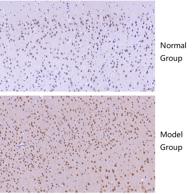
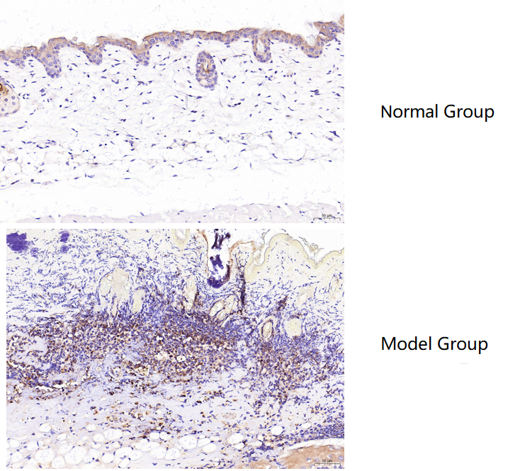
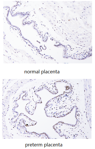
| Application | Immunohistochemistry |
| Sample | normal and preterm human placentas |
| Sample Processing Description | Clinically collected normal and preterm placentas were sectioned longitudinally (with amnion and chorion visible) and prepared as paraffin-embedded sections. |
| Other Reagents | Tris-EDTA Antigen Retrieval Solution, DAB Chromogenic Solution |
| Primary Antibody | Histone H3 (acetyl K27) Rabbit Monoclonal Antibody |
| Primary Incubation | 1:200, overnight at 4 ℃ |
| Secondary Antibody | Polymer anti-rabbit IgG-HRP IHC detection kit |
| Secondary Incubation | 45 min at 37℃ |
| Detection | Imaging system:Leica DM2500 |
| Results Summary | Histone H3 (acetyl K27) is the acetylated form of histone H3 at lysine 27 and serves as a sensitive indicator of epigenetic abnormalities in placental cells; IHC results showed higher expression in preterm placentas than in normal placentas. |