| SKU | M00207-1 |
|---|---|
| Application | Western Blot |
| Sample | huaman 293 cells |
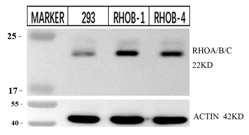
| Sample Processing Description | RHOB detection was performed using total protein extracted from normal 293 cells and 293 cells with RHOB overexpression. |
| Other Reagents | RIPA lysis buffer, Protease inhibitor, Electrophoresis buffer, Transfer buffer, Blocking buffer |
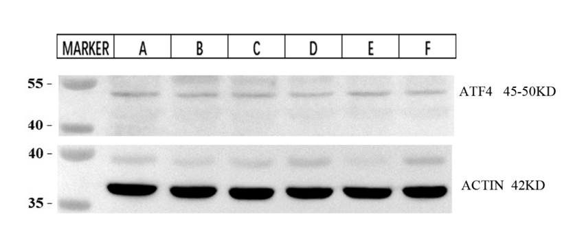
| Primary Antibody | ATF4 Rabbit Monoclonal Antibody |
| Primary Incubation | 1:2500, overnight at 4 ℃ |
| Secondary Antibody | HRP Goat Anti-Rabbit IgG |
| Secondary Incubation | 1:10000, 1 hour in room temperature |
| Detection | Substrate: ECL, Imaging system:ChemiDoc MP |
| Results Summary | We generated a stable RHOB overexpression cell line using 293 cells. The results show that the RHOB expression level in the transfected samples is significantly higher than in the normal 293 cells, indicating successful transfection. |