| SKU | M00024-1 |
|---|---|
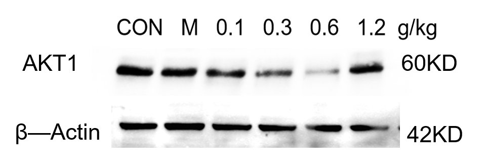
| Application | Western Blot |
| Sample | Mouse hippocampus tissue |
| Sample Processing Description | The mouse hippocampus was lysed with RIPA buffer containing a protease inhibitor cocktail. After protein quantification, samples were mixed with 5× protein loading buffer and heated for 10 minutes to denature. Load 5 μL of protein per lane and apply to SDS-PAGE. |
| Primary Antibody | Anti-HIF-1 alpha Rabbit Monoclonal Antibody |
| Primary Incubation | 1:1000, overnight at 4 ℃ |
| Secondary Antibody | HRP-conjugated Anti-Rabbit IgG Secondary Antibody |
| Secondary Incubation | 1 hour in room temperature |
| Detection | Substrate: Ultra-sensitive ECL luminescent reagent (Cat# AR1191), Imaging system:Tanon |
| Results Summary | The HIF1A antibody was used to detect the expression of the target protein in human uterine tissue. The Western blot results showed clear bands, and the antibody maintained good performance after reuse. It offers excellent cost-effectiveness, with a clear advantage over other antibodies with similar specifications. Highly recommended for use! |