Customer Testimonials

Peer Reviews from Real Labs

Browse peer-submitted results linked to specific products—ratings, images, and practical notes included. Contribute your own result to strengthen the evidence base and claim rewards after verification.

Video Interviews

See real workflows and decision points—what researchers tried, what worked, and why. Share your story in a short interview and claim rewards!

Get Interview Rewards

Filter by category, reactivity, and host to find the most relevant peer results.

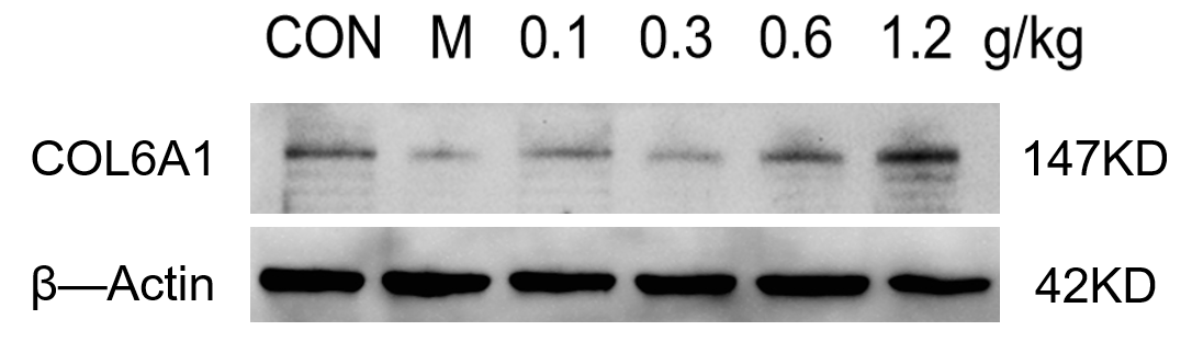
Western blot analysis was performed using the COL6A1 antibody to detect COL6A1 protein expression in the mouse hippocampus.

Excellent, submitted by on
SKU M02226
Application Western Blot
Sample Mouse hippocampus tissue
Sample Processing Description The mouse hippocampus was lysed in RIPA buffer supplemented with a protease inhibitor cocktail. After protein quantification, samples were mixed with 5× protein loading buffer and denatured by heating at 100°C for 10 minutes. Five microliters of each protein sample were loaded per lane onto SDS-PAGE.
Primary Antibody Anti-Collagen VI COL6A1 Rabbit Monoclonal Antibody
Primary Incubation overnight at 4 ℃
Secondary Antibody HRP-conjugated Anti-Rabbit IgG Secondary Antibody
Secondary Incubation 1 hour in room temperature
Detection Substrate: Ultra-sensitive ECL luminescent reagent (Cat# AR1191), Imaging system:ChemiDoc MP
Results Summary Western blot analysis was performed using the COL6A1 antibody to detect COL6A1 protein expression in the mouse hippocampus. Although minor non-specific bands were observed, they did not affect the trend analysis, indicating that the antibody is suitable for detecting the target protein in this tissue.

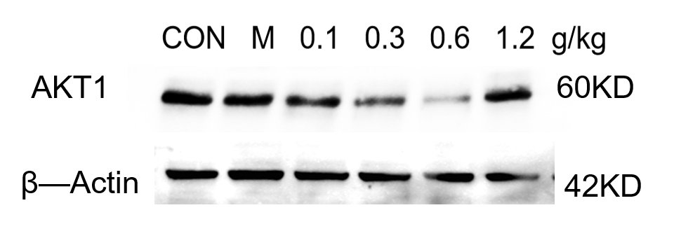
Western blot analysis of AKT1 protein in mouse hippocampus using the AKT1 antibody showed clear bands. The antibody is suitable for mouse hippocampal tissue samples.

Excellent, submitted by on
SKU M00024-1
Application Western Blot
Sample Mouse hippocampus tissue
Sample Processing Description The mouse hippocampus was lysed with RIPA buffer containing a protease inhibitor cocktail. After protein quantification, samples were mixed with 5× protein loading buffer and heated for 10 minutes to denature. Load 5 μL of protein per lane and apply to SDS-PAGE.
Primary Antibody Anti-AKT1 Monoclonal Antibody
Primary Incubation overnight at 4 ℃
Secondary Antibody HRP-conjugated Anti-Rabbit IgG Secondary Antibody
Secondary Incubation 1 hour in room temperature
Detection Substrate: Ultra-sensitive ECL luminescent reagent (Cat# AR1191), Imaging system: ChemiDoc MP (Bio-Rad)
Results Summary Western blot analysis of AKT1 protein in the mouse hippocampus using the AKT1 antibody showed clear bands. This antibody is suitable for mouse hippocampal tissue samples.

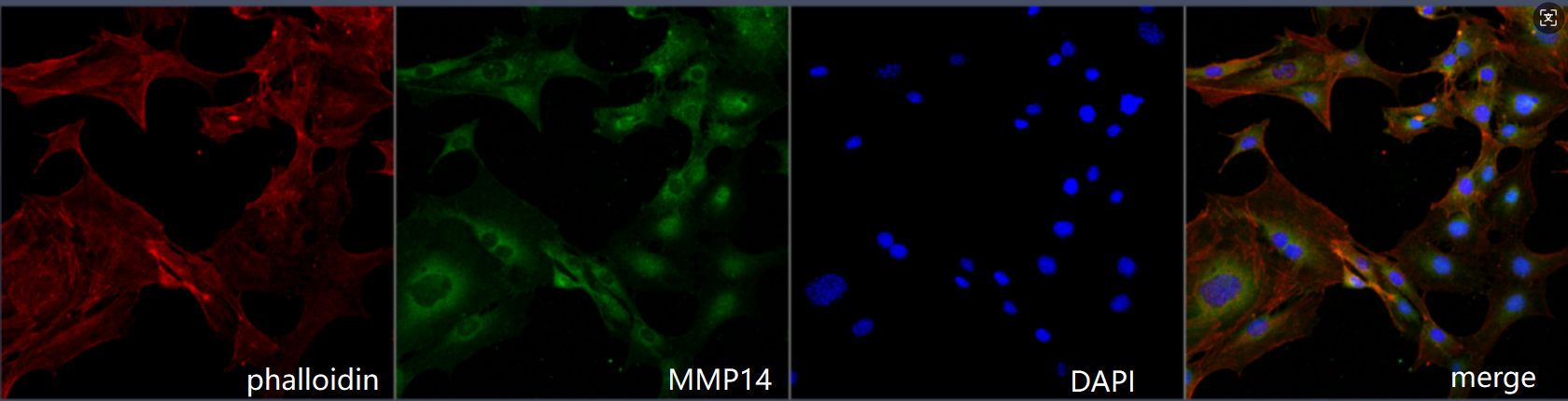
Highly prone to positive signals, with precise expression localization

Excellent, submitted by on
SKU M00656
Application Immunofluorenscence
Sample Mouse MC-8 cells
Sample Processing Description 4% paraformaldehyde for 15 minutes
Primary Antibody Anti-MMP14/Mt1 Mmp Rabbit Monoclonal Antibody
Primary Incubation 1:200, overnight at 4 ℃
Blocking Agent Goat serum
Secondary Antibody DyLight 488-conjugated Goat Anti-Rabbit IgG.
Secondary Incubation Incubate at room temperature for 1 hour
Detection Laser confocal microscopy
Results Summary I will purchase Boster products again and recommend them to my classmates and colleagues.

The antibody is highly efficient and specific, showing a clear target band with no non-specific bands.

Excellent, submitted by on
SKU M30929
Application Western Blot
Sample HEK293T
Primary Incubation 1:1000, overnight at 4 ℃
Blocking Agent 5% non-fat milk
Secondary Antibody Anti-Rabbit IgG Secondary Antibody, HRP-conjugated
Secondary Incubation Incubate at room temperature for 1 hour
Detection Signal was developed using ECL substrate on a Azure Biosystems c600.
Results Summary I will definitely purchase BosterBio products again and recommend them to my classmates and colleagues.

This antibody shows excellent consistency and reproducibility across batches, ensuring reliable and stable experimental results. Its performance remains consistent across different experiments, providing strong technical support for our research.

Excellent, submitted by on
SKU M03918
Application Western Blot
Sample Mouse brown adipose tissue
Sample Processing Description Tissue and cell proteins were extracted using RIPA buffer. After BCA quantification, 5× loading buffer was added, and the samples were boiled for 5 minutes for denaturation.
Primary Incubation The membrane was incubated with the PLIN1 primary antibodies (1:1000) overnight at 4 °C.
Secondary Antibody Goat Anti-Rabbit IgG Antibody (1:5000)
Secondary Incubation Incubate at room temperature for 1 hour
Other Reagents used Non-fat milk
Detection Signal was developed using ECL substrate
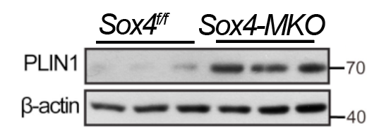
Results Summary This antibody shows excellent consistency and reproducibility across batches, ensuring reliable and stable experimental results. Its performance remains consistent across different experiments, providing strong technical support for our research.

The antibody is highly efficient and specific, showing a clear target band with no nonspecific bands.

Excellent, submitted by on
SKU M00220-1
Application Western Blot
Sample Mouse lung cancer tissue
Sample Processing Description Tissue samples were directly lysed in RIPA buffer, mixed with loading buffer at the appropriate ratio, and denatured by heating at 98 °C. Load 20 µL of protein sample per lane onto SDS-PAGE.
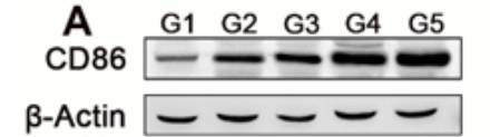
Primary Incubation The membrane was incubated with the CD86 primary antibodies (1:2000) overnight at 4 °C.
Secondary Antibody HRP-conjugated Goat Anti-Rabbit IgG Secondary Antibody
Secondary Incubation Incubate at room temperature for 1 hour
Other Reagents used 5% non-fat milk
Detection Signal was developed using ECL substrate on an Tanon system.
Results Summary The antibody is highly efficient and specific, showing a clear target band with no nonspecific bands. I will definitely continue using BosterBio products and will recommend them to my classmates and colleagues.

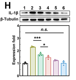
IL-6 and IL-1β levels in the pancreas are key for assessing the anti-inflammatory effect of this nanomedicine. Antibodies from two previous suppliers showed poor specificity, while BosterBio antibodies proved highly specific, potent, and cost-effective.

Excellent, submitted by on
SKU M00101-1
Application Western Blot
Sample Mouse pancreatic acinar cells
Sample Processing Description After centrifugation, collect the supernatant. Take a small portion for protein quantification using the BCA assay. Mix the remaining protein solution with an equal volume of loading buffer and denature in a 100°C water bath for 5 mins.
Primary Incubation The membrane was incubated with the IL-6 and IL-1β primary antibodies (1:1000) overnight at 4 °C.
Secondary Antibody HRP-conjugated Goat Anti-Rabbit IgG Secondary Antibody
Secondary Incubation Incubate at room temperature for 1 hour
Other Reagents used Protein-Free Blocking Buffer
Detection Signal was developed using ECL substrate on a ChemiDoc MP system.
Results Summary IL-6 and IL-1β levels in the pancreas are key for assessing the anti-inflammatory effect of this nanomedicine. Antibodies from two previous suppliers showed poor specificity, while BosterBio antibodies proved highly specific, potent, and cost-effective. In our subsequent experiments, our group continued to use BosterBio products, which proved to be highly reliable and provided solid support for the publication of several articles.

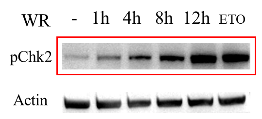
Works very well in Western blot for detecting pChk2 in human HeLa cells.

Excellent, submitted by on
SKU P00277-1
Application Western Blot
Sample Hela cell
Sample Processing Description After 12 h of viral infection, cells were lysed in RIPA buffer with PMSF and heated in 1× protein loading buffer for 10 min to denature proteins.
Primary Incubation The membrane was incubated with the CHEK2 (Phospho-T68) primary antibody (1:2000) overnight at 3 hours.
Secondary Antibody HRP-conjugated Goat Anti-Rabbit IgG Secondary Antibody
Secondary Incubation Incubate at room temperature for 1 hour
Other Reagents used None
Detection Signal was developed using ECL substrate on an ChemiDoc MP system.
Results Summary Works very well in Western blot for detecting pChk2 in human HeLa cells.

Excellent CD14 ELISA Kit From BosterBio for Barrier Dysfunction

Excellent, submitted by on

Source: Biocompare.com

SKU EK0695
Application ELISA
Starting Material Serum
Tips Follow the incubation times closely
Results Summary Absorbance was measured at 450 nm with an ELISA Reader. The CD14 concentration determined the gut permeability in mice treated with DSS.

"CD14 (sCD14), released by macrophages on stimulation with endotoxin, has been used as a marker of gut hyperpermeability. In our study with DSS, we measured the gut permeability to evaluate barrier dysfunction."

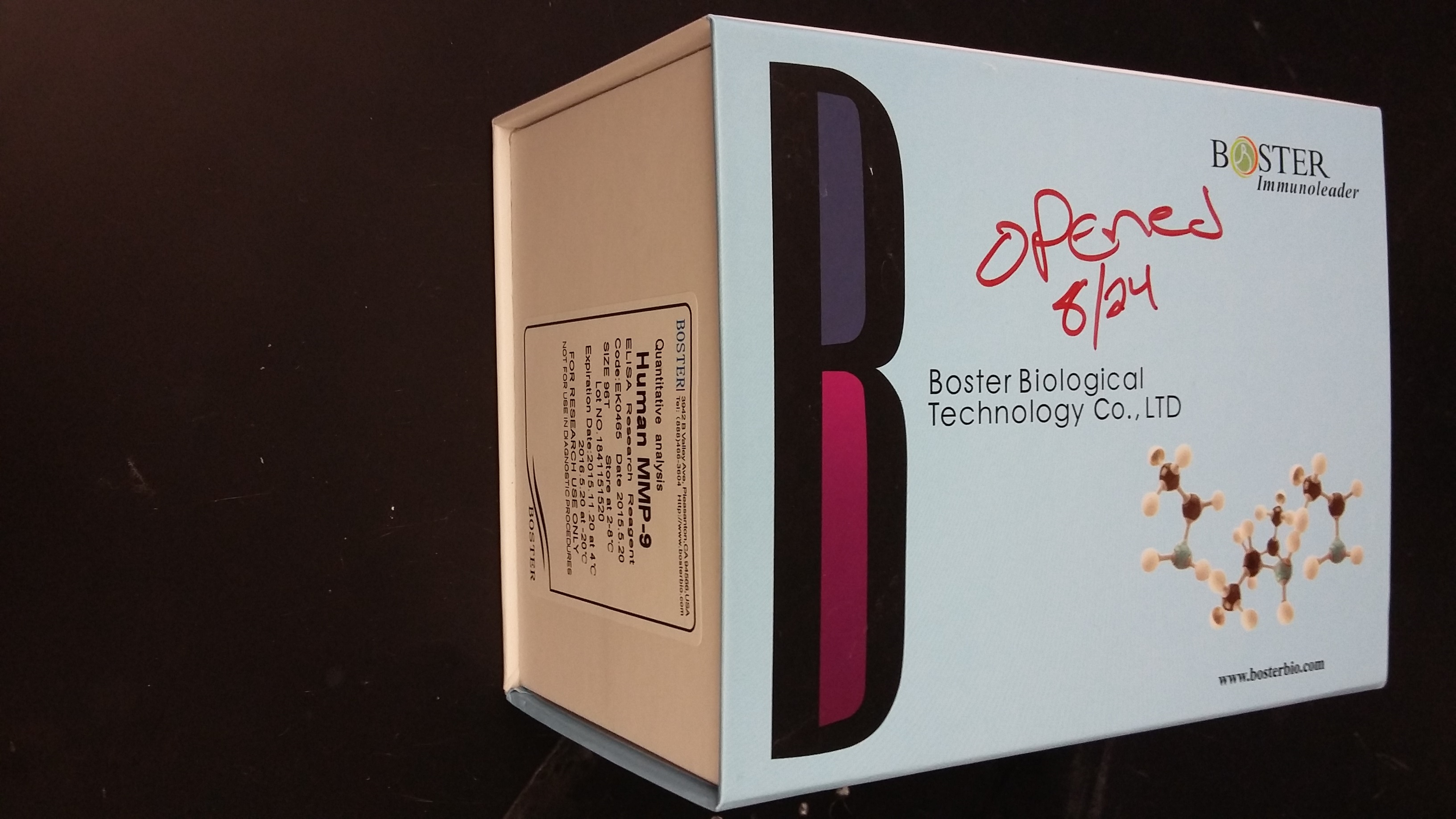
Successful MMP-9 measurements

Excellent, submitted by on

Source: Biocompare.com

SKU EK0465
Application Test the quantity of MMP-9 released from neutrophils after challenge with bacteria
Starting Material Human Neutrophils
Protocol Overview 1. collect supernatant from bacteria-challenged neutrophils2. Run samples on MMP-9 ELISA kit3. Read results on plate reader
Tips Frozen supernatants work just as well as fresh ones
Results Summary I highly recommend the Boster line of ELISA kits because of their cost and their effectiveness. I had one last round of experiments to run for a publication and we regrettably decided to purchase a lower cost kit from another company. I ran the same samples on two different plates from that company and even my controls were all over the place, resulting in a waste of time and money. This is when we purchased the Boster kit, and by only using one plate, I was able to see the real results of my experiment. My negative and positive controls had the necessary jump and my samples followed the expected trend.

"The instructions are clear, concise, and the process is more streamlined than other kits. I highly recommend investing in these kits compared to other, less expensive ones."