Customer Testimonials

Peer Reviews from Real Labs

Browse peer-submitted results linked to specific products—ratings, images, and practical notes included. Contribute your own result to strengthen the evidence base and claim rewards after verification.

Video Interviews

See real workflows and decision points—what researchers tried, what worked, and why. Share your story in a short interview and claim rewards!

Get Interview Rewards

Filter by category, reactivity, and host to find the most relevant peer results.

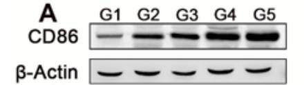
The antibody is highly efficient and specific, showing a clear target band with no nonspecific bands.

Excellent, submitted by on
SKU M00220-1
Application Western Blot
Sample Mouse lung cancer tissue
Sample Processing Description Tissue samples were directly lysed in RIPA buffer, mixed with loading buffer at the appropriate ratio, and denatured by heating at 98 °C. Load 20 µL of protein sample per lane onto SDS-PAGE.
Primary Incubation The membrane was incubated with the CD86 primary antibodies (1:2000) overnight at 4 °C.
Secondary Antibody HRP-conjugated Goat Anti-Rabbit IgG Secondary Antibody
Secondary Incubation Incubate at room temperature for 1 hour
Other Reagents used 5% non-fat milk
Detection Signal was developed using ECL substrate on an Tanon system.
Results Summary The antibody is highly efficient and specific, showing a clear target band with no nonspecific bands. I will definitely continue using BosterBio products and will recommend them to my classmates and colleagues.

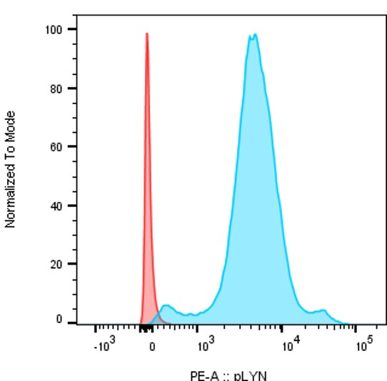
Anti-phospho Lyn (Y396) Rabbit Monoclonal Antibody

Excellent, submitted by on

Source: Biocompare.com

SKU P01424-1
Application Flow Cytometry
Sample B cells
Primary Incubation 0.25 µl/10^6 cells 50min RT
Blocking Agent FBS 5%
Secondary Incubation F(ab')2-Donkey anti-Rabbit IgG (H+L), PE 0.1 µl/10^6
Detection Flow Cytometry
Results Summary Staining of B cells from mice bone marrow after cytoplasmic permezbiliation. The cells were first fixed and permeabilized with intracellular fix and perm set from ebioscience and then stained with 0,25 µl/ million cells with pLYN antibody (P01424-1) during 50min After, a seconde staining was performed with 0,1µl/million cells of F(ab')2-Donkey anti-Rabbit IgG (H+L), PE, Secondary Antibody from invitrogen. In red secondary antibody only and in blue primary anti-phospho lyn + secondary antibody.