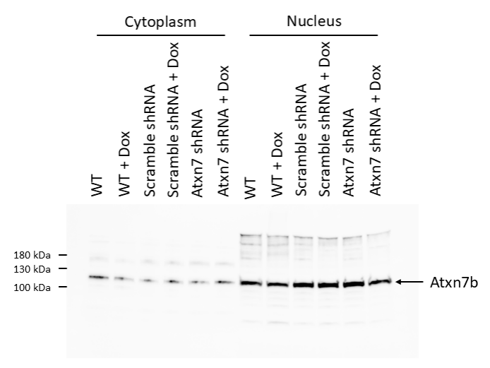
Customer Testimonials

Peer Reviews from Real Labs

Browse peer-submitted results linked to specific products—ratings, images, and practical notes included. Contribute your own result to strengthen the evidence base and claim rewards after verification.

Video Interviews

See real workflows and decision points—what researchers tried, what worked, and why. Share your story in a short interview and claim rewards!

Get Interview Rewards

Filter by category, reactivity, and host to find the most relevant peer results.

An shRNA targeting Atxn7 displayed a decrease in Atxn7 abondance showing specificity of the antibody.

Excellent, submitted by on
SKU DZ41648
Application Western Blot
Sample 3T3 Cell lysate
Sample Processing Description Nuclear and Cytoplasmic Extraction was performed on 3T3 cells with NE-PER kit by following the protocol and by adding protease and phosphatase inhibitors. Total protein was quantified with Qubit Protein Assay. Lysates were mixed with 4× Laemmli Sample Buffer and 2-mercaptoethanol, and then heated at 95°C for 5 min.
Primary Antibody Anti-Mouse Atxn7 Antibody
Primary Incubation Incubated in non-fat dry milk in TBST overnight at 4°C with antibody dilution at 1:1000 dilution
Secondary Antibody Goat anti-Rabbit IgG (H+L) Secondary Antibody, HRP
Secondary Incubation 1:4000 dilution in milk for 1 hour at room temperature
Other Reagents used SuperSignal West Pico PLUS Chemiluminescent Substrate
Detection Biorad ChemiDoc
Results Summary I was looking to target Atxn7 isoform 2, named as Atxn7b in the literature, and referenced as ENSMUST00000223714.2 on Ensembl. (It differs only on the C-terminus compared to Atxn7a, isoform 1). Atxn7 is mainly in the nucleus and is between 100-130 kDa. An shRNA targeting Atxn7 displayed a decrease in Atxn7 abondance showing specificity of the antibody.

This antibody is highly specific and efficient, suitable for detecting STAT3 protein in rat colon by Western blot, with clear results.

Excellent, submitted by on
SKU M00007
Application Western Blot
Sample rat colon tissue
Sample Processing Description RIPA lysis buffer with protease inhibitor PMSF (100:1) was used to lyse the sample for 10 minutes, followed by centrifugation at 12,000 rpm for 15 minutes. The supernatant was mixed with 5× loading buffer, denatured at 100°C for 10 minutes, and then loaded onto SDS-PAGE.
Other Reagents Blocking buffer
Primary Antibody Phospho-STAT3 (Y705) Rabbit Monoclonal Antibody
Primary Incubation 1:1000, overnight at 4 ℃
Secondary Antibody HRP Conjugated AffiniPure Goat Anti-Rabbit IgG (H+L)
Secondary Incubation 1:5000, 1 hour in room temperature
Detection Substrate: ECL, Imaging system:ChemiDoc MP
Results Summary The figure shows Western blot results of the target protein STAT3 and the loading control Actin in the colon of normal rats, model rats, low/medium/high dose Chinese medicine treatment groups, and Western medicine treatment group. No differences were observed between the groups. The target bands are clear and distinct, and the experimental results are satisfactory.

This antibody is highly efficient and specific, suitable for Western blot detection of STAT3 (Phospho-Y705) protein in rat colon tissue, with only minor nonspecific bands observed.

Excellent, submitted by on
SKU P00007-2
Application Western Blot
Sample rat colon tissue
Sample Processing Description RIPA lysis buffer with protease inhibitor PMSF (100:1) was used to lyse the sample for 10 minutes, followed by centrifugation at 12,000 rpm for 15 minutes. The supernatant was mixed with 5× loading buffer, denatured at 100°C for 10 minutes, and then loaded onto SDS-PAGE.
Other Reagents Blocking buffer
Primary Antibody Phospho-STAT3 (Y705) Rabbit Monoclonal Antibody
Primary Incubation 1:1000, overnight at 4 ℃
Secondary Antibody HRP Conjugated AffiniPure Goat Anti-Rabbit IgG (H+L)
Secondary Incubation 1:5000, 1 hour in room temperature
Detection Substrate: ECL, Imaging system:ChemiDoc MP
Results Summary The figure shows representative Western blot results of the target protein STAT3 (Phospho-Y705) and the internal control Actin in rat colon tissue from the normal control group, disease model group, low-, medium-, and high-dose traditional Chinese medicine–treated groups, and the western medicine–treated group. The target bands are clear and well defined, and the experimental results are satisfactory.

This antibody is highly efficient and specific, suitable for Western blot detection of SLC6A4 protein in rat colon tissue, with only slight nonspecific bands observed.

Excellent, submitted by on
SKU PB9438
Application Western Blot
Sample rat colon tissue
Sample Processing Description RIPA lysis buffer with protease inhibitor PMSF (100:1) was used to lyse the sample for 10 minutes, followed by centrifugation at 12,000 rpm for 15 minutes. The supernatant was mixed with 5× loading buffer, denatured at 100°C for 10 minutes, and then loaded onto SDS-PAGE.
Other Reagents Blocking buffer
Primary Antibody Serotonin transporter/SLC6A4 Antibody Picoband®
Primary Incubation 1:1000, overnight at 4 ℃
Secondary Antibody HRP Conjugated AffiniPure Goat Anti-Rabbit IgG (H+L)
Secondary Incubation 1:5000, 1 hour in room temperature
Detection Substrate: ECL, Imaging system:ChemiDoc MP
Results Summary The figure shows representative Western blot results of the target protein SLC6A4 and the internal control Actin in rat colon tissue from the normal control group, disease model group, low-, medium-, and high-dose traditional Chinese medicine–treated groups, and the western medicine–treated group. The target bands are clear and well defined, and the experimental results are satisfactory.

The antibody is highly specific and efficient, suitable for detecting ROCK2 protein in rat colon by Western blot, with only minimal non-specific bands.

Excellent, submitted by on
SKU PB9387
Application Western Blot
Sample rat colon tissue
Sample Processing Description RIPA lysis buffer with protease inhibitor PMSF (100:1) was used to lyse the sample for 10 minutes, followed by centrifugation at 12,000 rpm for 15 minutes. The supernatant was mixed with 5× loading buffer, denatured at 100°C for 10 minutes, and then loaded onto SDS-PAGE.
Other Reagents Blocking buffer
Primary Antibody ROCK2 Antibody Picoband®
Primary Incubation 1:1000, overnight at 4 ℃
Secondary Antibody HRP Conjugated AffiniPure Goat Anti-Rabbit IgG (H+L)
Secondary Incubation 1:5000, 1 hour in room temperature
Detection Substrate: ECL, Imaging system:ChemiDoc MP
Results Summary The figure shows the Western blot results of the target protein ROCK2 and the internal control Actin in rat colon across the following groups: normal, disease model, low/middle/high dose traditional Chinese medicine treatment, and Western medicine treatment. Expression was increased in the model group. Among the traditional Chinese medicine treatments, the high-dose group showed the best effect. The target bands are clear and distinct, and the experimental results are satisfactory.

This antibody is highly specific and efficient, suitable for detecting RHOA/B/C protein in rat colon by Western blot, with only minimal non-specific bands.

Excellent, submitted by on
SKU M00207-1
Application Western Blot
Sample rat colon tissue
Sample Processing Description RIPA lysis buffer with protease inhibitor PMSF (100:1) was used to lyse the sample for 10 minutes, followed by centrifugation at 12,000 rpm for 15 minutes. The supernatant was mixed with 5× loading buffer, denatured at 100°C for 10 minutes, and then loaded onto SDS-PAGE.
Other Reagents Blocking buffer
Primary Antibody Rho A + B + C Rabbit Monoclonal Antibody
Primary Incubation 1:1000, overnight at 4 ℃
Secondary Antibody HRP Conjugated AffiniPure Goat Anti-Rabbit IgG (H+L)
Secondary Incubation 1:5000, 1 hour in room temperature
Detection Substrate: ECL, Imaging system:ChemiDoc MP
Results Summary The figure shows the Western blot results of the target protein RHOA/B/C and the internal control Actin in rat colon tissue across the following groups: normal, disease model, low/middle/high dose traditional Chinese medicine treatment, and Western medicine treatment. The target bands are clear and distinct, and the experimental results are satisfactory.

This antibody is highly efficient and specific, suitable for Western blot detection of PTEN protein in rat colon tissue, with only minor nonspecific bands.

Excellent, submitted by on
SKU M00006-2
Application Western Blot
Sample rat colon tissue
Sample Processing Description RIPA lysis buffer with protease inhibitor PMSF (100:1) was used to lyse the sample for 10 minutes, followed by centrifugation at 12,000 rpm for 15 minutes. The supernatant was mixed with 5× loading buffer, denatured at 100°C for 10 minutes, and then loaded onto SDS-PAGE.
Other Reagents Blocking buffer
Primary Antibody PTEN Rabbit Monoclonal Antibody
Primary Incubation 1:1000, overnight at 4 ℃
Secondary Antibody HRP Conjugated AffiniPure Goat Anti-Rabbit IgG (H+L)
Secondary Incubation 1:5000, 1 hour in room temperature
Detection Substrate: ECL, Imaging system:ChemiDoc MP
Results Summary The figure shows a schematic of Western blot results for the target protein PTEN and the internal control Actin in rat colon across different groups. Expression was increased in the model group. Among the low, medium, and high doses of the herbal treatment, the low-dose group showed the best effect. The target bands are clear, and the experimental results are satisfactory.

This antibody is highly efficient and specific, suitable for Western blot detection of MUC6 protein in rat colon tissue, with only minor nonspecific bands.

Excellent, submitted by on
SKU A02143
Application Western Blot
Sample rat colon tissue
Sample Processing Description RIPA lysis buffer with protease inhibitor PMSF (100:1) was used to lyse the sample for 10 minutes, followed by centrifugation at 12,000 rpm for 15 minutes. The supernatant was mixed with 5× loading buffer, denatured at 100°C for 10 minutes, and then loaded onto SDS-PAGE.
Other Reagents Blocking buffer
Primary Antibody Gastric Mucin/MUC6 Antibody Picoband®
Primary Incubation 1:1000, overnight at 4 ℃
Secondary Antibody HRP Conjugated AffiniPure Goat Anti-Rabbit IgG (H+L)
Secondary Incubation 1:5000, 1 hour in room temperature
Detection Substrate: ECL, Imaging system:ChemiDoc MP
Results Summary The figure shows the Western blot results of the target protein MUC6 and the internal control Actin in rat colon tissue across different groups. The target bands are clear and well defined, and the experimental results are satisfactory.

This antibody is highly efficient and specific, suitable for Western blot detection of JAK2 protein in rat colon tissue, with only minor nonspecific bands.

Excellent, submitted by on
SKU M00027
Application Western Blot
Sample rat colon tissue
Sample Processing Description RIPA lysis buffer with protease inhibitor PMSF (100:1) was used to lyse the sample for 10 minutes, followed by centrifugation at 12,000 rpm for 15 minutes. The supernatant was mixed with 5× loading buffer, denatured at 100°C for 10 minutes, and then loaded onto SDS-PAGE.
Other Reagents Blocking buffer
Primary Antibody JAK2 Monoclonal Antibody
Primary Incubation 1:1000, overnight at 4 ℃
Secondary Antibody HRP Conjugated AffiniPure Goat Anti-Rabbit IgG (H+L)
Secondary Incubation 1:5000, 1 hour in room temperature
Detection Substrate: ECL, Imaging system:ChemiDoc MP
Results Summary The figure shows the Western blot results of the target protein JAK2 and the internal control Actin in rat colon tissue across the following groups: normal, disease model, low/middle/high dose traditional Chinese medicine treatment, and Western medicine treatment. The target bands are clear and well defined, and the experimental results are satisfactory.

This antibody is highly efficient and specific, suitable for Western blot detection of CD44 protein in rat colon tissue, with only minor nonspecific bands.

Excellent, submitted by on
SKU A00052
Application Western Blot
Sample rat colon tissue
Sample Processing Description RIPA lysis buffer with protease inhibitor PMSF (100:1) was used to lyse the sample for 10 minutes, followed by centrifugation at 12,000 rpm for 15 minutes. The supernatant was mixed with 5× loading buffer, denatured at 100°C for 10 minutes, and then loaded onto SDS-PAGE.
Other Reagents Blocking buffer
Primary Antibody Cyclin A2/CCNA2 Antibody Picoband®
Primary Incubation 1:1000, overnight at 4 ℃
Secondary Antibody HRP Conjugated AffiniPure Goat Anti-Rabbit IgG (H+L)
Secondary Incubation 1 hour in room temperature
Detection Substrate: ECL, Imaging system:ChemiDoc MP
Results Summary The figure shows a schematic of Western blot results for the target protein CCNA2 and the internal control Actin in rat colon across different groups. Expression was increased in the model group. Among the low, medium, and high doses of the herbal treatment, the low-dose group showed the best effect. The target bands are clear, and the experimental results are satisfactory.