| SKU | M00101-1 |
|---|---|
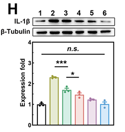
| Application | Western Blot |
| Sample | Mouse pancreatic acinar cells |
| Sample Processing Description | After centrifugation, collect the supernatant. Take a small portion for protein quantification using the BCA assay. Mix the remaining protein solution with an equal volume of loading buffer and denature in a 100°C water bath for 5 mins. |
| Primary Incubation | The membrane was incubated with the IL-6 and IL-1β primary antibodies (1:1000) overnight at 4 °C. |
| Secondary Antibody | HRP-conjugated Goat Anti-Rabbit IgG Secondary Antibody |
| Secondary Incubation | Incubate at room temperature for 1 hour |
| Other Reagents used | Protein-Free Blocking Buffer |
| Detection | Signal was developed using ECL substrate on a ChemiDoc MP system. |
| Results Summary | IL-6 and IL-1β levels in the pancreas are key for assessing the anti-inflammatory effect of this nanomedicine. Antibodies from two previous suppliers showed poor specificity, while BosterBio antibodies proved highly specific, potent, and cost-effective. In our subsequent experiments, our group continued to use BosterBio products, which proved to be highly reliable and provided solid support for the publication of several articles. |