| SKU | DZ41317 |
|---|---|
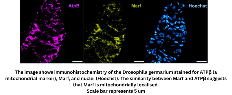
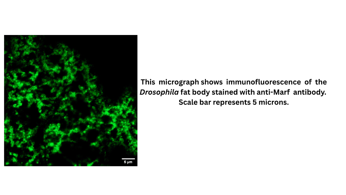
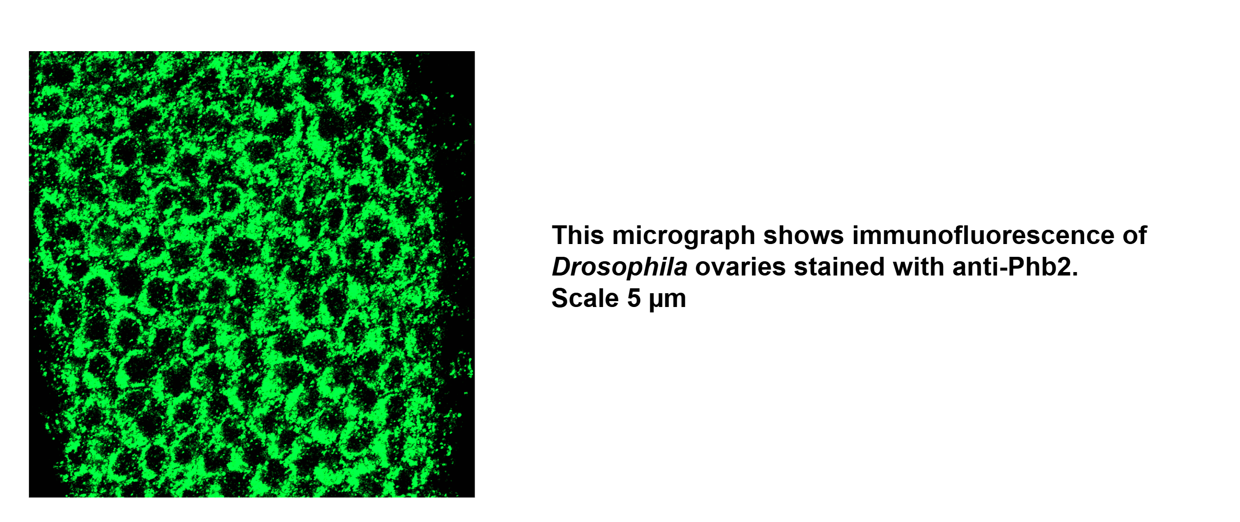
| Application | Immunofluorescence |
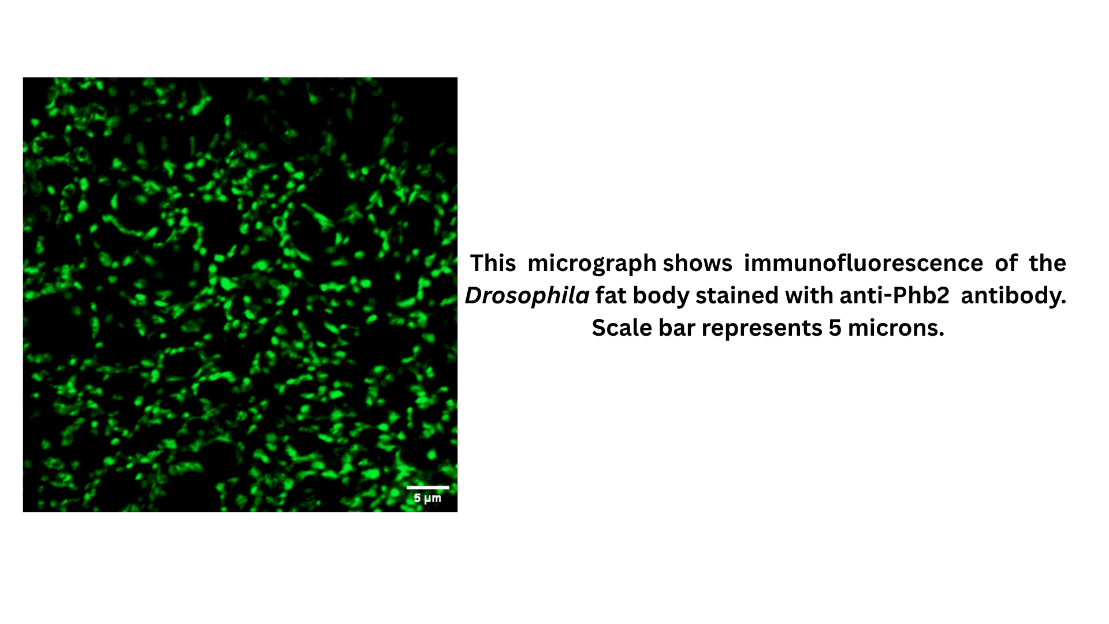
| Sample | Drosophila Fat body and Ovaries |
| Sample Processing Description | Dissection, Fixation in 4%PFA, Permeabilization in PBS-TX, Blocking, Primary, Washes,Secondary, Washes,Nuclei Staining, Slide Preparation |
| Primary Antibody | Anti-Fruit fly Phb2 Antibody |
| Primary Incubation | 1:200 in 2%BSA and 0.5% PBS-TX for Ovaries, 1:100 in 2%BSA and 0.1% PBS-TX for Fat body |
| Secondary Antibody | 1:1000 Anti-Rabbit/Mouse Alexa Fluor 488/Cy3/Cy5 |
| Secondary Incubation | 1 hour at room temperature |
| Detection | Zeiss LSM 900 Confocal Microscope with Airyscan 2 |
| Results Summary | Drosophila fatbody showed good staining of Phb2. |