| SKU | M00024-1 |
|---|---|
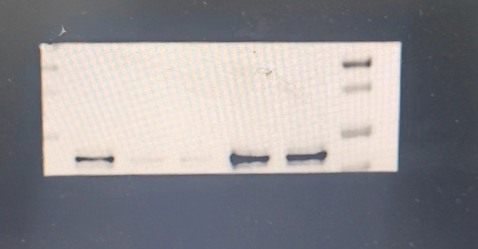
| Application | Western Blot |
| Sample | Mouse hippocampus tissue |
| Sample Processing Description | The mouse hippocampus was lysed with RIPA buffer containing a protease inhibitor cocktail. After protein quantification, samples were mixed with 5× protein loading buffer and heated for 10 minutes to denature. Load 5 μL of protein per lane and apply to SDS-PAGE. |
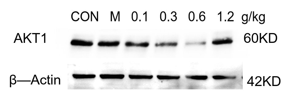
| Primary Antibody | Anti-AKT1 Monoclonal Antibody |
| Primary Incubation | overnight at 4 ℃ |
| Secondary Antibody | HRP-conjugated Anti-Rabbit IgG Secondary Antibody |
| Secondary Incubation | 1 hour in room temperature |
| Detection | Substrate: Ultra-sensitive ECL luminescent reagent (Cat# AR1191), Imaging system: ChemiDoc MP (Bio-Rad) |
| Results Summary | Western blot analysis of AKT1 protein in the mouse hippocampus using the AKT1 antibody showed clear bands. This antibody is suitable for mouse hippocampal tissue samples. |