| SKU | M04247 |
|---|---|
| Application | Western Blot |
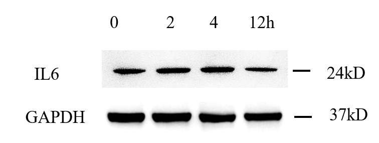
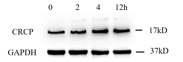
| Sample | Human mcf-7 cells |
| Sample Processing Description | RIPA lysis buffer supplemented with a protease inhibitor cocktail was added to the cell culture dish, and the cells were lysed on ice for 30 min. The lysates were then centrifuged, and the supernatants were collected. Protein concentration was determined, and samples were adjusted to equal concentrations. Subsequently, 5× protein loading buffer was added, and the samples were denatured at 100 °C for 10 min. A total of 15 μL of protein sample was loaded per lane for SDS-PAGE electrophoresis. |
| Other Reagents | Blocking buffer |
| Primary Antibody | CRCP Rabbit Monoclonal Antibody |
| Primary Incubation | 1:500, overnight at 4 ℃ |
| Secondary Antibody | HRP Conjugated AffiniPure Goat Anti-Rabbit IgG (H+L) |
| Secondary Incubation | 1:2000, 1 hour in room temperature |
| Detection | Substrate: ECL, Imaging system:ChemiDoc MP |
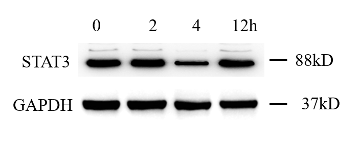
| Results Summary | As shown in the figure, the expression levels of CRCP protein in MCF-7 cells at different drug treatment time points are presented. The Western blot results obtained with this antibody are clear; although there are a few faint nonspecific bands above, they do not affect result interpretation. It can be observed that the expression of the target protein in MCF-7 cells increases with longer drug treatment times. |