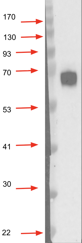
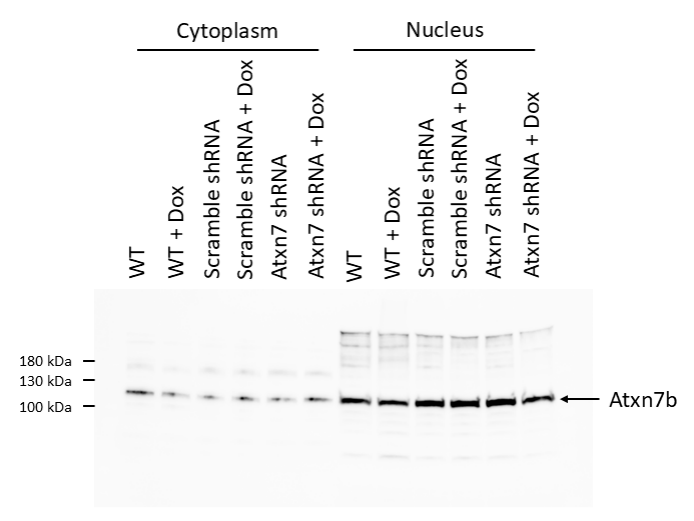
Customer Testimonials

Peer Reviews from Real Labs

Browse peer-submitted results linked to specific products—ratings, images, and practical notes included. Contribute your own result to strengthen the evidence base and claim rewards after verification.

Video Interviews

See real workflows and decision points—what researchers tried, what worked, and why. Share your story in a short interview and claim rewards!

Get Interview Rewards

Filter by category, reactivity, and host to find the most relevant peer results.

WB analysis using Anti-GH antibody (A00851-2) in mouse hippocampal tissue revealed a specific band at the expected molecular weight with negligible non-specific signals.

Excellent, submitted by on
SKU A00851-2
Application Western Blot
Sample mouse hippocampal tissue
Sample Processing Description Total protein was extracted from the left hippocampus of normal mouse brain.
Other Reagents RIPA lysis buffer, Protease inhibitor, Electrophoresis buffer, Transfer buffer, Blocking buffer
Primary Antibody GH1 Antibody Picoband®
Primary Incubation 1:4000, overnight at 4 ℃
Secondary Antibody HRP-conjugated goat anti-rabbit IgG
Secondary Incubation 1:10000, 1h in RT
Detection Substrate: ECL substrate; Image system: ChemiDoc MP
Results Summary Growth hormone (GH) serves as a central integrative signal that coordinates growth, metabolism, and tissue repair in response to changes in nutritional status. It is not only the primary driver of linear growth during puberty but also plays a critical role throughout life in maintaining muscle mass, bone strength, and metabolic flexibility. In this study, hippocampal tissues from two normal mouse brains were used to evaluate the performance of the GH antibody. The results showed a band at the expected position with good specificity, indicating that the antibody performs well in WB applications.

The ELAVL1 antibody (A00736-1) produced a clear specific band at the expected size in WB using rat colon and colon cancer tissue samples, consistent with the expected expression pattern.

Excellent, submitted by on
SKU A00736-1
Application Western Blot
Sample HepG2 subcutaneous xenograft in nude mice
Sample Processing Description ① normal rat colon tissue; ② rat colon cancer model tissue; total protein extracted.
Other Reagents RIPA lysis buffer, Protease inhibitor, Electrophoresis buffer, Transfer buffer, Blocking buffer
Primary Antibody HuR/ELAVL1 Antibody Picoband®
Primary Incubation 1:2000, overnight at 4 ℃
Secondary Antibody HRP-conjugated goat anti-rabbit IgG
Secondary Incubation 1:10000, 30 min in 37℃
Detection Substrate: ECL substrate; Image system: ChemiDoc MP
Results Summary HUR protein is an RNA-binding protein that plays multiple key roles in gene expression regulation. It is essential in maintaining cellular homeostasis, stress response, inflammation, and the development and progression of diseases, especially cancer. In normal tissues, it is expressed at moderate levels, maintaining a delicate balance of cell proliferation, stress response, inflammation, and neural functions, while in tumors it is highly overexpressed, driving malignant progression. The experimental results are consistent with these observations.

WB results showed that Cingulin/CGN (A02373-1) was downregulated in the rat colon model group and restored after Chinese medicine treatment, with the high-dose group showing the best effect, and the target bands were clear and specific.

Excellent, submitted by on
SKU A02373-1
Application Western Blot
Sample rat colon tissue
Sample Processing Description Rat colon tissues were lysed in RIPA buffer containing PMSF (100:1) for 10 min, centrifuged at 12,000 rpm for 15 min, and the supernatant was mixed with 5× loading buffer, denatured at 100°C for 10 min, and then loaded onto SDS-PAGE.
Other Reagents5% non-fat milk
Primary Antibody Cingulin/CGN Antibody
Primary Incubation 1:1000, overnight at 4 ℃
Secondary Antibody HRP Conjugated AffiniPure Goat Anti-Rabbit IgG (H+L) (BA1054)
Secondary Incubation 1:5000, 1 h in RT
Detection Substrate: ECL substrate, Image system:ChemiDoc MP
Results Summary The figure shows WB results of Cingulin/CGN and the internal control Actin in rat colon across different groups; expression was decreased in the model group and restored after Chinese medicine treatment, with the high-dose group showing the best effect, and the target bands were clear and specific, indicating satisfactory results.

In the IF experiment using Anti-IBA-1 antibody (Cat# M01394-4), the antibody clearly and specifically labeled microglial cells in mouse cerebral infarction tissue, showing distinct staining and well-defined cellular morphology.

Excellent, submitted by on
SKU M01394-4
Application Immunofluorescence
Sample mouse brain tissue
Sample Processing Description Mouse cerebral infarction model; brain tissues were collected, fixed in formaldehyde for 48 hours, and then sagittally paraffin-embedded.
Other ReagentsGoat serum, DAPI Staining Solution, Antifade fluorescence mounting medium.
Primary Antibody Iba1 Rabbit Monoclonal Antibody
Primary Incubation 1:100, overnight at 4 ℃
Secondary Antibody Goat Anti-Rabbit IgG (H+L) Secondary Antibody, Fluoro594 Conjugated (BA1142, Boster)
Secondary Incubation 45 min at 37℃
Detection Imaging system:Leica DMi3000
Results Summary IBA-1 is a well-established marker of microglia in the nervous system. Microglia are the primary immune effector cells in the central nervous system and respond rapidly to CNS injury by proliferating, upregulating or re-expressing MHC antigens, migrating, and adopting a phagocyte-like morphology. In this study, IBA-1 was used to label microglia in cerebral infarction samples, showing clear staining and accurate cellular morphology.

One major band was detected at the expected size (84 kDa), while multiple minor bands were detected in the egg lysate under the condition tested.

Excellent, submitted by on
SKU DZ41739
Application Western Blot
Sample egg lysate
Sample Processing Description Egg lysate was prepared and loaded onto a 10% polyacrylamide gel for electrophoresis.
Primary Antibody Anti-Purple sea urchin LOC581356 Antibody
Primary Incubation 1:2000, incubated overnight at 4°C.
Secondary Antibody Goat anti-rabbit IgG-HRP
Secondary Incubation 2 hour at room temperature.
Detection Chemiluminescent detection.
Results Summary One major band was detected at the expected size (84 kDa), while multiple minor bands were detected in the egg lysate under the condition tested.

A single band was detected at the expected size of 60kDa in the gastrula stage lysate.

Excellent, submitted by on
SKU DZ41738
Application Western Blot
Sample Embryo lysate
Sample Processing Description Embryonic lysate was prepared and loaded onto a 10% polyacrylamide gel for electrophoresis.
Primary Antibody Anti-Purple sea urchin LOC584590 Antibody
Primary Incubation 1:2000, incubated overnight at 4°C.
Secondary Antibody Goat anti-rabbit IgG-HRP
Secondary Incubation 2 hour at room temperature.
Detection Chemiluminescent detection.
Results Summary A single band was detected at the expected size of 60kDa in the gastrula stage lysate.

An shRNA targeting Atxn7 displayed a decrease in Atxn7 abondance showing specificity of the antibody.

Excellent, submitted by on
SKU DZ41648
Application Western Blot
Sample 3T3 Cell lysate
Sample Processing Description Nuclear and Cytoplasmic Extraction was performed on 3T3 cells with NE-PER kit by following the protocol and by adding protease and phosphatase inhibitors. Total protein was quantified with Qubit Protein Assay. Lysates were mixed with 4× Laemmli Sample Buffer and 2-mercaptoethanol, and then heated at 95°C for 5 min.
Primary Antibody Anti-Mouse Atxn7 Antibody
Primary Incubation Incubated in non-fat dry milk in TBST overnight at 4°C with antibody dilution at 1:1000 dilution
Secondary Antibody Goat anti-Rabbit IgG (H+L) Secondary Antibody, HRP
Secondary Incubation 1:4000 dilution in milk for 1 hour at room temperature
Other Reagents used SuperSignal West Pico PLUS Chemiluminescent Substrate
Detection Biorad ChemiDoc
Results Summary I was looking to target Atxn7 isoform 2, named as Atxn7b in the literature, and referenced as ENSMUST00000223714.2 on Ensembl. (It differs only on the C-terminus compared to Atxn7a, isoform 1). Atxn7 is mainly in the nucleus and is between 100-130 kDa. An shRNA targeting Atxn7 displayed a decrease in Atxn7 abondance showing specificity of the antibody.

Western blot analysis of Drosophila ovaries from three fly strains showed comparable Phb2 protein levels across all samples. Consistent bands at 30 kDa confirmed expected molecular weight, with β-Actin used as a loading control to verify equal protein loa

Excellent, submitted by on
SKU DZ41317
Application Western Blot
Sample Drosophila Ovaries
Sample Processing Description Dissection, Homogenization, Sample boiling in 1X Lamelli, SDS-PAGE, Standard Western Blotting
Primary Antibody Anti-Fruit fly Phb2 Antibody
Primary Incubation 1:1000 in 5% BSA and 0.1% PBST. Either 2 hours at RT or Overnight at 4 Degrees
Secondary Antibody 1:10000 Anti-rabbit/mouse IgG horseradish peroxidase-conjugated
Secondary Incubation 1 hour at room temperature
Detection Biorad ChemiDoc
Results Summary Western blot analysis of Drosophila ovaries from three fly strains showed comparable Phb2 protein levels across all samples. Consistent bands at 30 kDa confirmed expected molecular weight, with β-Actin used as a loading control to verify equal protein loading.

Drosophila fatbody showed good staining of Phb2.

Excellent, submitted by on
SKU DZ41317
Application Immunofluorescence
Sample Drosophila Fat body and Ovaries
Sample Processing Description Dissection, Fixation in 4%PFA, Permeabilization in PBS-TX, Blocking, Primary, Washes,Secondary, Washes,Nuclei Staining, Slide Preparation
Primary Antibody Anti-Fruit fly Phb2 Antibody
Primary Incubation 1:200 in 2%BSA and 0.5% PBS-TX for Ovaries, 1:100 in 2%BSA and 0.1% PBS-TX for Fat body
Secondary Antibody 1:1000 Anti-Rabbit/Mouse Alexa Fluor 488/Cy3/Cy5
Secondary Incubation 1 hour at room temperature
Detection Zeiss LSM 900 Confocal Microscope with Airyscan 2
Results Summary Drosophila fatbody showed good staining of Phb2.

This was only optimization experiment and still needs validation through necessary experiments.

Excellent, submitted by on
SKU DZ41315
Application Immunofluorescence
Sample Drosophila Fat body and Ovaries
Sample Processing Description Dissection, Fixation in 4%PFA, Permeabilization in PBS-TX, Blocking, Primary, Washes,Secondary, Washes,Nuclei Staining, Slide Preparation
Primary Antibody Anti-Fruit fly Marf Antibody
Primary Incubation 1:200 in 2%BSA and 0.5% PBS-TX for Ovaries, 1:100 in 2%BSA and 0.1% PBS-TX for Fat body
Secondary Antibody 1:1000 Anti-Rabbit/Mouse Alexa Fluor 488/Cy3/Cy5
Secondary Incubation 1 hour at room temperature
Detection Zeiss LSM 900 Confocal Microscope with Airyscan 2
Results Summary This was only optimization experiment and still needs validation through necessary experiments.