Click image to see more details
-

-
-
-
-
+1
Product Info Summary
| SKU: | A01252-2 |
|---|---|
| Size: | 100 μg/vial |
| Reactive Species: | Human |
| Host: | Rabbit |
| Application: | ELISA, Flow Cytometry, IF, IHC, ICC |
Customers Who Bought This Also Bought

Product info
Product Name
Anti-MDC1 Antibody
SKU/Catalog Number
A01252-2
Size
100 μg/vial
Form
Lyophilized
Description
Boster Bio Anti-MDC1 Antibody catalog # A01252-2. Tested in ELISA, Flow Cytometry, IF, IHC, ICC applications. This antibody reacts with Human.
Storage & Handling
Store at -20˚C for one year from date of receipt. After reconstitution, at 4˚C for one month. It can also be aliquotted and stored frozen at -20˚C for six months. Avoid repeated freeze-thaw cycles.
Cite This Product
Anti-MDC1 Antibody (Boster Biological Technology, Pleasanton CA, USA, Catalog # A01252-2)
Host
Rabbit
Contents
Each vial contains 4mg Trehalose, 0.9mg NaCl, 0.2mg Na2HPO4, 0.05mg NaN3.
Clonality
Polyclonal
Isotype
Rabbit IgG
Immunogen
E. coli-derived human MDC1 recombinant protein (Position: A534-Q626).
Cross-reactivity
No cross-reactivity with other proteins.
Reactive Species
A01252-2 is reactive to MDC1 in Human
Calculated molecular weight
226.7 kDa
Background of MDC1
Mediator of DNA damage checkpoint protein 1 is a 2080 amino acid long protein that in humans is encoded by the MDC1 gene. The protein encoded by this gene contains an N-terminal forkhead domain, two BRCA1 C-terminal (BRCT) motifs and a central domain with 13 repetitions of an approximately 41-amino acid sequence. The encoded protein is required to activate the intra-S phase and G2/M phase cell cycle checkpoints in response to DNA damage. This nuclear protein interacts with phosphorylated histone H2AX near sites of DNA double-strand breaks through its BRCT motifs, and facilitates recruitment of the ATM kinase and meiotic recombination 11 protein complex to DNA damage foci.
Antibody Validation
Boster validates all antibodies on WB, IHC, ICC, Immunofluorescence, and ELISA with known positive control and negative samples to ensure specificity and high affinity, including thorough antibody incubations.
Application & Images
Applications
A01252-2 is guaranteed for ELISA, Flow Cytometry, IF, IHC, ICC Boster Guarantee
Recommend Dilution
| Application | Dilution | Species |
|---|---|---|
| Immunohistochemistry (Paraffin-embedded Section) | 0.5-1μg/ml | |
| Immunocytochemistry/Immunofluorescence | 2μg/ml | |
| Flow Cytometry (Fixed) | 1-3μg/1x106 cells | |
| ELISA | 0.1-0.5μg/ml |
Tested application
Use TE buffer pH 9.0 for antigen retrieval; (*) citrate buffer pH 6.0 is an alternative.
Validation Images & Assay Conditions
Click image to see more details
IHC analysis of MDC1 using anti-MDC1 antibody (A01252-2).
MDC1 was detected in paraffin-embedded section of human colon cancer tissue. Heat mediated antigen retrieval was performed in citrate buffer (pH6, epitope retrieval solution) for 20 mins. The tissue section was blocked with 10% goat serum. The tissue section was then incubated with 1μg/ml rabbit anti-MDC1 Antibody (A01252-2) overnight at 4°C. Biotinylated goat anti-rabbit IgG was used as secondary antibody and incubated for 30 minutes at 37°C. The tissue section was developed using Strepavidin-Biotin-Complex (SABC)(Catalog # SA1022) with DAB as the chromogen.

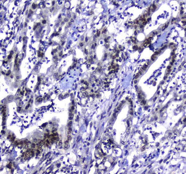
Click image to see more details
IHC analysis of MDC1 using anti-MDC1 antibody (A01252-2).
MDC1 was detected in paraffin-embedded section of human lung cancer tissue. Heat mediated antigen retrieval was performed in citrate buffer (pH6, epitope retrieval solution) for 20 mins. The tissue section was blocked with 10% goat serum. The tissue section was then incubated with 1μg/ml rabbit anti-MDC1 Antibody (A01252-2) overnight at 4°C. Biotinylated goat anti-rabbit IgG was used as secondary antibody and incubated for 30 minutes at 37°C. The tissue section was developed using Strepavidin-Biotin-Complex (SABC)(Catalog # SA1022) with DAB as the chromogen.

Click image to see more details
IHC analysis of MDC1 using anti-MDC1 antibody (A01252-2).
MDC1 was detected in paraffin-embedded section of human testis tissue. Heat mediated antigen retrieval was performed in citrate buffer (pH6, epitope retrieval solution) for 20 mins. The tissue section was blocked with 10% goat serum. The tissue section was then incubated with 1μg/ml rabbit anti-MDC1 Antibody (A01252-2) overnight at 4°C. Biotinylated goat anti-rabbit IgG was used as secondary antibody and incubated for 30 minutes at 37°C. The tissue section was developed using Strepavidin-Biotin-Complex (SABC)(Catalog # SA1022) with DAB as the chromogen.

Click image to see more details
IF analysis of MDC1 using anti-MDC1 antibody (A01252-2).
MDC1 was detected in immunocytochemical section of A431 cells. Enzyme antigen retrieval was performed using IHC enzyme antigen retrieval reagent (AR0022) for 15 mins. The cells were blocked with 10% goat serum. And then incubated with 2μg/mL rabbit anti-MDC1 Antibody (A01252-2) overnight at 4°C. DyLight®488 Conjugated Goat Anti-Rabbit IgG (BA1127) was used as secondary antibody at 1:100 dilution and incubated for 30 minutes at 37°C. The section was counterstained with DAPI. Visualize using a fluorescence microscope and filter sets appropriate for the label used.

Click image to see more details
Flow Cytometry analysis of 293T cells using anti-MDC1 antibody (A01252-2).
Overlay histogram showing 293T cells stained with A01252-2 (Blue line). To facilitate intracellular staining, cells were fixed with 4% paraformaldehyde and permeabilized with permeabilization buffer. The cells were blocked with 10% normal goat serum. And then incubated with rabbit anti-MDC1 Antibody (A01252-2,1μg/1x106 cells) for 30 min at 20°C. DyLight®488 conjugated goat anti-rabbit IgG (BA1127, 5-10μg/1x106 cells) was used as secondary antibody for 30 minutes at 20°C. Isotype control antibody (Green line) was rabbit IgG (1μg/1x106) used under the same conditions. Unlabelled sample without incubation with primary antibody and secondary antibody (Red line) was used as a blank control.
Specific Publications For Anti-MDC1 Antibody (A01252-2)
Loading publications
Recommended Resources
Here are featured tools and databases that you might find useful.
- Boster's Pathways Library
- Protein Databases
- Bioscience Research Protocol Resources
- Data Processing & Analysis Software
- Photo Editing Software
- Scientific Literature Resources
- Research Paper Management Tools
- Molecular Biology Software
- Primer Design Tools
- Bioinformatics Tools
- Phylogenetic Tree Analysis
Customer Reviews
Have you used Anti-MDC1 Antibody?
Share your experimental results or join a short interview to earn up to $1,000 in product credits or other rewards.
0 Reviews For Anti-MDC1 Antibody
Customer Q&As
Have a question?
Find answers in Q&As, reviews.
Can't find your answer?
Submit your question
1 Customer Q&As for Anti-MDC1 Antibody
Question
We are currently using anti-MDC1 antibody A01252-2 for human tissue, and we are well pleased with the IHC results. The species of reactivity given in the datasheet says human. Is it possible that the antibody can work on dog tissues as well?
Verified Customer
Verified customer
Asked: 2020-01-17
Answer
The anti-MDC1 antibody (A01252-2) has not been validated for cross reactivity specifically with dog tissues, but there is a good chance of cross reactivity. We have an innovator award program that if you test this antibody and show it works in dog you can get your next antibody for free. Please contact me if I can help you with anything.
Boster Scientific Support
Answered: 2020-01-17
