Click image to see more details
-

-
-
-
-
+4
Product Info Summary
| SKU: | BA1003 |
|---|---|
| Size: | 0.5ml |
| Reactive Species: | Rabbit |
| Host: | Goat |
| Application: | ELISA, IHC |
Product info
Product Overview
| Product Name | Goat Anti-Rabbit IgG (H+L) Secondary Antibody, Biotin Conjugate |
|---|---|
| Synonyms | Biotin-conjugated Goat Anti-Rabbit IgG; Goat Anti-Rabbit IgG Biotinylated Antibody; Biotinylated Goat Anti-Rabbit IgG Secondary Antibody; Goat Anti-Rabbit IgG Secondary Antibody, Biotin-labeled |
| Description | Goat Anti-Rabbit IgG (H+L) Secondary Antibody, Biotin Conjugate, for indirect sensitive immunodetection and/or quantification of low-abundance target proteins through ELISA or IHC by using reporter-labeled biotin-binding signal amplification systems. |
| Reagent Type | Biotin conjugated secondary antibody |
| Conjugate | Biotin |
| Host | Goat |
| Target Species | Rabbit |
| Antibody Class | IgG |
| Clonality | Polyclonal |
| Immunogen | Whole molecule rabbit IgG |
| Purification | Immunoaffinity chromatography, solid-phase adsorbed with human serum proteins |
| Specificity | Rabbit IgG specific; no cross-reactivity with human/bovine/mouse IgG |
| Form Supplied | Liquid: concentrated buffered stock solution |
| Formulation | 0.5 mg biotin-conjugated secondary antibody
0.01 M PBS (PH 7.4) 50% glycerol |
| Pack Size | 0.5 ml |
| Concentration | 1 mg/ml |
| Application | ELISA, IHC |
| Storage | At -20˚C for one year from date of receipt. Avoid repeated freezing and thawing. |
| Shipping | Ships with gel ice. |
| Precautions | FOR RESEARCH USE ONLY. NOT FOR DIAGNOSTIC OR CLINICAL USE |
Assay Information
| Sample Type | Human primary-antibody-probed: SDS-PAGE separated-, membrane-immobilized-proteins from cell/tissue lysates, formalin-fixed paraffin-embedded (FFPE) tissue sections on slides |
|---|---|
| Assay Type | Immunoanalytical |
| Technique | Indirect immunodetection of target protein via reporter-labeled biotin-binding detection systems |
| Assay Purpose | Protein detection/quantification |
| Equipment Needed | WB/Dot blot/ELISA/IHC instrumentation; Reporter signal detectors: X-ray film cassette; a charge-coupled device (CCD) imager; Spectrophotometer; fluorescent or electron microscope |
Main Advantages
| Specificity | High signal-to-noise ratio |
|---|---|
| High Signal Amplification | Multiple secondary antibodies can bind to a single primary antibody; Multiple reporter molecules localize to a single biotin via avidin/streptavidin bridges |
| Fast | Fewer processing steps - no need to add a substrate; Less optimization required compared to enzymatic detection; Generates strong signals in a relatively short time span; Fluorescence can be observed directly |
| Quantifiable | Allows quantification of detected signal |
| Easy to Use | Supplied in a workable liquid format |
| Flexible | Biotin- (Strept)Avidin system can be coupled with various types of reporters (enzymes, fluorochromes, fluorophores, chromophores, etc.); One type of labeled secondary antibody can be used to recognize different types of primary antibodies of the target organism specific to a particular antigen |
| Compatible | Biotin does not interfere with catalysis or antibody binding |
Background
Most commonly, secondary antibodies are generated by immunizing the host animal with a pooled population of immunoglobulins from the target species. The host antiserum is then purified through immunoaffinity chromatography to remove all host serum proteins, except the specific antibody of interest. Purified secondary antibodies are further solid phase adsorbed with other species serum proteins to minimize cross-reactivity in tissue or cell preparations, and are then modified with antibody fragmentation, label conjugation, etc., to generate highly specific reagents. Secondary antibodies can be conjugated to a large number of labels, including enzymes, biotin, and fluorescent dyes/proteins. Here, the antibody provides the specificity to locate the protein of interest, and the label generates a detectable signal. The label of choice depends upon the experimental application.
Biotinylated antibodies are widely used in systems where signal amplification is desired. Often 15-20 biotin moieties are coupled to a single IgG secondary antibody. Biotin binds avidin, streptavidin, or neutravidin with a high degree of affinity and specificity. In immunoassays avidin/streptavidin-biotin binding is used as a bridge between antibodies and reporters like enzymes (HRP, AP), fluorophores, chromophores, etc. Both avidin and streptavidin are tetrameric proteins capable of binding 4 biotin groups to each molecule of avidin or streptavidin, thus amplifying the signal intensity and detection sensitivity by increasing the concentration of reporters at the antigenic site. Two main biotin-binding detection systems have been widely utilized: Avidin-Biotin Complex (ABC) and Labeled Streptavidin Biotin (LSAB) methods. In the ABC method free avidin (or streptavidin) is used as a bridge/link between the biotinylated antibody and а biotinylated reporter molecule, resulting in three reporter molecules coupled to the biotinylated antibody. The LSAB method employs a reporter-labeled streptavidin (avidin or neutravidin can alternatively be used) to detect the bound biotinylated-secondary antibody on the tissue section, blotting membrane or ELISA plate, improving the sensitivity of detection by 8-fold. The LSAB method is used when the avidin-biotin-enzyme complex in the ABC method becomes too large to penetrate the tissue.
Product Images
Validation Images & Assay Conditions
Click image to see more details
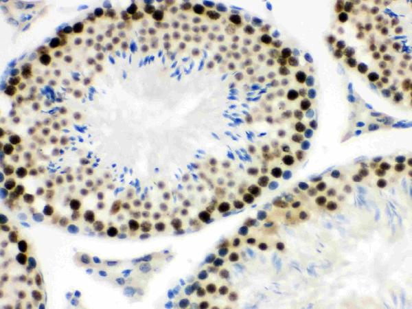
MNAT1 was detected in paraffin-embedded sections of mouse testis tissues using rabbit anti-MNAT1 Antigen Affinity purified polyclonal antibody (Catalog # PB9614) at 1 μg/mL. Biotin Conjugated Goat Anti-rabbit IgG (H+L) secondary antibody (Catalog # BA1003) was used to detect the primary antibody at 10μg/mL.

Click image to see more details
Nucleophosmin was detected in paraffin-embedded sections of human intestinal cancer tissues using rabbit anti-Nucleophosmin Antigen Affinity purified polyclonal antibody (Catalog # PB9341) at 1 μg/mL. Biotin Conjugated Goat Anti-rabbit IgG (H+L) secondary antibody (Catalog # BA1003) was used to detect the primary antibody at 10μg/mL.

Click image to see more details
SLC10A1 was detected in paraffin-embedded sections of mouse liver tissues using rabbit anti-SLC10A1 Antigen Affinity purified polyclonal antibody (Catalog # PB9745) at 1 μg/mL. Biotin Conjugated Goat Anti-rabbit IgG (H+L) secondary antibody (Catalog # BA1003) was used to detect the primary antibody at 10μg/mL.

Click image to see more details
Aquaporin 1 was detected in paraffin-embedded sections of mouse kidney tissues using rabbit anti-Aquaporin 1 Antigen Affinity purified polyclonal antibody (Catalog # PB9473) at 1 μg/mL. Biotin Conjugated Goat Anti-rabbit IgG (H+L) secondary antibody (Catalog # BA1003) was used to detect the primary antibody at 10μg/mL.

Click image to see more details
ABCB11 was detected in paraffin-embedded sections of rat liver tissues using rabbit anti-ABCB11 Antigen Affinity purified polyclonal antibody (Catalog # PB9414) at 1 μg/mL. Biotin Conjugated Goat Anti-rabbit IgG (H+L) secondary antibody (Catalog # BA1003) was used to detect the primary antibody at 10μg/mL.

Click image to see more details
HLA A was detected in paraffin-embedded sections of human intestinal cancer tissues using rabbit anti-HLA A Antigen Affinity purified polyclonal antibody (Catalog # PB9376) at 1 μg/mL. Biotin Conjugated Goat Anti-rabbit IgG (H+L) secondary antibody (Catalog # BA1003) was used to detect the primary antibody at 10μg/mL.

Click image to see more details
RbAp48 was detected in paraffin-embedded sections of mouse liver tissues using rabbit anti-RbAp48 Antigen Affinity purified polyclonal antibody (Catalog # PB9797) at 1 μg/mL. Biotin Conjugated Goat Anti-rabbit IgG (H+L) secondary antibody (Catalog # BA1003) was used to detect the primary antibody at 10μg/mL.

Click image to see more details
Aquaporin 2 was detected in paraffin-embedded sections of rat kidney tissues using rabbit anti-Aquaporin 2 Antigen Affinity purified polyclonal antibody (Catalog # PB9474) at 1 μg/mL. Biotin Conjugated Goat Anti-rabbit IgG (H+L) secondary antibody (Catalog # BA1003) was used to detect the primary antibody at 10μg/mL.
Specific Publications For BA1003
Loading publications
Customer Reviews
Have you used Goat Anti-Rabbit IgG (H+L) Secondary Antibody, Biotin Conjugated?
Submit a review and receive an Amazon gift card.
- $30 for a review with an image
0 Reviews For Goat Anti-Rabbit IgG (H+L) Secondary Antibody, Biotin Conjugated
Customer Q&As
Have a question?
Find answers in Q&As, reviews.
Can't find your answer?
Submit your question
1 Customer Q&As for Goat Anti-Rabbit IgG (H+L) Secondary Antibody, Biotin Conjugated
Question
Would either BA1054 or BA1003 secondary antibody work for M00139 for flow cytometry?
Verified customer
Asked: 2019-11-25
Answer
We don't recommend the Goat Anti-Rabbit IgG (H+L) Secondary Antibody, HRP Conjugate (BA1054) and Goat Anti-Rabbit IgG (H+L) Secondary Antibody, Biotin Conjugate (BA1003) for M00139 for flow cytometry.
Boster Scientific Support
Answered: 2019-11-25
