Click image to see more details
Product Info Summary
| SKU: | AZQ5U3E8 |
|---|---|
| Size: | 100 μg/vial |
| Reactive Species: | Zebrafish |
| Host: | Rabbit |
| Application: | IHC |
Customers Who Bought This Also Bought

Product info
Product Name
Anti-Zebrafish COPE Antibody
SKU/Catalog Number
AZQ5U3E8
Size
100 μg/vial
Form
Lyophilized
Description
Boster Bio Anti-Zebrafish-COPE-Antibody catalog # AZQ5U3E8. Tested in IHC applications. This antibody reacts with Zebrafish.
Storage & Handling
At -20°C for one year from date of receipt. After reconstitution, at 4°C for one month. It can also be aliquotted and stored frozen at -20°C for six months. Avoid repeated freezing and thawing.
Cite This Product
Anti-Zebrafish COPE Antibody (Boster Biological Technology, Pleasanton CA, USA, Catalog # AZQ5U3E8)
Host
Rabbit
Contents
Each vial contains 4 mg Trehalose, 0.9 mg NaCl, 0.2 mg Na2HPO4.
Clonality
Polyclonal
Immunogen
E.coli-derived zebrafish COPE recombinant protein (Position: E72-A300).
Reactive Species
AZQ5U3E8 is reactive to COPE in Zebrafish
Background of COPE
Coatomer subunit epsilon is a protein that in humans is encoded by the COPE gene. The product of this gene is an epsilon subunit of coatomer protein complex. Coatomer is a cytosolic protein complex that binds to dilysine motifs and reversibly associates with Golgi non-clathrin-coated vesicles. It is required for budding from Golgi membranes, and is essential for the retrograde Golgi-to-ER transport of dilysine-tagged proteins. Coatomer complex consists of at least the alpha, beta, beta', gamma, delta, epsilon and zeta subunits. Alternatively spliced transcript variants encoding different isoforms have been identified.
Antibody Validation
Boster validates all antibodies on WB, IHC, ICC, Immunofluorescence, and ELISA with known positive control and negative samples to ensure specificity and high affinity, including thorough antibody incubations.
Application & Images
Applications
AZQ5U3E8 is guaranteed for IHC Boster Guarantee
Recommend Dilution
| Application | Dilution | Species |
|---|---|---|
| Immunohistochemistry(Paraffin-embedded Section) | 2-5 μg/ml | Zebrafish |
Tested application
Use TE buffer pH 9.0 for antigen retrieval; (*) citrate buffer pH 6.0 is an alternative.
Validation Images & Assay Conditions
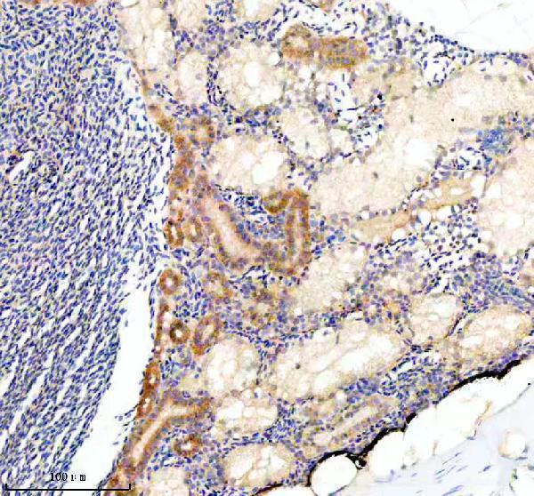
Click image to see more details
IHC analysis of COPE using anti-COPE antibody (AZQ5U3E8).
COPE was detected in a paraffin-embedded section of zebrafish kidney tissue. Heat mediated antigen retrieval was performed in EDTA buffer (pH 8.0, epitope retrieval solution). The tissue section was blocked with 10% goat serum. The tissue section was then incubated with 2 μg/ml rabbit anti-COPE Antibody (AZQ5U3E8) overnight at 4°C. Peroxidase Conjugated Goat Anti-rabbit IgG was used as secondary antibody and incubated for 30 minutes at 37°C. The tissue section was developed using HRP Conjugated Rabbit IgG Super Vision Assay Kit (Catalog # SV0002) with DAB as the chromogen.

Click image to see more details
IHC analysis of COPE using anti-COPE antibody (AZQ5U3E8).
COPE was detected in a paraffin-embedded section of zebrafish brain tissue. Heat mediated antigen retrieval was performed in EDTA buffer (pH 8.0, epitope retrieval solution). The tissue section was blocked with 10% goat serum. The tissue section was then incubated with 2 μg/ml rabbit anti-COPE Antibody (AZQ5U3E8) overnight at 4°C. Peroxidase Conjugated Goat Anti-rabbit IgG was used as secondary antibody and incubated for 30 minutes at 37°C. The tissue section was developed using HRP Conjugated Rabbit IgG Super Vision Assay Kit (Catalog # SV0002) with DAB as the chromogen.

Click image to see more details
IHC analysis of COPE using anti-COPE antibody (AZQ5U3E8).
COPE was detected in a paraffin-embedded section of zebrafish colon tissue. Heat mediated antigen retrieval was performed in EDTA buffer (pH 8.0, epitope retrieval solution). The tissue section was blocked with 10% goat serum. The tissue section was then incubated with 2 μg/ml rabbit anti-COPE Antibody (AZQ5U3E8) overnight at 4°C. Peroxidase Conjugated Goat Anti-rabbit IgG was used as secondary antibody and incubated for 30 minutes at 37°C. The tissue section was developed using HRP Conjugated Rabbit IgG Super Vision Assay Kit (Catalog # SV0002) with DAB as the chromogen.
Specific Publications For Anti-Zebrafish COPE Antibody (AZQ5U3E8)
Loading publications
Recommended Resources
Here are featured tools and databases that you might find useful.
- Boster's Pathways Library
- Protein Databases
- Bioscience Research Protocol Resources
- Data Processing & Analysis Software
- Photo Editing Software
- Scientific Literature Resources
- Research Paper Management Tools
- Molecular Biology Software
- Primer Design Tools
- Bioinformatics Tools
- Phylogenetic Tree Analysis
Customer Reviews
Have you used Anti-Zebrafish COPE Antibody?
Share your experimental results or join a short interview to earn up to $1,000 in product credits or other rewards.
0 Reviews For Anti-Zebrafish COPE Antibody
Customer Q&As
Have a question?
Find answers in Q&As, reviews.
Can't find your answer?
Submit your question
