Click image to see more details
Product Info Summary
| SKU: | AZQ90WW1 |
|---|---|
| Size: | 100 μg/vial |
| Reactive Species: | Zebrafish |
| Host: | Rabbit |
| Application: | IHC |
Customers Who Bought This Also Bought

Product info
Product Name
Anti-Zebrafish NANOS3 Antibody
SKU/Catalog Number
AZQ90WW1
Size
100 μg/vial
Form
Lyophilized
Description
Boster Bio Anti-Zebrafish NANOS3 Antibody catalog #AZQ90WW1. Tested in IHC applications. This antibody reacts with Zebrafish.
Storage & Handling
At -20°C for one year from date of receipt. After reconstitution, at 4°C for one month. It can also be aliquotted and stored frozen at -20°C for six months. Avoid repeated freezing and thawing.
Cite This Product
Anti-Zebrafish NANOS3 Antibody (Boster Biological Technology, Pleasanton CA, USA, Catalog # AZQ90WW1)
Host
Rabbit
Contents
Each vial contains 4 mg Trehalose, 0.9 mg NaCl, 0.2 mg Na2HPO4.
Clonality
Polyclonal
Immunogen
E.coli-derived zebrafish NANOS3 recombinant protein (Position: M1-W159)
Reactive Species
AZQ90WW1 is reactive to NANOS3 in Zebrafish
Background of NANOS3
Nanos is a zinc-finger containing, RNA-binding protein that has been implicated in germ cell development in both invertebrates and vertebrates. In Drosophila, Nanos represses apoptosis during development to ensure proper germ-line development. Unlike Nanos1 whose expression in mice is dispensable, the Nanos2 and Nanos3 proteins are required for germ cell development. Nanos2-null primordial germ cells (PGCs) die only in the male gonads and show no defects in females, while Nanos3-null PGCs are lost during the migration stage regardless of sex. Nanos2 and Nanos3 have distinct expression patterns during embryo development, suggesting that these two proteins do not have redundant functions. However, expression of Nanos2 can at least partially replace Nanos3 function in a Nanos3-null background. Nanos3 expression can not rescue Nanos2-null defects.
Antibody Validation
Boster validates all antibodies on WB, IHC, ICC, Immunofluorescence, and ELISA with known positive control and negative samples to ensure specificity and high affinity, including thorough antibody incubations.
Application & Images
Applications
AZQ90WW1 is guaranteed for IHC Boster Guarantee
Recommend Dilution
| Application | Dilution | Species |
|---|---|---|
| Immunohistochemistry(Paraffin-embedded Section) | 2-5 μg/ml | Zebrafish |
Tested application
Use TE buffer pH 9.0 for antigen retrieval; (*) citrate buffer pH 6.0 is an alternative.
Validation Images & Assay Conditions
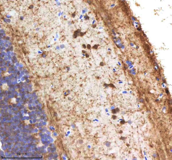
Click image to see more details
IHC analysis of NANOS3 using anti-NANOS3 antibody (AZQ90WW1).
NANOS3 was detected in a paraffin-embedded section of zebrafish brain tissue. Heat mediated antigen retrieval was performed in EDTA buffer (pH 8.0, epitope retrieval solution). The tissue section was blocked with 10% goat serum. The tissue section was then incubated with 2 μg/ml rabbit anti-NANOS3 Antibody (AZQ90WW1) overnight at 4°C. Peroxidase Conjugated Goat Anti-rabbit IgG was used as secondary antibody and incubated for 30 minutes at 37°C. The tissue section was developed using HRP Conjugated Rabbit IgG Super Vision Assay Kit (Catalog # SV0002) with DAB as the chromogen.

Click image to see more details
IHC analysis of NANOS3 using anti-NANOS3 antibody (AZQ90WW1).
NANOS3 was detected in a paraffin-embedded section of zebrafish pancreas tissue. Heat mediated antigen retrieval was performed in EDTA buffer (pH 8.0, epitope retrieval solution). The tissue section was blocked with 10% goat serum. The tissue section was then incubated with 2 μg/ml rabbit anti-NANOS3 Antibody (AZQ90WW1) overnight at 4°C. Peroxidase Conjugated Goat Anti-rabbit IgG was used as secondary antibody and incubated for 30 minutes at 37°C. The tissue section was developed using HRP Conjugated Rabbit IgG Super Vision Assay Kit (Catalog # SV0002) with DAB as the chromogen.

Click image to see more details
IHC analysis of NANOS3 using anti-NANOS3 antibody (AZQ90WW1).
NANOS3 was detected in a paraffin-embedded section of zebrafish ovary tissue. Heat mediated antigen retrieval was performed in EDTA buffer (pH 8.0, epitope retrieval solution). The tissue section was blocked with 10% goat serum. The tissue section was then incubated with 2 μg/ml rabbit anti-NANOS3 Antibody (AZQ90WW1) overnight at 4°C. Peroxidase Conjugated Goat Anti-rabbit IgG was used as secondary antibody and incubated for 30 minutes at 37°C. The tissue section was developed using HRP Conjugated Rabbit IgG Super Vision Assay Kit (Catalog # SV0002) with DAB as the chromogen.
Specific Publications For Anti-Zebrafish NANOS3 Antibody (AZQ90WW1)
Loading publications
Recommended Resources
Here are featured tools and databases that you might find useful.
- Boster's Pathways Library
- Protein Databases
- Bioscience Research Protocol Resources
- Data Processing & Analysis Software
- Photo Editing Software
- Scientific Literature Resources
- Research Paper Management Tools
- Molecular Biology Software
- Primer Design Tools
- Bioinformatics Tools
- Phylogenetic Tree Analysis
Customer Reviews
Have you used Anti-Zebrafish NANOS3 Antibody?
Share your experimental results or join a short interview to earn up to $1,000 in product credits or other rewards.
0 Reviews For Anti-Zebrafish NANOS3 Antibody
Customer Q&As
Have a question?
Find answers in Q&As, reviews.
Can't find your answer?
Submit your question
