This website uses cookies to ensure you get the best experience on our website.


- Table of Contents
Plan chromogenic AAMP IHC in paraffin sections using the catalog antibody at 2–5 μg/mL (datasheet PB9123). Assess cytoplasmic staining and use smooth muscle as a tissue reference (HPA tissue IHC).

Expected localisation, validated protocols, controls and antibodies — the at-a-glance facts below, then the full design guide.
| Expected localisation | Cytoplasmic in tissue (HPA tissue IHC); membrane also annotated (UniProt) | |
| Staining pattern | Cytoplasmic staining, most abundant in smooth muscle (HPA tissue IHC) | |
| Antigen retrieval | EDTA pH 8.0 HIER, heat-mediated (datasheet PB9123) | |
| Positive control | Adipose tissue+4 more · see all | |
| Negative control | Skeletal muscle |
| Fixation | Keep paraffin-section fixation consistent across specimens. (selected-SKU IHC image PB9123) | |
| Caveat | Smooth muscle content can shift apparent tissue signal (HPA tissue IHC) | |
| Regulation | Expression in activated T cells is reported (UniProt) | |
| Isoform / epitope | 0 annotated isoforms; one 1–434 chain (UniProt) |
Compare the catalog antibody’s IHC-P protocol (datasheet PB9123) with the published glioma IHC protocol (PMC9341442).
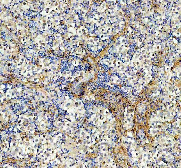
| Sample | Paraffin-embedded human testis cancer tissue; fixative not specified (datasheet PB9123) |
| Fixation | Image fixative and duration unreported (datasheet PB9123); verify before use. |
| Sectioning | 4–5 µm sections on charged slides (standard) |
| Deparaffinisation | Xylene, graded ethanol series to water (standard) |
| Antigen retrieval | Heat retrieval: EDTA pH 8.0 (datasheet PB9123); 20 min, 95–100 °C (standard) |
| Peroxidase block | 3% H2O2, 10 min, room temperature (standard) |
| Blocking | 10% goat serum (datasheet PB9123) |
| Primary antibody | Rabbit anti-AAMP, 2-5μg/ml (datasheet PB9123) |
| Primary incubation | Overnight at 4 °C (datasheet PB9123) |
| Detection | HRP-conjugated secondary, DAB chromogen (datasheet PB9123) |
| Counterstain | Hematoxylin, blue, dehydrate and mount (standard) |
| Expected result | AAMP-positive staining in adipocytes of adipose tissue (HPA tissue IHC: High). HPA tissue profile: General cytoplasmic expression, most abundant in smooth muscle. No signal in the no-primary control. |
AAMP staining in paraffin sections is expected mainly in the cytoplasm, with strong signal in smooth muscle cells and several other cell populations (HPA: tissue IHC, Approved; High in smooth muscle). Membrane-associated staining can also fit its annotation, although AAMP has no annotated transmembrane segment (UniProt Q13685: cell membrane, cytoplasm; topology). Interpret intensity by cell type: HPA reports low tissue specificity and only medium consistency between antibody staining and RNA expression (HPA: tissue IHC).
| Strong cytoplasmic staining in smooth muscle cells, with cellular outlines still discernible. | This matches the most abundant compartment and cell type in the tissue profile (HPA: general cytoplasmic expression; High in smooth muscle cells). Use the distribution across cells as well as intensity when judging a positive section. |
| Cytoplasmic signal in adipocytes or alveolar cells, or signal in the named ciliated cell populations. | These can be expected positives (HPA: High in adipocytes, alveolar cells, and specified ciliated cells). Cilia-associated staining is documented in fallopian tube and nasopharynx; do not require every listed cell type to show the same pattern. |
| Predominantly nuclear staining, without a convincing cytoplasmic or membrane-associated pattern. | This does not match the reported IHC profile or UniProt locations (HPA: general cytoplasmic expression; UniProt Q13685: cytoplasm, cell membrane). Consider nonspecific staining or a detection artefact before assigning it to AAMP. |
| Strong staining of skeletal myocytes, particularly when adjacent expected positive cells are weak. | Treat this as discordant with the cell-level reference, rather than proof of cross-reactivity (HPA: skeletal muscle myocytes Not detected). UniProt reports protein expression in skeletal muscle, so compare the exact stained cell population and antibody controls. |
| Diffuse color across cells, stroma, and blank areas, or no staining in a suitable positive section. | A pattern that ignores cell boundaries is hard to score as AAMP; absent signal in smooth muscle cells conflicts with a reported high-staining reference (HPA: general cytoplasmic expression; High in smooth muscle). Either finding needs control review. |
| Cell population and tissue choice | Smooth muscle cells provide a strong reference, while adipocytes, alveolar cells, ovarian stromal cells, and named ciliated populations are also reported High (HPA: tissue IHC). Sample and score the specified cells; whole-tissue intensity can obscure cell-level differences. |
| Reference-negative interpretation | Skeletal myocytes are reported Not detected by HPA, but UniProt lists skeletal muscle among tissues with protein expression (HPA: tissue IHC; UniProt Q13685: tissue specificity). A negative myocyte result is a useful comparison, not proof that the whole tissue lacks AAMP. |
| Compartment and protein annotation | The cytoplasmic tissue profile agrees with UniProt's cytoplasm annotation; membrane-associated staining is also plausible (HPA: tissue IHC; UniProt Q13685: cell membrane, cytoplasm). No transmembrane segment is annotated, so do not demand a sharp membrane-only rim (UniProt Q13685: topology). |
| IHC evidence strength | The tissue profile is Approved, with medium consistency between staining and RNA data; the listed IHC antibody CAB017717 is Approved (HPA: tissue IHC reliability; antibody validation). These labels support use as a reference but do not establish specificity for every cell or slide condition. |
| Processing and epitope limits | UniProt lists one 1–434 chain, no signal peptide or propeptide, no annotated glycosylation sites, and no isoforms (UniProt Q13685: processing, glycosylation, isoforms). The supplied sources give no epitope position or AAMP-specific fixation sensitivity; neither can explain a failed section here. |
| IF/ICC Q: Should its pattern match chromogenic tissue IHC? | A: Judge IF/ICC on its own page. HPA reports mainly microtubules, plus plasma membrane, cytosol, cytokinetic bridge, and primary cilium locations in ICC-IF (HPA: subcellular). Those observations do not require every paraffin tissue section to resolve those structures. |
| Situation | Likely cause | Next action |
|---|---|---|
| No signal in a section expected to contain smooth muscle cells. | The result conflicts with a High cell-level reference, but the slide alone cannot identify the failed step (HPA: smooth muscle cells High). | Confirm that smooth muscle cells are present and identifiable; review the positive control, antibody dilution, retrieval, detection reagents, and counterstain using the applicable IHC workflow. Treat retrieval checks as general FFPE practice, not an AAMP-specific fixation claim. |
| Strong nuclear-only color. | The dominant compartment is discordant with the reported cytoplasmic tissue pattern and UniProt locations (HPA: tissue IHC; UniProt Q13685: subcellular location). | Inspect the no-primary control and detection background, then compare with a section containing a known positive cell population (HPA: smooth muscle cells High). Do not score nuclear-only staining as confirmed AAMP. |
| Strong color in skeletal myocytes. | HPA reports skeletal myocytes as Not detected, although UniProt reports expression in skeletal muscle without assigning it to myocytes (HPA: tissue IHC; UniProt Q13685: tissue specificity). | Verify myocyte identity and compare staining with neighboring cells and a no-primary control. If strong myocyte signal persists, record the discordance and check antibody specificity before scoring it as AAMP. |
| Color appears throughout cells, stroma, or empty areas. | Diffuse deposit lacks the cell-linked cytoplasmic distribution used for interpretation; nonspecific binding or endogenous detection activity are general IHC possibilities (HPA: tissue IHC profile; standard IHC practice). | Compare with no-primary and detection controls; review blocking, wash steps, chromogen development, and endogenous enzyme blocking where relevant. Score only interpretable cellular staining after background is controlled. |
| Patchy staining within an otherwise suitable section. | Variation in section handling or reagent coverage is a general IHC possibility; the supplied sources do not establish AAMP-specific fixation sensitivity (standard IHC practice; HPA: tissue IHC scope). | Check whether positive cells occur in the weak area, then inspect section integrity, reagent coverage, and the positive control. Repeat the stain if these checks leave the local absence uninterpretable. |
| Weak staining is seen in a tissue outside the listed High populations. | AAMP has low tissue specificity, and HPA records several Low cell-level results; intensity alone does not define a new positive or negative tissue class (HPA: RNA specificity; tissue IHC Low entries). | Identify and score the stained cell type, compare its compartment with the cytoplasmic reference, and document the result alongside controls (HPA: tissue IHC profile). Avoid extending the reference to an unlisted cell population without validation. |


Comprehensive Human Protein Atlas IHC scoring per tissue (reliability: Approved — Medium consistency between antibody staining and RNA expression data.). Rows are taken directly from the HPA tissue chart — click any row's HPA link to view the source.
| Tissue | Cell type | Level | Evidence | Source |
|---|---|---|---|---|
| Adipose tissue | Adipocytes | High | Protein (IHC) | HPA → |
| Breast | Adipocytes | High | Protein (IHC) | HPA → |
| Bronchus | Ciliated cells (cell body) | High | Protein (IHC) | HPA → |
| Fallopian tube | Ciliated cells (cilia axoneme) | High | Protein (IHC) | HPA → |
| Lung | Alveolar cells | High | Protein (IHC) | HPA → |
| Tissue | Cell type | Level | Evidence | Source |
|---|---|---|---|---|
| Skeletal muscle | Myocytes | Not detected | Protein (IHC) | HPA → |
Troubleshoot AAMP staining in paraffin sections by checking retrieval, cell identity, subcellular pattern and detection controls (datasheet PB9123; HPA tissue IHC; UniProt Q13685).
PB9123 has a human paraffin-section IHC figure (catalog image caption); M07409 lists IF/ICC and Human, Mouse, Rat reactivity, with no supplied IF figure (catalog applications/reactivity/images).
PB9123 will render with its IHC figure from a paraffin-embedded human testis cancer section (catalog image caption). M07409 lists IF/ICC and Human, Mouse, Rat reactivity, but has no supplied IF figure (catalog applications/reactivity/images).
Which to pick: Choose PB9123 for tissue IHC: its own image caption documents EDTA pH 8.0 retrieval and 2 μg/ml antibody on a human paraffin section; the fixative is unreported (PB9123 image caption). Choose M07409 for IF/ICC because those applications and a 1:50–1:200 dilution are listed, although no IF image is supplied (M07409 catalog applications/dilution/images). Both list Human, Mouse and Rat reactivity for cross-species planning, but PB9123’s stated paraffin-section IHC dilution is for Human (catalog reactivity/dilution).