AATF / Protein AATF · IHC design guide

Design Immunohistochemistry for AATF

Plan chromogenic IHC on paraffin sections with the catalog antibody at 1–2 μg/mL (datasheet A03945-3). Assess granular cytoplasmic staining (HPA tissue IHC) while treating localisation cautiously: UniProt annotates AATF in the nucleus and nucleolus (UniProt), and HPA rates its tissue localisation uncertain (HPA tissue IHC).

Evidence assembled Oct 2026 · For research use; verify linked source records and product datasheet before use
Immunohistochemistry protocol sheet for AATF (IHC for AATF): expected localisation Granular cytoplasm (HPA tissue IHC); nucleus/nucleolus (UniProt), antibody A03945-3, validated IHC image, and IHC protocol steps
Printable AATF IHC protocol sheet — expected localisation Granular cytoplasm (HPA tissue IHC); nucleus/nucleolus (UniProt), antibody A03945-3, controls and protocol steps. Open the full AATF IHC guide →

AATF Immunohistochemistry Experimental Design Guide

Expected localisation, validated protocols, controls and antibodies — the at-a-glance facts below, then the full design guide.

Must know before staining
Expected localisation Granular cytoplasm (HPA tissue IHC); nucleus/nucleolus (UniProt)
Staining pattern Ubiquitous granular cytoplasmic staining (HPA tissue IHC)
Antigen retrieval EDTA pH 8.0 HIER, heat-mediated (datasheet A03945-3)
Positive control ⓘ Adipose tissue+4 more · see all
Negative control ⓘ None in HPA (detected in all 43 tissues); use no-primary + isotype controls
Important caveats
Reasons your staining may differ from the expected pattern.
Fixation Keep fixation consistent across paraffin sections (standard IHC practice; not target-specific)
Caveat Tissue localisation conflicts with external data (HPA tissue IHC)
Regulation Ubiquitous; no inducer specified (UniProt)
Isoform / epitope No annotated isoforms; chain 2–560, no transmembrane span (UniProt)
Section 1

Recommended AATF IHC & IF Protocols

Compare the catalog antibody’s IHC-P protocol (datasheet: A03945-3) with two published AATF tissue-staining protocols (PMC11794051; PMC6168685).

Recommended immunohistochemistry (IHC-P) protocol parameters
SampleParaffin-embedded mouse cerebellum tissue; fixative not specified (datasheet A03945-3)
FixationImage fixative and duration unreported (datasheet A03945-3); verify before use.
Sectioning4–5 µm sections on charged slides (standard)
DeparaffinisationXylene, graded ethanol series to water (standard)
Antigen retrievalHeat retrieval: EDTA pH 8.0 (datasheet A03945-3); 20 min, 95–100 °C (standard)
Peroxidase block3% H2O2, 10 min, room temperature (standard)
Blocking10% goat serum (datasheet A03945-3)
Primary antibodyRabbit anti-AATF, 1-2 μg/ml (datasheet A03945-3)
Primary incubationOvernight at 4 °C (datasheet A03945-3)
DetectionHRP-conjugated secondary, DAB chromogen (datasheet A03945-3)
CounterstainHematoxylin, blue, dehydrate and mount (standard)
Expected resultAATF-positive staining in adipocytes of adipose tissue (HPA tissue IHC: High). HPA tissue profile: Ubiquitous granular cytoplasmic expression. No signal in the no-primary control.
💡Decision noteStart with heat-mediated EDTA pH 8.0 retrieval for the catalog antibody (datasheet: A03945-3). Use citrate retrieval when following either published protocol (PMC11794051; PMC6168685).
Section 2

What Is the Expected AATF Staining Pattern?

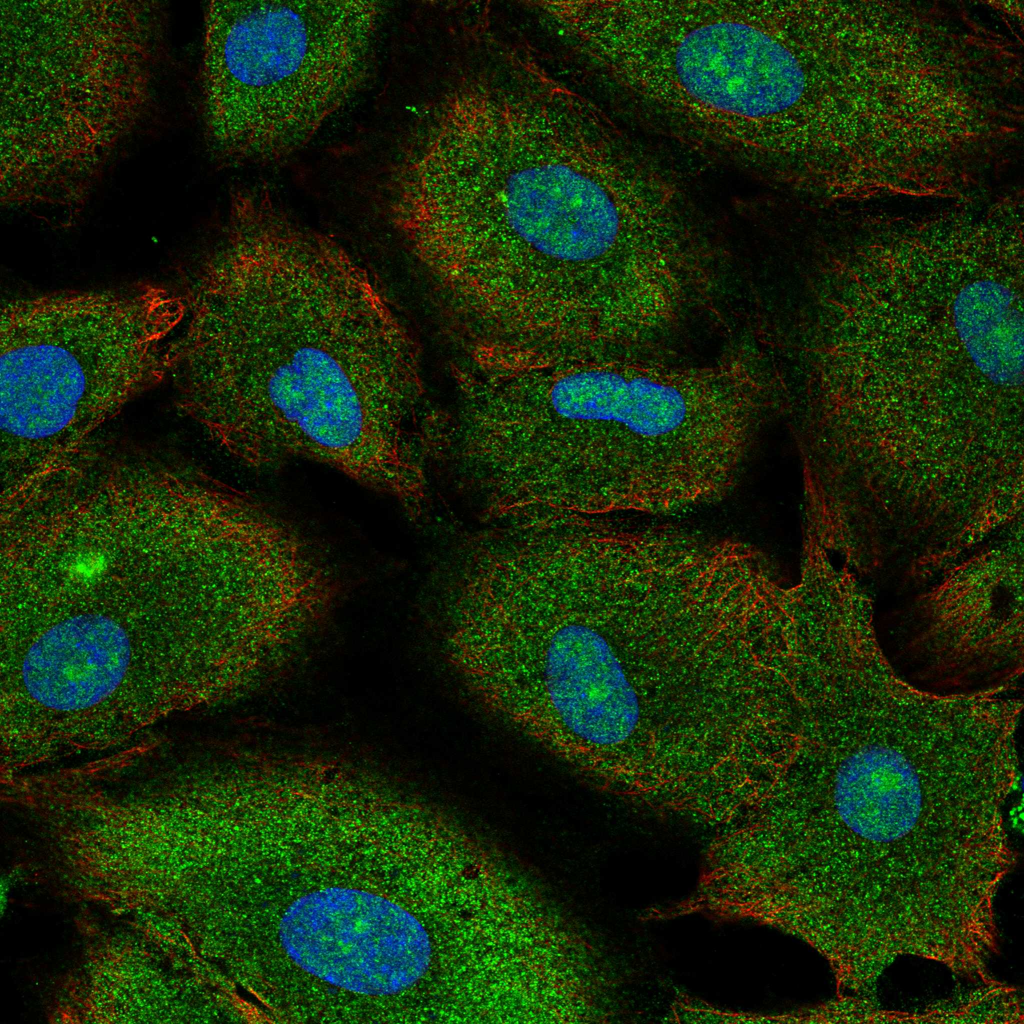
AATF is assigned to the nucleus and nucleolus (UniProt Q9NY61), and HPA ICC-IF supports mainly nucleolar staining with additional nucleoplasmic staining (HPA: subcellular). In tissue, examine glandular, respiratory epithelial and neuronal cells among the reported high-staining populations (HPA: tissue IHC). HPA also reports ubiquitous granular cytoplasmic IHC staining, but rates its tissue IHC localisation Uncertain because external data contradict it (HPA: tissue IHC). AATF has no transmembrane segment (UniProt Q9NY61 topology).

What am I looking at on my slide?
Distinct nucleolar staining within identifiable cell nuclei, with possible weaker nucleoplasmic staining.This matches the supported ICC-IF localisation and UniProt assignment (HPA: subcellular; UniProt Q9NY61). On a chromogenic section, use the counterstain and cell boundaries to judge the compartment; intensity alone cannot establish specificity (general IHC practice).
Granular cytoplasmic staining dominates, with little convincing nuclear signal.HPA describes this tissue IHC pattern but labels its localisation Uncertain because external data contradict it (HPA: tissue IHC). It conflicts with the nuclear and nucleolar assignment (UniProt Q9NY61). Record it as unresolved; review staining controls and antibody validation before calling it AATF-specific.
Strong staining is confined to an unexpected cell population, or parathyroid glandular cells stain strongly.AATF is ubiquitously expressed, so an unlisted cell population is not automatically negative (UniProt Q9NY61). HPA reports low staining in parathyroid glandular cells (HPA: tissue IHC). A strong discordant result warrants checks for cross-reactivity or endogenous detection activity, without proving either cause (general IHC practice).
Diffuse colour covers nuclei, cytoplasm and surrounding tissue without clear cellular boundaries.This cannot resolve the expected nuclear or nucleolar compartment (UniProt Q9NY61; HPA: subcellular). Inspect the negative detection control and section background; excess primary antibody, incomplete blocking or endogenous detection activity are general IHC possibilities, not reported AATF-specific effects (general IHC practice).
No interpretable staining appears in a section containing an HPA high-staining population.Examples include breast glandular cells, bronchial respiratory epithelial cells and caudate neuronal cells (HPA: tissue IHC). Check section quality and the staining run before inferring absence of AATF. HPA tissue IHC reliability is Uncertain, so its reported intensity is a reference observation, not a guaranteed positive-control result (HPA: tissue IHC).
💡Expected AATF appearanceA convincing positive shows clear nuclear staining, preferably enriched in nucleoli, in identifiable cells; compare intensity within the section and with controls (UniProt Q9NY61; HPA: subcellular; general IHC practice). Strong granular cytoplasmic staining alone is an unresolved IHC pattern, not a confirmed positive (HPA: tissue IHC, Uncertain).
How each factor affects the staining
Compartment evidenceUniProt assigns AATF to nucleus and nucleolus, and HPA ICC-IF supports mainly nucleolar plus nucleoplasmic localisation (UniProt Q9NY61; HPA: subcellular). HPA tissue IHC instead describes granular cytoplasm and rates that localisation Uncertain (HPA: tissue IHC). Keep these observations distinct when scoring sections.
Tissue and cell contextUniProt describes ubiquitous expression and high levels in brain, heart, kidney, placenta and thymus (UniProt Q9NY61). HPA reports high staining in several listed cell populations and low staining in parathyroid glandular cells (HPA: tissue IHC). These are different evidence types; neither defines an absolute negative tissue.
Antibody validationThe supplied HPA status is IHC Uncertain for HPA004940 and ICC Supported for HPA075963; neither entry supplies an IHC Enhanced result (HPA: antibodies). An ICC-supported localisation does not independently validate a chromogenic IHC result. Record the antibody used and interpret a discordant compartment cautiously.
Topology and processingAATF has no transmembrane segment, no annotated signal peptide or propeptide, and a reported protein chain spanning residues 2–560 (UniProt Q9NY61). These annotations support evaluating intracellular staining. They do not identify the antibody epitope or establish how paraffin processing affects signal.
IF/ICC Q: What localisation should a separate IF/ICC guide expect?A: Mainly nucleoli, with additional nucleoplasm (HPA: subcellular). HPA lists ICC-IF images from A-549, U-251MG and U2OS and rates HPA075963 ICC Supported (HPA: subcellular; HPA: antibodies). This is localisation evidence, not an IHC protocol or an IHC validation claim.
Why is my staining missing, weak or wrong?
SituationLikely causeNext action
Cytoplasmic granules are the only prominent signal.This resembles the HPA tissue IHC profile, whose localisation is rated Uncertain and conflicts with external data (HPA: tissue IHC).Score nuclear and cytoplasmic compartments separately; examine controls and seek independent confirmation before assigning the granules to AATF (general IHC practice).
A reported high-staining population shows no signal.The staining run, detection or section may have failed; the HPA high call is an observation, not a guaranteed control outcome (HPA: tissue IHC; general IHC practice).Confirm tissue identity and section integrity, review the run control, then check the catalog antibody's IHC-P instructions for the validated conditions (general IHC practice).
Strong diffuse staining obscures cell boundaries.Background from antibody concentration, blocking or detection chemistry can prevent compartment assessment (general IHC practice).Compare a negative detection control, review blocking and detection steps, and adjust the antibody within its validated IHC-P guidance (general IHC practice).
Colour persists when the primary antibody is omitted.The signal can arise from endogenous detection activity or another reagent-dependent background source (general IHC practice).Check the detection chemistry and apply its appropriate endogenous-activity control or blocking step before interpreting cellular staining (general IHC practice).
Parathyroid glandular cells stain as strongly as a reported high-staining population.HPA reports low parathyroid glandular staining, although its tissue IHC reliability is Uncertain (HPA: tissue IHC).Verify tissue identity and compare matched controls; treat the intensity mismatch as a specificity question rather than proof of cross-reactivity (general IHC practice).
Results vary between sections or staining runs.Section quality, retrieval or reagent handling can affect routine IHC consistency; no AATF-specific fixation or retrieval effect is supplied (general IHC practice).Compare section quality and run controls, then follow the catalog antibody's validated IHC-P conditions without assuming a target-specific retrieval requirement (general IHC practice).

Sample controls for AATF IHC & IF

🧪Run adipose tissue first and look for nuclear staining in adipocytes (HPA: adipocytes High; UniProt Q9NY61: nucleus and nucleolus). HPA detects AATF in all 43 scored tissues, so use no-primary and isotype controls instead of a negative tissue; no validated antigen-negative cell type is identified within the positive slide, although cells without nuclear chromogen can be recorded for review (HPA: no negative tissues; UniProt Q9NY61: nucleus and nucleolus).
Positive control tissue: Adipose tissue (Adipocytes, HPA High)
Negative control tissue: None in HPA: AATF is detected in all 43 scored tissues. Use a no-primary (secondary-only) and an isotype control instead.
ICC-IF cell lines (HPA subcellular resource): HPA ICC-IF images show AATF in A-549, U-251MG, U2OS, with annotated localisation: Nucleoli (supported) (HPA subcellular).
Technical controls: Include a no-primary, secondary-only slide and a rabbit IgG isotype control matched to the rabbit catalog antibody; confirm specificity with AATF-knockout material or immunizing-peptide competition if the peptide is known (caption: rabbit anti-AATF). Quench endogenous peroxidase and check adipose tissue for background around empty lipid vacuoles before scoring nuclear signal (HPA: adipocytes High; standard IHC practice).
⚠️Feasibility: No target-specific fixation window or fixation effect is reported, and the selected A03945-3 paraffin-section caption does not state the fixative (caption: fixative unreported). The reported IHC used heat retrieval in EDTA at pH 8.0; whether retrieval is required across other specimens is unreported (caption: EDTA heat retrieval). Frozen-section or IF ease relative to IHC-P is unestablished; for IF/ICC, nucleolar signal with additional nucleoplasmic signal provides a localization check, while adipocyte lipid vacuoles complicate interpretation of the IHC slide (HPA: nucleoli supported, nucleoplasm approved; HPA: adipocytes High; standard histology practice).

HPA tissue IHC evidence for AATF

Comprehensive Human Protein Atlas IHC scoring per tissue (reliability: Uncertain — Subcellular location is contradicted by external data. Pending external verification.). Rows are taken directly from the HPA tissue chart — click any row's HPA link to view the source.

Positive expression · recommended positive controls

TissueCell typeLevelEvidenceSource
Adipose tissue Adipocytes High Protein (IHC) HPA →
Adrenal gland Glandular cells High Protein (IHC) HPA →
Appendix Glandular cells High Protein (IHC) HPA →
Breast Glandular cells High Protein (IHC) HPA →
Bronchus Respiratory epithelial cells High Protein (IHC) HPA →

Undetected expression · recommended negative controls

TissueCell typeLevelEvidenceSource
None in HPA: AATF is detected in all 43 scored tissues. Use a no-primary (secondary-only) and an isotype control instead.
Section 3

Advanced AATF IHC Tips

Use the catalog antibody’s tissue staining conditions as a starting point, and interpret AATF staining in light of the reported disagreement about its location.

What retrieval conditions should I start with for AATF in paraffin sections?
Start with heat-mediated retrieval in EDTA at pH 8.0 (datasheet A03945-3). The catalog antibody stained a paraffin-embedded mouse cerebellum section after this retrieval, followed by 10% goat serum blocking, 2 μg/ml primary antibody overnight at 4°C, and HRP/DAB detection (datasheet A03945-3). If staining is weak, first check retrieval heating, cooling, and section integrity against a concurrently processed positive control (standard IHC practice). Change retrieval conditions one variable at a time, then compare nuclear and nucleolar signal with diffuse cytoplasmic staining, since the reported locations disagree (UniProt Q9NY61; HPA subcellular; HPA tissue IHC).
How should I troubleshoot fixation when AATF staining is weak or patchy?
The selected tissue-IHC caption identifies a paraffin-embedded mouse cerebellum section but does not state its fixative, so AATF-specific fixation sensitivity is unknown (datasheet A03945-3). Record the fixative, fixation interval, tissue thickness, and processing history for each specimen before comparing staining intensity (standard IHC practice). If a batch stains unevenly, compare sections processed together and inspect morphology for poorly preserved or excessively hardened regions (standard IHC practice). Keep retrieval and antibody incubation constant while evaluating processing differences; the documented starting conditions are EDTA at pH 8.0 and 2 μg/ml primary overnight at 4°C (datasheet A03945-3).
Should AATF staining appear in nucleoli or throughout the cytoplasm?
Expect nuclear staining with a nucleolar emphasis when assessing the proposed cellular location of AATF (UniProt Q9NY61; HPA subcellular). The tissue-IHC profile instead reports ubiquitous granular cytoplasmic expression and assigns the staining uncertain reliability because external location data contradict it (HPA tissue IHC). Score nucleolar, other nuclear, and cytoplasmic signal separately rather than combining them into one positive category (standard IHC practice; HPA subcellular; HPA tissue IHC). Use morphology and a nuclear counterstain to establish compartment boundaries, and treat a predominantly cytoplasmic pattern as requiring independent confirmation before assigning it to AATF (standard IHC practice; UniProt Q9NY61; HPA tissue IHC).
Could an AATF isoform or modified epitope explain different staining patterns?
The supplied record lists 0 isoforms and no annotated domains, so it provides no named isoform to explain divergent IHC patterns (UniProt Q9NY61). AATF has reported acetylation and phosphorylation sites, including phosphoserines at residues 61, 63, 150, and 155, but the catalog antibody’s epitope is not supplied here (UniProt Q9NY61; datasheet A03945-3). Do not assign a staining difference to a specific modification without epitope and modification-sensitive validation (standard IHC practice). Compare serial sections using the same retrieval and detection settings, then seek independent evidence if the nuclear and granular cytoplasmic patterns remain discordant (standard IHC practice; HPA subcellular; HPA tissue IHC).
How can I check an AATF IHC pattern with multiplex immunofluorescence?
Treat IF as a separate antibody validation experiment: the selected catalog evidence documents paraffin-section chromogenic IHC, while HPA reports nucleolar and additional nucleoplasmic localization from ICC/IF (datasheet A03945-3; HPA subcellular). Pair AATF with a marker for the cell population being evaluated, such as a neuronal-cell marker when examining the reported caudate signal, and include a nuclear stain for compartment assignment (HPA tissue IHC; standard IF practice). Choose a fluorophore channel with low tissue autofluorescence and inspect an unstained section in that channel (standard IF practice). Because AATF lacks a transmembrane segment and is reported within the nucleus, assess permeabilisation that permits antibody access to the nuclear compartment while preserving nucleoli (UniProt Q9NY61; HPA subcellular; standard IF practice).
What should I check when AATF DAB staining is widespread or granular?
Start with a no-primary control and inspect pigment, section edges, damaged areas, and residual peroxidase activity before calling widespread DAB signal positive (standard IHC practice). The documented chromogenic example used a peroxidase-conjugated secondary and DAB, with 10% goat serum blocking and 2 μg/ml primary antibody (datasheet A03945-3). Check peroxidase blocking as a general HRP workflow step, and compare antibody dilution or blocking changes on matched sections (standard IHC practice). Interpret granular cytoplasmic staining cautiously: HPA reports that pattern in tissue IHC but marks its reliability uncertain because location data conflict with it (HPA tissue IHC; HPA subcellular).
How should I quantify AATF staining across tissue sections? ⚠ ANSWER MARKED FOR VERIFICATION
Define the scored cell population and compartment before measuring AATF, since nucleolar and nucleoplasmic localization conflicts with the uncertain granular cytoplasmic tissue-IHC profile (UniProt Q9NY61; HPA subcellular; HPA tissue IHC). For chromogenic sections, report the percentage of positive cells and an intensity-weighted H-score for each compartment; cell density per mm² can supplement these when populations vary (standard IHC practice). Normalize positive counts to the number of evaluable cells in the defined population, and density to the analyzed viable tissue area (standard IHC practice). Keep threshold, counterstain, imaging, and region-selection rules constant across batches, and report cytoplasmic scores separately pending validation (standard IHC practice; HPA tissue IHC).
When is an apparent AATF-positive cell likely to be an artefact?
A nucleolar or nucleoplasmic pattern is consistent with reported AATF localization, whereas predominantly granular cytoplasmic tissue staining carries an explicit reliability warning (UniProt Q9NY61; HPA subcellular; HPA tissue IHC). Check whether the signal lies in the intended cell population: HPA reports high staining in cerebellar molecular-layer cells and caudate neuronal cells, alongside broad tissue expression (HPA tissue IHC). Exclude edge accentuation, necrotic regions, pigment, and staining retained in the no-primary control before scoring cells (standard IHC practice). If DAB persists without primary antibody, investigate endogenous enzyme activity; if compartment disagreement persists under controlled staining, require independent confirmation before interpreting it biologically (standard IHC practice; HPA tissue IHC).
Boster reagents

Best AATF / Protein AATF IHC Antibodies

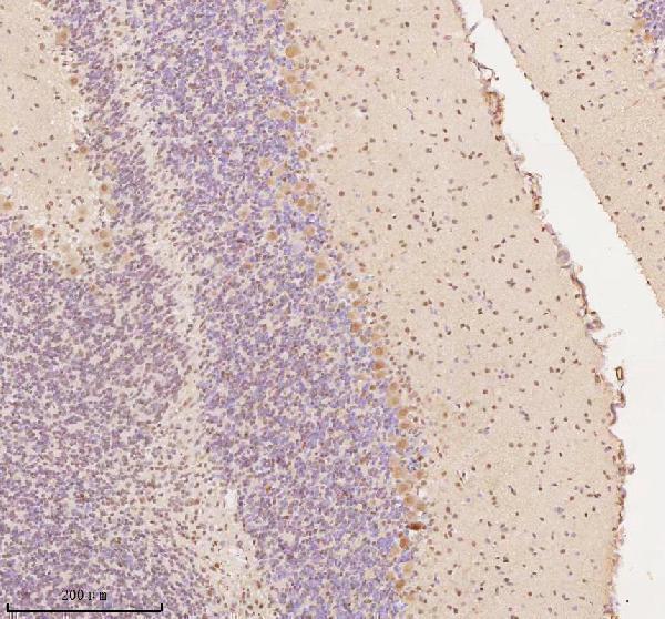
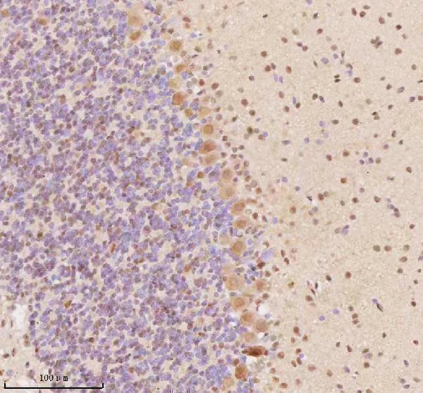
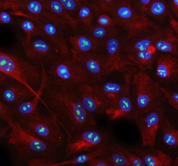
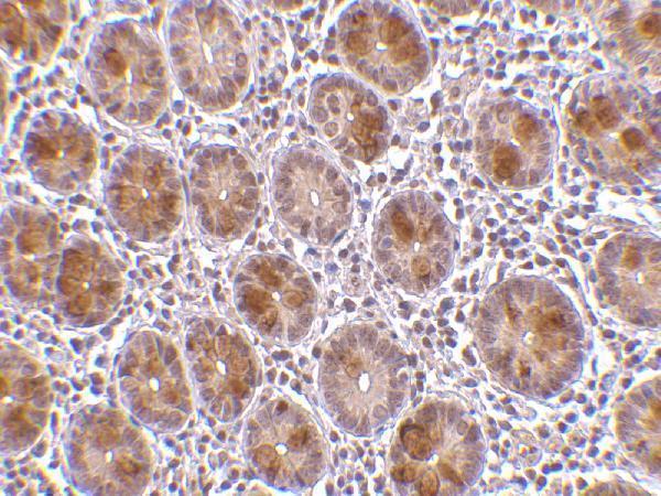
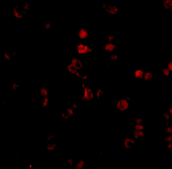
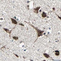
Catalog antibodies A03945 and A03945-3 have IHC images from human small intestine and mouse cerebellum, respectively; IF images cover human small intestine, A549 cells, and rat brain (catalog image captions).

Real IHC data IHC analysis of AATF using anti-AATF antibody (A03945-3). AATF was detected in a paraffin-embedded section of mouse cerebellum tissue. Heat mediated antigen retrieval was performed in EDTA buffer (pH 8.0, epitope retrieval solution). The tissue section was blocked with 10% goat serum. The tissue section was then incubated with 2 μg/ml rabbit anti-AATF Antibody (A03945-3) overnight at 4°C. Peroxidase Conjugated Goat Anti-rabbit IgG was used as secondary antibody and incubated for 30 minutes at 37°C. The tissue section was developed using HRP Conjugated Rabbit IgG Super Vision Assay Kit (Catalog # SV0002) with DAB as the chromogen.
Anti-AATF Antibody ®
Cat # A03945-3
Real IHC data Immunohistochemistry of AATF in human small intestine tissue with AATF antibody at 10 μg/mL.
Anti-Protein AATF AATF Antibody
Cat # A03945

A03945 will render with human small-intestine IHC at 10 μg/mL; its applications include IHC-P and IF (A03945 image caption and catalog applications). A03945-3 will render with mouse-cerebellum paraffin-section IHC at 2 μg/mL; its applications include IHC, ICC, and IF (A03945-3 image caption and catalog applications).

Which to pick: For tissue IHC, choose A03945 for the pictured human small-intestine example or A03945-3 for the pictured mouse-cerebellum paraffin-section example; the fixative is unreported for both (catalog IHC image captions). For IF/ICC, A03945-3 has images from A549 cells and rat-brain paraffin sections, while A03945 has a human small-intestine IF image (catalog IF image captions). For cross-species planning, A03945-3 lists human, mouse, and rat reactivity, whereas A03945 lists human and mouse; neither catalog entry reports clonality, and the pictured rat result is IF rather than IHC (catalog reactivity, clone fields, and IF image captions).

Each figure is that product's own IHC / IF validation image from its datasheet.

References

  1. UniProt Consortium. UniProt entry Q9NY61 (AATF_HUMAN, Protein AATF).
  2. Human Protein Atlas. AATF tissue IHC expression (reliability: Uncertain).
  3. Human Protein Atlas. AATF subcellular location (ICC-IF): Mainly localized to the nucleoli. In addition localized to the nucleoplasm..
  4. Human Protein Atlas. AATF antibody validation summary (2 antibodies).
  5. Apoptosis antagonizing transcription factor expression and its validation as a potential diagnostic and prognostic biomarker in oral squamous cell carcinoma. Frontiers in oncology 2025 — PMC11794051.
  6. The PI3K/AKT axis modulates AATF activity in Wilms' tumor cells. FEBS open bio 2018 — PMC6168685.
  7. The Oncogenic and Immunological Roles of Apoptosis Antagonistic Transcription Factors in Human Tumors: A Pan-Cancer Analysis. Oxidative medicine and cellular longevity 2022 — PMC9581705.
  8. AATF and SMARCA2 are associated with thyroid volume in Hashimoto's thyroiditis patients. Scientific reports 2020 — PMC7000742.
  9. PubMed PMID:11027528 — UniProt-cited evidence.
  10. PubMed PMID:10783144 — UniProt-cited evidence.
  11. PubMed PMID:14702039 — UniProt-cited evidence.