This website uses cookies to ensure you get the best experience on our website.
- Table of Contents
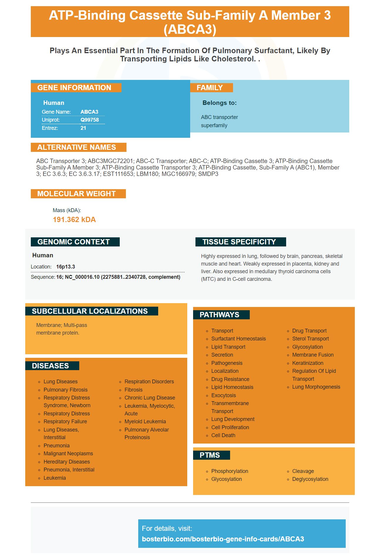
Facts about ATP-binding cassette sub-family A member 3.
Human | |
---|---|
Gene Name: | ABCA3 |
Uniprot: | Q99758 |
Entrez: | 21 |
Belongs to: |
---|
ABC transporter superfamily |
ABC transporter 3; ABC3MGC72201; ABC-C transporter; ABC-C; ATP-binding cassette 3; ATP-binding cassette sub-family A member 3; ATP-binding cassette transporter 3; ATP-binding cassette, sub-family A (ABC1), member 3; EC 3.6.3; EC 3.6.3.17; EST111653; LBM180; MGC166979; SMDP3
Mass (kDA):
191.362 kDA
Human | |
---|---|
Location: | 16p13.3 |
Sequence: | 16; NC_000016.10 (2275881..2340728, complement) |
Highly expressed in lung, followed by brain, pancreas, skeletal muscle and heart. Weakly expressed in placenta, kidney and liver. Also expressed in medullary thyroid carcinoma cells (MTC) and in C-cell carcinoma.
Membrane; Multi-pass membrane protein.
This article will discuss Cellular dysfunction, Early Apoptosis Markers and Targeted Therapy. Boster Bio: The ABCA3 Marker's Top Uses will be reviewed. This book is essential reading for anyone who is interested in the ABCA3 gene. It's a useful marker of genes for researchers working in the field of cancer research, because it is crucial for researchers to comprehend how a cell's apoptosis process works.
Because of its role in regulating immune and inflammatory responses The ABCA3 gene may be a crucial candidate for diagnosing pulmonary conditions. The gene can be expressed by various types of cells, including SOX2+ cells as well as conducting airways cells. These cells are sensitive to various types of insults and are capable of generating different types of disease. By identifying ABCA3 mutations, scientists are able to further study these diseases and develop new treatments.
A mutation in the ABCA3 gene may cause disruptions in lipid transport. Traditional structure-function analysis relies upon the expression of in vitro cells and the assessment of lipid transport and trafficking. However, this low-throughput approach is unable to identify functionally defective mutations. Future strategies for this gene would benefit from the generation of 3D crystal structures and creation of high-throughput expression and screening assays.
Two sites are responsible for mutations in the ABCA3 gene: R280C and L43L. Both mutations are located in a limited way, and the NBD-labeled lipids were detected in vesicles shaped like lamellar body. The mutants were also detected in cells transfected with the pUB6HA/ABCA3.
In mouse models, the ABCA3 gene has been modified. Mice that do not have the ABCA3 gene suffer from respiratory failure, alveolar leakage, and inflammation. This could be due in part to AT2 cell dysfunction. Furthermore, the ABCA3 gene deletion causes a decrease of activity in other genes, like XBP1 and IL-33.
ABCA3 mutations affect a variety of the folding of proteins, their trafficking, and synthesizing. After the synthesis of proteins, newly synthesized proteins are folded within the ER lumen and require molecular chaperones to keep them folded in the correct way. When misfolded proteins build up in the ER there is a stress response.
is initiated increases ER protein folding capability and blocks general protein translation.Recent advancements in ABCA3 biosynthesis have been made. In addition, this gene is responsible for alveolar epithelial cell dysregulation. In the context of disease presentation variation, environmental and modifier genes were also examined. An improved ABCA3 biology could result in better therapies for cell dysfunction. The ABCA3 marker is not as powerful in its potential.
The ABCA3 marker gene is a brand new gene
has numerous potential applications. It has been associated with several disorders, including Tangier syndrome and chronic idiopathic Lupus. Certain mutations, like the R43L mutation, induce an ER stress and apoptosis. Certain mutations may cause chronic ILD or even fatal neonatal respiratory distress syndrome. Another mutation, L101P, has been connected to a trafficking defect. However there is no information discovered regarding its function in ER stress.Numerous studies have revealed the importance of ER stress in the development of COPD. Certain studies have revealed an abnormal microenvironment in COPD patients
increase susceptibility to ER stress. ABCA3 is particularly useful in this regard, since it can be used in the detection of a patient's condition. Inflammatory pulmonary disease is an atypical symptom of COPD. It is essential to detect ER stress to manage and tre/p>it.Moreover, ABCA3 has been shown to increase in levels of ER stress when stimulated by airborne pollutants such as aqueous smoke extract. It isn't known if it causes Apoptosis. Cells with the L188Q mutation showed higher levels of apoptosis exposed to tunicamycin and bleomycin.
The ABCA3 marker is most effective in the context of the process of apoptosis. Apoptosis can be activated in A549 cells due to stress in the ER, and ABCA3 mutations can cause this process to occur. YFP fluorescence was used to determine the amount of transfected cells aswell in determining the presence of markers of apoptosis.
Moreover, ABCA3 mutations can result in trafficking and folding defects. The ER lumen is the area where newly synthesized protein molecules are folded. This requires molecular chaperones in order to ensure proper folding. ER stress is caused by the accumulation of misfolded proteins. The unfolded protein response.(UPR) promotes ER protein folding capacity and blocks general protein translation.
Analyzing the surface sensitivity of phospminimum 100 cells and multiple independent experiments were conducted.
The methods used to study the apoptosis markers differ. The methods used to determine which proteins are apoptotic>are essential for inhibiting protease activity, because otherwise the PARP-1 protein could be cleaved independently of apoptosis. The results were consistent when with urine buffers
contain concentrated urine and dense.denaturing conditions. The aspiration of cells should be done on the ice. The cells should be heated to a temperature of 65°C after aspiration until electrophoresis.Using HL-60 cells as models, apoptotic>markers can be easily identified by looking at the time-course of events
trigger apoptosis. CDNAs are labeled from cells tare induced are hybridized to microarrays with genes tare tare associated with apoptosis. For example If a cell's death has been induced by VP-16, the cells thave died mighp>have different levels of VP-16 in comparison to the control group.L. pneumophila caused DNA fragmentation in U937>macrophages, based on the same study. Similarly, ActD causes DNA fragmentation in L. pneumophila-infected macrophages. Using the method, the researchers were able to detect cells t
are apoptotic>in both the late and early phases of infection. The findings of the study indicated a strong dose-dependent induction of apoptosis within the two cell lines.After UA treatment the HEPG2, SNU-449 and HUVECs showed morphological modifications which were similar to the control group. The cells were able to show vacuolization and condensation of chromatin in HCC and SNU-449. The UA treatment did not significantly inhibit the autophagy process. To understand the mechanism that drives the autophagic>effects of UA more research is required.
The presence of the apoptotic>marker, PI, is a distinguishing feature between the early and late indicators of apoptosis. The latter is more sensitive than the former and consequently more specific. It detects apoptotic>cells t
Targeted therapy using the ABCA3 gene marker was developed using a PubMed search in November 2017. We found 119 articles
contained a relevant mutation of ABCA3. We considered the papers only when they were written in English and reported on specific patients. There were 59 papers were thoroughly reviewed. Ten were excluded because they did not contain genetic>mutations, or contained only identical, non-disease-causing mutations.The ABCA3 gene is mutated and mutations are classified according to experimentally discovered outcomes. Atypical protein synthesis may be caused by frameshift or nonsense.mutations. Extracellular domains are believed to play a role in intracellular trafficking. Missense.mutations are classified as Type II (CFTR) mutations. NBD2 mutations can cause changes in intracellular trafficking of proteins. ABCA3 is a perfect candidate for targeted therapy.
ABCA3 mutations can be linked to ER stress and apoptosis of lung tissue. It is unclear w
impact ABCA3 mutations have on the process of apoptosis. Further studies are needed to establish the importance of apoptosis and ER stress in lung tissue. Furthermore, ABCA3 variants could be related to disease progression. At present, there is no treatment specific to ABCA3 mutations. However, targeting the gene in those suffering from lung disease may be an effective option.The expression of the ABCA3 gene was found to be in inverse connected to the MLF level in patients with CL. When macrophages infected were treated with L. V. panamensis strain, ABCA3 knockdown decreased parasite survival. ABCA3 is also found in the membranes of resting macrophages and intracellular compartments within cells t
are infected.ABCA4 is a photoreceptor cell-specific ATP binding cassette transporter. ABCA4 was also known as ABCCR prior to its current name. This gene mutation is related with Stargardt disease and fundus flavimaculatus. Single-strand conformation polymorphism analysis has found 19 mutations in the ABCA4 gene.
PMID: 8706931 by Klugbauer N., et al. Primary structure of a novel ABC transporter with a chromosomal localization on the band encoding the multidrug resistance-associated protein.
PMID: 9027511 by Connors T.D., et al. The cloning of a human ABC gene (ABC3) mapping to chromosome 16p13.3.