This website uses cookies to ensure you get the best experience on our website.
- Table of Contents
2 Citations 7 Q&As
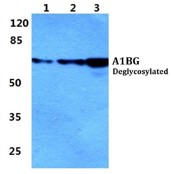
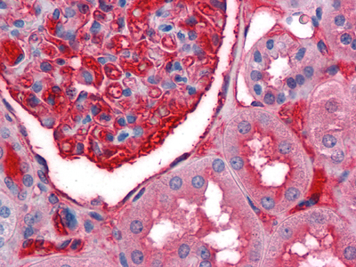
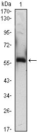
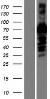
Facts about Alpha-1B-glycoprotein.
Human | |
---|---|
Gene Name: | A1BG |
Uniprot: | P04217 |
Entrez: | 1 |
Belongs to: |
---|
No superfamily |
A1B; A1BG; ABG; alpha 1BGlycoprotein; alpha 1B-Glycoprotein; GAB
Mass (kDA):
54.254 kDA
Human | |
---|---|
Location: | 19q13.43 |
Sequence: | 19; NC_000019.10 (58345183..58353492, complement) |
Plasma.
Secreted.
If you're looking for a gene expression test that measures a specific protein, Boster Bio's A1BG Marker could be perfect for your needs. AQP3 plays a role in cell volume regulation, cell migration and invasion. It also regulates hydrogen-peroxide transport and downstream signals. It also promotes intracellular ATP and is a transcription factor.
AQP3 is a membrane protein involved in the permeability of hydrogen peroxide and mediates H2O2-dependent mucosal responses. These results may help explain AQP3's protective effect on intestinal mucosal tissues, which are vital in the inflammatory response and epithelial destruction. AQP3 modulates cell volume in different tissues, including the colon.
AQP3 is found in the plasma membranes of intestinal cells. It is vital for cell proliferation and migration which both require ATP. It may also play an important role in glycerol transportation. Further studies are needed to confirm the mechanism by which AQP3 regulates cells volume. The IMCD cell lines used in this study were cultivated at 300 and 600 mosmol/kg. AQP3 expression has been observed. Secondary Alexa-Fluor-488-labeled antibodies were used to detect AQP signals.
Moreover, AQP3 contributes to the wound healing process. It aids in the uptake and transport of H2O2, an important signaling molecule. Inhibiting this enzyme reduced the number actin-containing, lamellopodial projections. The wound's size was decreased by inhibition of H2O2 signaling, suggesting that AQP3 is crucial for epithelial wound closure.
The aqueous humoral protein (AQP5) plays an important role in the regulation of cell migration and invasion in human cells. Mutations in AQP5 reduce the expression transcription factors that regulate cell movement and invasion, such Ecadherin and Wnt/bcatenin. AQP5 is also known to inhibit cancer cell EMT.
This protein is secreted by eukaryotic cells to determine if AQP5 is involved with the regulation of cell movement. AQP5 protein expression is increased in tumor cell invasion. Knockdown inhibits this process. These results can be confirmed by Western blotting or RTPCR. The AQP5 siRNA is a valuable tool to aid in inflammatory research.
CDDP was used in hypertonic conditions to increase AQP5 expression. However, AQP5 mRNA expression was induced when cells were exposed to a low amount of CDDP. A high-dose CDDP reduced AQP5 mRNA expression. Further, the presence of AQP5 was associated with the reduction of hyperosmosis and prolonged treatment time.
AQP3 acts as a signaling protein in cytosol and transports glycerol over the cytomembrane. AQP3 plays an important role in cell proliferation, invasion, and migration. It is overexpressed among many cancers, including lung and colorectal squamous cells carcinomas. Jiang et al. Jiang et al. reported that AQP3 expression was positively correlated to tumor size and lymph node metastaticis.
AQP3 is involved with many aspects of tumor growth including migration, proliferation, invasion, and migration. Knockdown experiments revealed that AQP3 knockdown significantly reduced invasion, migration, and proliferation. This protein is essential in these processes due to its ability regulate HIF-1a transcription factor, a master regulator of hypoxia. Hypoxic conditions cause the protein to accumulate, and knocking it down inhibits tumor metastasis.
MiR-874-mediated lung carcinoma has revealed the function of AQP3. MiR-874 targets AQP3 mRNA 3'-UTR, and inhibits NSCLC cellular proliferation and movement. Inversely, miR-874 also had a reduced effect on NSCLC cells when AQP3 was upregulated. However, the mechanism of AQP3 regulatory is still not fully understood.
Antibodies against AQP5 can be developed in rabbit and mouse models. Aquaporin-5 can be interacted with by the BosterBio Anti-pNF5 AQP5 marker. The protein includes six transmembranedomains. It also has tandem repeat structure. Researchers believe that water movement is dependent on the tight turn structure. The mud loach AQP1b gene consists five exons as well as four introns.
Molecular analysis is a great tool to identify proteins in eukaryotic tissues. By detecting proteins with AQP5, researchers will be able to determine which proteins are causing inflammation in patients. This type of research is ideal for AQP5. Here are some of the reasons it's so valuable. Once you've decided that this gene is important for your research, you'll be able to identify the proteins that cause inflammation in patients.
Hypoxia is a factor that regulates the AQP proteins. In lung cancer, AQP3 is linked to AQP5 and AQP3. AQP3 is also linked to tumor osmoregulation. This influences the growth and development of HTR8/Svneo line cells. AQP3 regulates the expression of EMT proteins, and promotes intracellular production of ATP.
Breast cancer is often associated with aquaporin-5 (5 (AQP5)). This protein activates Ras signaling pathways to regulate cell migration in cultured cancer cells. Both AQP5 expression and Ras activation are associated with increased cell migration and cancer metastasis. This study also identified a candidate ROS-sensitive intracellular protein, AQP3, that affects cell migration. The candidate protein belongs to the src kinase family. It also contains free cysteines, which may change its function.
AQP4 was localized at the rear of migrating cells. The study promoted the osmotic engine model, in which water enters the cell from the leading edge and exits at the trailing edge. Although AQP4 may be involved in this process, other studies using kidney cells are needed. Further studies are needed to determine whether AQP4 is endogenously expressed in kidney cells.
AQP3 & AQP4 can be found at the plasma membranes in migrating cells. Hyper osmolality results in cells developing polarization. This is where the front end of a cell faces the direction for migration. This osmotic gradient encourages the formation protrusions at cells' plasma membranes. The osmotic gradient generates local water flow. It also adds an extra protrusive force.
Boster Bio created a mouse model of Tilapia that can be used in freshwater or seawater. They stained the gill chloride cells of the mice with anti-AQP3 antibodies, and a control antibody that did NOT react to AQP3. Both samples showed AQP3 immunoreactions centered at the basolateral membrane. The immunoreactions on mitochondria were not detectable.
AQP3 is thought to play a crucial role in wound heal, as it promotes the growth and migration of epidermal tissue. Boster Bio has made it an integral part of their products. However, its exact function is not known. Many researchers are still studying the mechanisms by which AQP3 regulates implantation. For the moment, it remains unclear whether AQP3 can be used to improve fertility.
Aquaporins, which are integral pore proteins, regulate water and other small molecular compounds across a membrane. AQP3 is highly expressed in the plasma membranes of keratinocytes, where it serves as a water-glycerol transporter. Deficient mice exhibit reduced skin elasticity. This suggests a possible role for AQP3 when embryos are conceived and implantation occurs.
AQPs are molecules which regulate the volume and function of cancer cells. They are also associated with angiogenesis, cellular dissociation, and cell division. AQPs play a crucial role in cell migration and invasion, in addition to their role as tumor regulators. Plasma membrane expression of AQP1 (and AQP3) is a hallmark of cancer cells. They also alter cellular signaling pathways to promote tumor development and growth. Uncontrolled cell proliferation and cell migration are hallmarks of cancer.
Studies have shown that AQP3 (and AQP4) are expressed at the plasma membranes in colon cancer cells. Treatment with H2O2 causes a rise in AQP3 and SUM159, indicating that the latter cells have a lower sensitivity to oxidative stress. Furthermore, AQP expression is correlated with the aggressiveness of invasive cancer cells. AQP2 & AQP3 are responsible for transporting glycerol (from the plasma membrane) to the cells. This glycerol is used as an energy source in glycolytic pathway and as a building blocks for phospholipids. Moreover, AQP3 and AQP4 are implicated in cancer progression.
Infections account for the third leading cause worldwide of cancer. Aquaporin functions often are regulated by virus. A polymorphism in AQP3 may make you more vulnerable to Epstein-Barr disease. Myeloradiculitis can be exacerbated by the presence of an anti AQP4 antibody. AQP3 protein expression is regulated by UVA exposure.
PMID: 15221005 by Yamada S., et al. Expression profiling and differential screening between hepatoblastomas and the corresponding normal livers: identification of high expression of the PLK1 oncogene as a poor-prognostic indicator of hepatoblastomas.
PMID: 3458201 by Ishioka N., et al. Amino acid sequence of human plasma alpha 1B-glycoprotein: homology to the immunoglobulin supergene family.
High expression of CD68 TAM in patients with neuroblastoma was associated with poorer overall response after autologous transplant. Conversely, high expression of iNOS and CD68-expressing TAMs was not associated with poor overall response. These findings suggest that TAMs may play an important role in the progression of the disease and its progression-free survival.
In addition, CD163 TAMs correlated with poor overall and progression-free survival. This study also demonstrated that the two markers can be used together to predict survival. When analyzed separately, they are weak prognostic indicators that could be incorporated into a single prognostic tool such as the international staging system. Moreover, combined analysis of the two markers is more sensitive and powerful than a single marker.
Although TAMs produce Th1 and Th2 cytokines, the function of these cells is not yet clear. In addition, they may also activate tumor-promoting programs, particularly in the absence of the IFN-g receptor. As a result, the combined signaling from the receptors is required for TAM functional differentiation. However, synthetic ligands are available that polarize the NKT response towards Th1 or Th2.
Among all these factors, the M1/M2 ratio of TAMs is the most significant independent prognostic factor for MM. Based on the M1/M2 ratio, this nomogram can help physicians stratify patients for therapies, customize follow-up and counsel patients. The data in this study suggest that tumor-associated CD68 TAMs may be associated with longer progression-free survival after autologous transplant.
The role of TAMs in the immune response is not completely understood, but they are crucial for the tumor's progression. TAMs secrete various soluble mediators, such as IL-6 and VEGF. In addition, TAMs also attract immunosuppressive cells to the tumor site. This leads to a pro-inflammatory environment, which may facilitate cancer metastasis.
In addition to the role of TAMs in tumor-associated inflammation, TAMs can also recruit and activate POSTN, a potential therapeutic target. TnC is another protein that binds to many different ECM proteins, probably playing a structural role. Its expression has been associated with poor outcomes in iCCA. OPN is a glycosylated phosphoprotein normally involved in bone remodeling, immune regulation, and vascularization.
*More publications can be found for each product on its corresponding product page