ABCB6 / ATP-binding cassette sub-family B member 6 · IHC design guide

Design Immunohistochemistry for ABCB6

Start ABCB6 IHC-P at 0.5–1 μg/ml (datasheet: PA1723), using epididymal or gallbladder glandular cells as positive controls (HPA tissue IHC). Interpret the granular cytoplasmic tissue pattern (HPA tissue IHC) in light of ABCB6’s multiple membrane locations (UniProt).

Evidence assembled Oct 2026 · For research use; verify linked source records and product datasheet before use
Immunohistochemistry protocol sheet for ABCB6 (IHC for ABCB6): expected localisation Granular cytoplasmic staining in most tissues (HPA tissue IHC), antibody PA1723, validated IHC image, and IHC protocol steps
Printable ABCB6 IHC protocol sheet — expected localisation Granular cytoplasmic staining in most tissues (HPA tissue IHC), antibody PA1723, controls and protocol steps. Open the full ABCB6 IHC guide →

ABCB6 Immunohistochemistry Experimental Design Guide

Expected localisation, validated protocols, controls and antibodies — the at-a-glance facts below, then the full design guide.

Must know before staining
Expected localisation Granular cytoplasmic staining in most tissues (HPA tissue IHC)
Staining pattern Granular cytoplasm; strong glandular staining in epididymis and gallbladder (HPA tissue IHC)
Antigen retrieval Citrate pH 6.0 HIER, 95–98 °C, 20 min (rule: cytoplasmic / membrane antigen)
Positive control ⓘ Epididymis+4 more · see all
Negative control ⓘ Lymph node+2 more · see all
Important caveats
Reasons your staining may differ from the expected pattern.
Fixation Keep fixation consistent across paraffin sections (standard IHC practice; not target-specific)
Caveat Membrane trafficking may shift the observed pattern (UniProt)
Regulation Staining regulation is not established in this record (UniProt)
Isoform / epitope 2 isoforms; map the lumenal or cytoplasmic epitope (UniProt)
Section 1

Recommended ABCB6 IHC & IF Protocols

Compare the catalog antibody’s IHC-P protocol with 3 published ABCB6 IHC workflows (PMC3818219; PMC9818642; PMC7079220).

Recommended immunohistochemistry (IHC-P) protocol parameters
SampleTissue sections; selected-image fixative not specified (standard IHC workflow)
FixationImage fixative and duration unreported (datasheet PA1723); verify before use.
Sectioning4–5 µm sections on charged slides (standard)
DeparaffinisationXylene, graded ethanol series to water (standard)
Antigen retrievalHeat-induced epitope retrieval in citrate buffer, pH 6.0, 20 min at 95–98 °C (standard rule: cytoplasmic / membrane antigen)
Peroxidase block3% H2O2, 10 min, room temperature (standard)
Blocking10% normal serum of the secondary host, 30 min, room temperature (standard)
Primary antibodyRabbit anti-ABCB6, 0.5-1μg/ml (datasheet PA1723)
Primary incubationOvernight at 4 °C (standard)
DetectionHRP-polymer secondary, DAB chromogen 5–10 min (standard)
CounterstainHematoxylin, blue, dehydrate and mount (standard)
Expected resultABCB6-positive staining in glandular cells of epididymis (HPA tissue IHC: High). HPA tissue profile: Cytoplasmic expression with a granular pattern in most tissues. No signal in the no-primary control.
💡Decision noteStart with citrate pH 6.0 HIER at 95–98 °C for 20 min (page retrieval); EDTA pH 8.0 was used for skin (PMC3818219).
Section 2

What Is the Expected ABCB6 Staining Pattern?

ABCB6 is an 11-transmembrane protein found in several membranes, including the plasma membrane and intracellular organelles (UniProt Q9NP58 topology and subcellular location). In paraffin-section IHC, expect granular cytoplasmic staining in many tissues (HPA tissue IHC). HPA reports high staining in epididymal and gallbladder glandular cells and late spermatids; its tissue IHC assessment is Approved, with medium consistency against RNA data (HPA tissue IHC).

What am I looking at on my slide?
Granular cytoplasmic stain in epididymal or gallbladder glandular cells, or late spermatids (HPA tissue IHC).This matches HPA's tissue pattern and High cell-level staining. Judge intensity within the specified cells; a tissue label alone does not identify every cell as positive (HPA tissue IHC).
Predominantly nuclear staining, with little granular cytoplasmic signal.This does not match the reported tissue IHC pattern (HPA tissue IHC). Check the counterstain and controls before assigning it to ABCB6; HPA's nucleoplasmic ICC-IF finding does not establish a predominantly nuclear paraffin-section pattern (HPA subcellular ICC-IF).
Strong staining in germinal center cells, parathyroid glandular cells, or chondrocytes.HPA reports these cell populations as Not detected in tissue IHC (HPA tissue IHC). Unexpected chromogen may reflect cross-reactivity or endogenous detection activity (general IHC practice); investigate before scoring it as ABCB6.
Diffuse color across cells, stroma, and empty areas, without cell-specific granules.The distribution is inconsistent with HPA's granular cytoplasmic tissue pattern (HPA tissue IHC). Background can arise from nonspecific antibody binding or detection reagents (general IHC practice); it cannot identify an ABCB6-positive cell.
No signal in the selected positive-control cells.A blank run in a population reported as High by HPA, such as gallbladder glandular cells, does not establish that all specimens lack ABCB6 (HPA tissue IHC). Review control quality and the staining workflow (general IHC practice).
💡Expected ABCB6 appearanceCall a result positive when the appropriate cells show discrete granular cytoplasmic chromogen, potentially High in HPA-listed glandular cells or late spermatids; diffuse field-wide or predominantly nuclear color is suspect (HPA tissue IHC).
How each factor affects the staining
IHC evidence and controlsHPA rates tissue IHC Approved but reports medium staining-to-RNA consistency; its Not detected cells are useful comparison areas, not proof of biological absence (HPA tissue IHC).
Membrane topologyUniProt records 11 transmembrane segments and a cytoplasmic C-terminal region (UniProt Q9NP58 topology). The supplied evidence gives no antibody epitope, so it cannot specify an ABCB6 retrieval condition.
Protein formsUniProt lists 2 isoforms, one glycosylation site at residue 6, and no signal peptide or propeptide (UniProt Q9NP58). Their effects on this IHC antibody's staining are unreported.
Antibody validationOne listed HPA antibody has IHC Approved and ICC Supported status; the other has ICC Supported status with no IHC status reported (HPA antibodies). ICC support alone does not validate a paraffin-section IHC result.
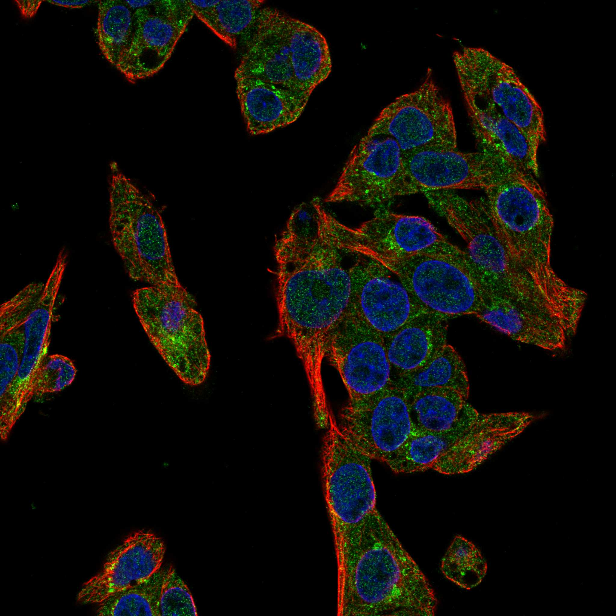
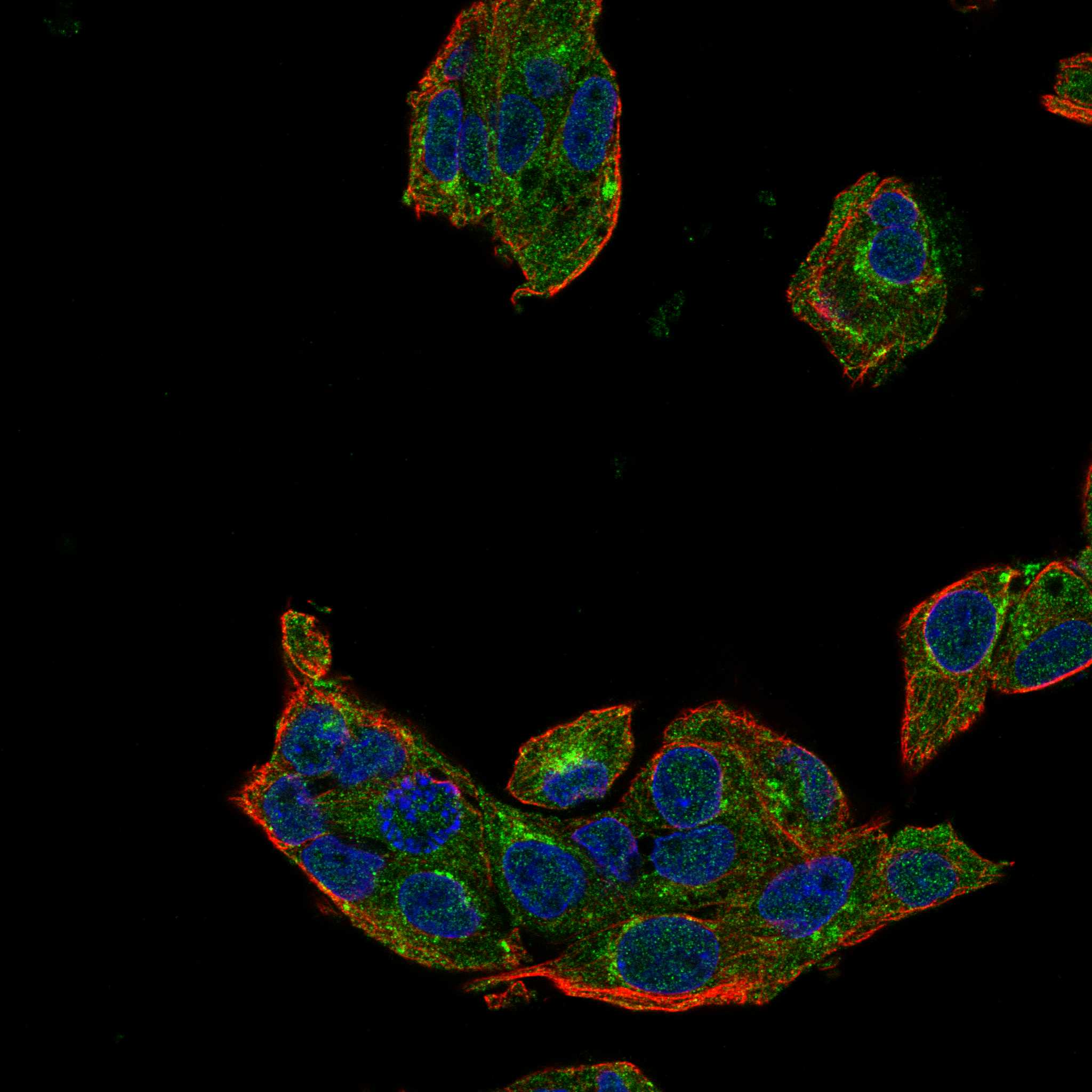
IF/ICC Q: where should ABCB6 appear?A: Mainly at the Golgi apparatus (supported); HPA also lists plasma membrane (supported) and mitochondria and nucleoplasm (approved) in ICC-IF (HPA subcellular ICC-IF). This is an imaging reference for the separate IF/ICC guide, not an IHC protocol.
Why is my staining missing, weak or wrong?
SituationLikely causeNext action
Positive-control tissue is blank.The expected High cell populations did not stain (HPA tissue IHC); the failed step cannot be identified from a blank slide alone.Check tissue and section quality, reagent delivery, retrieval, primary dilution, and detection with routine run controls (general IHC practice). Reassess the named cell population.
Only a smooth nuclear signal is visible.Predominantly nuclear tissue staining conflicts with HPA's granular cytoplasmic IHC profile (HPA tissue IHC).Compare with the no-primary control and counterstain, then inspect whether cytoplasmic granules are actually present (general IHC practice). Do not transfer the ICC-IF nucleoplasm call directly to tissue IHC (HPA subcellular ICC-IF).
HPA-listed negative cell populations stain strongly.HPA reports germinal center cells, parathyroid glandular cells, and chondrocytes as Not detected (HPA tissue IHC). Cross-reactivity or endogenous detection activity is possible (general IHC practice).Run a no-primary control and check the detection system's endogenous-activity block where applicable (general IHC practice). Compare staining within the exact cell population (HPA tissue IHC).
Most of the section has diffuse chromogen.Widespread haze obscures the granular cytoplasmic pattern reported by HPA (HPA tissue IHC); nonspecific binding or residual detection reagent may contribute (general IHC practice).Inspect the no-primary control; review blocking, primary dilution, washing, and chromogen development using the chosen detection system's instructions (general IHC practice).
Signal appears at a cell edge or as intracellular puncta.UniProt places ABCB6 at the plasma membrane and several intracellular membranes; HPA tissue IHC describes an overall granular cytoplasmic pattern (UniProt Q9NP58 subcellular location; HPA tissue IHC).Assess whether the signal also follows the expected cells and granular pattern. Do not assign a particular organelle from chromogenic morphology alone (general IHC practice).
A low-staining tissue looks negative.HPA lists some cells as Low, including lung macrophages and rectal glandular cells (HPA tissue IHC); weak signal may be difficult to distinguish from background (general IHC practice).Compare with a listed High cell population in the same run and score only interpretable cell-specific staining (HPA tissue IHC; general IHC practice).

Sample controls for ABCB6 IHC & IF

🧪Run epididymis first and require staining in its glandular cells (HPA: High in epididymis glandular cells); use parathyroid glandular cells as the tissue negative (HPA: Not detected in parathyroid glandular cells). The supplied HPA row does not identify an internal negative cell type in epididymis; any cells scored as negative should show only background-level chromogen relative to the no-primary control (HPA: epididymis glandular cells High; standard IHC practice).
Positive control tissue: Epididymis (Glandular cells, HPA High)
Negative control tissue: Lymph node (HPA Not detected)
ICC-IF cell lines (HPA subcellular resource): HPA ICC-IF images show ABCB6 in Hep-G2, U-251MG, U2OS, Sperm, with annotated localisation: Golgi apparatus (supported) (HPA subcellular).
Technical controls: Include a no-primary, secondary-only control; a control immunoglobulin matched to the primary antibody’s host species and class or isotype, with clonality considered; and ABCB6-knockout tissue or peptide competition when a validated blocking peptide is available (standard IHC practice). For chromogenic epididymis sections, quench endogenous peroxidase for HRP detection and assess endogenous biotin if using avidin–biotin detection (standard IHC practice).
⚠️Feasibility: A target-specific fixation window or fixation effect is unreported, and the selected PA1723 IHC(P) caption does not state a fixative (caption: fixative not stated). Retrieval dependence is unreported; optimize antigen retrieval against the positive and negative tissue controls rather than infer it from ABCB6 topology (UniProt Q9NP58 topology; HPA: epididymis High, parathyroid gland Not detected). The supplied evidence does not establish that frozen sections or IF are easier than IHC-P, or document an epididymis-specific artefact (caption: IHC(P) only; HPA: epididymis glandular cells High).

HPA tissue IHC evidence for ABCB6

Comprehensive Human Protein Atlas IHC scoring per tissue (reliability: Approved — Medium consistency between antibody staining and RNA expression data.). Rows are taken directly from the HPA tissue chart — click any row's HPA link to view the source.

Positive expression · recommended positive controls

TissueCell typeLevelEvidenceSource
Epididymis Glandular cells High Protein (IHC) HPA →
Gallbladder Glandular cells High Protein (IHC) HPA →
Testis Elongated or late spermatids High Protein (IHC) HPA →
Adipose tissue Adipocytes Medium Protein (IHC) HPA →
Adrenal gland Glandular cells Medium Protein (IHC) HPA →

Undetected expression · recommended negative controls

TissueCell typeLevelEvidenceSource
Lymph node Germinal center cells Not detected Protein (IHC) HPA →
Parathyroid gland Glandular cells Not detected Protein (IHC) HPA →
Soft tissue Chondrocytes Not detected Protein (IHC) HPA →
Section 3

Advanced ABCB6 IHC Tips

These questions address paraffin section chromogenic IHC for ABCB6; the immunofluorescence entry covers the separate IF/ICC application.

How should I retrieve ABCB6 antigen when staining is weak?
Start with citrate buffer at pH 6.0, heat-induced retrieval at 95–98 °C for 20 min (page retrieval setting for IHC-P). ABCB6 has 11 transmembrane segments and a cytoplasmic C-terminal region, making the antibody’s epitope position relevant to accessibility (UniProt Q9NP58 topology). If staining is weak, compare a modestly longer citrate treatment on matched sections while holding antibody dilution and detection conditions constant (standard IHC practice). Assess tissue morphology alongside staining, because excessive heating can damage sections (standard IHC practice). Include an epididymal glandular-cell positive control and a no-primary control to help separate retrieval failure from nonspecific chromogen (HPA: High in epididymal glandular cells; standard IHC practice).
Can fixation explain variable ABCB6 staining between paraffin blocks?
The PA1723 IHC(P) caption identifies human mammary cancer tissue but does not state its fixative, so ABCB6-specific fixation sensitivity is unknown (PA1723 tissue-IHC caption). Record the fixative and fixation duration for each block before comparing staining across cases (standard IHC practice). Process matched sections together, using the same pH 6.0 citrate retrieval and 20 min heating time to limit technical variation (page retrieval setting; standard IHC practice). Compare internal morphology and positive-control staining before attributing a difference to ABCB6 abundance (standard IHC practice). The reported granular cytoplasmic tissue pattern describes observed staining; it does not establish a fixation effect (HPA tissue IHC: granular cytoplasmic profile).
Which staining compartments are plausible for ABCB6 in tissue sections?
Expect chiefly granular cytoplasmic staining in routine tissue IHC, as reported across most tissues (HPA tissue IHC: granular cytoplasmic profile). ABCB6 is also assigned to the plasma membrane, Golgi, mitochondria and endolysosomal membranes, so distinct membranous or punctate patterns can merit investigation (UniProt Q9NP58 subcellular locations). Score cytoplasmic granules and convincing membrane staining separately, and check whether each pattern occurs in intact cells rather than damaged edges (standard IHC practice). The Golgi is the main supported location in the subcellular IF record, but that cell-imaging result does not specify a required chromogenic tissue pattern (HPA subcellular: Golgi supported). Treat isolated diffuse nuclear chromogen cautiously and compare it with controls and morphology (standard IHC practice).
How can epitope placement affect ABCB6 IHC interpretation?
ABCB6 has 2 listed isoforms, but the supplied record does not map the catalog antibody’s epitope to either one (UniProt Q9NP58 isoforms; supplied antibody evidence). Its 11 transmembrane segments end near residue 550, followed by a cytoplasmic region through residue 842 (UniProt Q9NP58 topology). A glycosylation site is annotated at residue 6; that annotation alone cannot predict this antibody’s staining response to retrieval (UniProt Q9NP58 glycosylation; standard IHC interpretation). Request the immunogen sequence or epitope mapping before assigning a negative section to loss of a particular isoform (standard antibody-validation practice). Until then, describe staining as ABCB6-reactive and use orthogonal evidence when an isoform-specific conclusion matters (standard antibody-validation practice).
How should I assess ABCB6 in a separate multiplex IF experiment?
For the separate IF/ICC application, pair ABCB6 with a glandular epithelial marker in a tissue where glandular cells are expected to stain, such as gallbladder (HPA: High in gallbladder glandular cells). Choose a far-red channel for the weaker signal when tissue autofluorescence is prominent, and examine an unstained section in every channel (standard IF practice). Permeabilisation should match the mapped epitope: a cytoplasmic C-terminal epitope requires access across cell membranes, whereas an exposed surface epitope may not (UniProt Q9NP58 topology; standard IF practice). Titrate permeabilisation on matched samples and inspect whether organelle patterns remain intact (standard IF practice). The supplied PA1723 image documents IHC(P), so establish IF performance independently with appropriate controls (PA1723 tissue-IHC caption; standard antibody-validation practice).
How do I reduce granular background without erasing ABCB6 signal?
First compare the stained section with a no-primary control and inspect whether granules align with intact cell cytoplasm (standard IHC practice). A granular cytoplasmic pattern is reported for ABCB6 in most tissues, so removing every punctum would also remove a plausible signal (HPA tissue IHC: granular cytoplasmic profile). Titrate the primary antibody and shorten chromogen development one variable at a time, keeping the positive-control section in the same run (standard IHC practice). Apply the routine endogenous peroxidase block before chromogenic detection, then check whether persistent color appears where antibody was omitted (standard chromogenic IHC practice). Use HPA-listed germinal-center cells with undetected staining as a comparison, while recognizing that tissue controls do not replace reagent controls (HPA: Not detected in lymph-node germinal-center cells; standard IHC practice).
What scoring method captures ABCB6 staining across heterogeneous tissue? ⚠ ANSWER MARKED FOR VERIFICATION
Define the cell population and compartment before scoring, because ABCB6 tissue staining is commonly granular cytoplasmic (HPA tissue IHC: granular cytoplasmic profile). For a cellular readout, record the percentage of positive cells and an H-score from 0–300 using intensity categories 0–3 (standard IHC scoring practice). If the biological question concerns sparse positive cells, report their density per mm² of viable tissue as an additional measure (standard IHC scoring practice). Normalize counts to the eligible cell population or viable tissue area, and exclude folds, edges and necrosis consistently (standard IHC scoring practice). Keep retrieval, imaging and scoring thresholds fixed across cases, and report cytoplasmic and membrane scores separately when both patterns are reproducible (standard IHC practice).
When is an apparent ABCB6-positive section convincing?
A convincing result places staining in intact cells with a reproducible granular cytoplasmic or plausible membrane pattern (HPA tissue IHC: granular cytoplasmic profile; UniProt Q9NP58 subcellular locations). Compare with a positive-control population such as epididymal glandular cells and an HPA-listed undetected population such as germinal-center cells (HPA: High in epididymal glandular cells; HPA: Not detected in lymph-node germinal-center cells). Review staining at section edges and in necrosis separately, since both can create misleading deposits (standard IHC practice). Check no-primary and endogenous peroxidase controls before assigning brown signal to ABCB6 (standard chromogenic IHC practice). Interpret unexpected nuclear-only or uniformly diffuse staining cautiously and seek independent confirmation before making a compartment-specific claim (HPA tissue IHC: granular cytoplasmic profile; standard antibody-validation practice).
Boster reagents

Best ABCB6 / ATP-binding cassette sub-family B member 6 IHC Antibodies

PA1723 is human-reactive and has a real IHC(P) image of human mammary cancer tissue (catalog: reactivity; PA1723 image caption). ICC is listed, but no IF image is supplied (catalog: applications; IF images).

Real IHC data Anti-ABCB6 antibody, PA1723, IHC(P) IHC(P): Human Mammary Cancer Tissue
Anti-ABCB6 Antibody ®
Cat # PA1723

PA1723 has an IHC(P) image from human mammary cancer tissue (PA1723 image caption). PA1723 is listed for human ICC, but the catalog supplies no IF image or IF sample (catalog: applications, reactivity, IF images).

Which to pick: Choose PA1723 for human paraffin-section IHC: its own image caption identifies IHC(P) on human mammary cancer tissue; the fixative is unreported (PA1723 image caption). For IF/ICC, PA1723 lists ICC and a 0.5–1 μg/ml IF dilution, but supplies no IF image (catalog: applications, IF dilution, IF images). No cross-species option is documented: PA1723 lists human reactivity only, and its clonality is unspecified (catalog: reactivity, clone).

Each figure is that product's own IHC / IF validation image from its datasheet.

References

  1. UniProt Consortium. UniProt entry Q9NP58 (ABCB6_HUMAN, ATP-binding cassette sub-family B member 6).
  2. Human Protein Atlas. ABCB6 tissue IHC expression (reliability: Approved).
  3. Human Protein Atlas. ABCB6 subcellular location (ICC-IF): Mainly localized to the Golgi apparatus. In addition localized to the nucleoplasm, plasma membrane, cytosol, mitochondria, mid piece and principal piece..
  4. Human Protein Atlas. ABCB6 antibody validation summary (2 antibodies).
  5. Understanding the role of iron/heme metabolism in the anti‑inflammatory effects of natural sulfur molecules against lipopolysaccharide‑induced inflammation. Molecular medicine reports 2025 — PMC12046942.
  6. Novel mutations of ABCB6 associated with autosomal dominant dyschromatosis universalis hereditaria. PloS one 2013 — PMC3818219.
  7. Protoporphyrin IX (PpIX) Fluorescence during Meningioma Surgery: Correlations with Histological Findings and Expression of Heme Pathway Molecules. Cancers 2023 — PMC9818642.
  8. Elevated Glutathione Peroxidase 2 Expression Promotes Cisplatin Resistance in Lung Adenocarcinoma. Oxidative medicine and cellular longevity 2020 — PMC7079220.
  9. PubMed PMID:10837493 — UniProt-cited evidence.
  10. PubMed PMID:11955620 — UniProt-cited evidence.
  11. PubMed PMID:14702039 — UniProt-cited evidence.