ABCC2 / ATP-binding cassette sub-family C member 2 · IHC design guide

Design Immunohistochemistry for ABCC2

Plan chromogenic IHC-P for ABCC2 using the catalog antibody at 2–5 μg/mL (datasheet A00974-1). Assess membranous staining in hepatocytes and kidney proximal-tubule microvilli, with adipocytes as a tissue-level negative reference (HPA tissue IHC).

Evidence assembled Oct 2026 · For research use; verify linked source records and product datasheet before use
Immunohistochemistry protocol sheet for ABCC2 (IHC for ABCC2): expected localisation Membranous in liver, kidney, gallbladder and intestines (HPA tissue IHC), antibody A00974-1, validated IHC image, and IHC protocol steps
Printable ABCC2 IHC protocol sheet — expected localisation Membranous in liver, kidney, gallbladder and intestines (HPA tissue IHC), antibody A00974-1, controls and protocol steps. Open the full ABCC2 IHC guide →

ABCC2 Immunohistochemistry Experimental Design Guide

Expected localisation, validated protocols, controls and antibodies — the at-a-glance facts below, then the full design guide.

Must know before staining
Expected localisation Membranous in liver, kidney, gallbladder and intestines (HPA tissue IHC)
Staining pattern Hepatocyte and proximal-tubule microvillar staining (HPA tissue IHC)
Antigen retrieval EDTA pH 8.0 HIER, heat-mediated (datasheet A00974-1)
Positive control ⓘ Kidney+4 more · see all
Negative control ⓘ Adipose tissue+4 more · see all
Important caveats
Reasons your staining may differ from the expected pattern.
Fixation Keep formalin fixation consistent across paraffin sections. (standard IHC practice; not target-specific)
Caveat Endogenous peroxidase may add liver DAB background (standard IHC practice)
Regulation Tissue-dependent; highest in liver (UniProt)
Isoform / epitope No annotated isoforms; epitope side matters across 17 transmembrane segments (UniProt)
Section 1

Recommended ABCC2 IHC & IF Protocols

The catalog antibody protocol is paired with published chromogenic IHC examples for papillary renal neoplasms (PMC11551816; PMC12805615).

Recommended immunohistochemistry (IHC-P) protocol parameters
SampleParaffin-embedded human liver cancer tissue; fixative not specified (datasheet A00974-1)
FixationImage fixative and duration unreported (datasheet A00974-1); verify before use.
Sectioning4–5 µm sections on charged slides (standard)
DeparaffinisationXylene, graded ethanol series to water (standard)
Antigen retrievalHeat retrieval: EDTA pH 8.0 (datasheet A00974-1); 20 min, 95–100 °C (standard)
Peroxidase block3% H2O2, 10 min, room temperature (standard)
Blocking10% goat serum (datasheet A00974-1)
Primary antibodyRabbit anti-ABCC2, 2-5μg/ml (datasheet A00974-1)
Primary incubationOvernight at 4 °C (datasheet A00974-1)
DetectionHRP-conjugated secondary, DAB chromogen (datasheet A00974-1)
CounterstainHematoxylin, blue, dehydrate and mount (standard)
Expected resultABCC2-positive staining in proximal tubules (microvilli) of kidney (HPA tissue IHC: High). HPA tissue profile: High membranous expression in liver, kidney, gallbladder and intestines. No signal in the no-primary control.
💡Decision noteStart with heat-mediated EDTA retrieval at pH 8.0 (datasheet A00974-1); the cited renal IHC excerpts do not specify retrieval conditions (PMC11551816; PMC12805615).
Section 2

What Is the Expected ABCC2 Staining Pattern?

ABCC2 is an apical membrane transporter in polarized cells, so paraffin-section IHC should emphasize hepatocyte canalicular surfaces, kidney proximal-tubule microvilli, and intestinal enterocyte microvilli (UniProt Q92887 localization; HPA tissue IHC). HPA reports high staining in liver and kidney and medium staining in intestinal enterocytes (HPA tissue IHC). Its tissue IHC reliability is Enhanced, reflecting agreement between staining and RNA data; its 17 transmembrane segments support a membrane-centered interpretation (HPA tissue IHC; UniProt Q92887 topology).

What am I looking at on my slide?
Crisp canalicular staining between hepatocytes, with little diffuse cytoplasmic color (HPA: High in hepatocytes; UniProt Q92887 localization).This fits ABCC2 at the apical surface involved in hepatobiliary excretion (UniProt Q92887 function). Judge the pattern at canalicular boundaries; overall brown color alone cannot establish the correct compartment (standard IHC interpretation).
Strong proximal-tubule microvillar staining, or moderate enterocyte microvillar staining (HPA: High in kidney proximal-tubule microvilli; Medium in intestinal enterocyte microvilli).These are expected epithelial patterns (HPA tissue IHC; UniProt Q92887 localization). Evaluate the luminal edge and identify the cell type before scoring; comparable intensity across every tissue is unnecessary (HPA tissue IHC).
Predominantly nuclear, basolateral, or uniformly cytoplasmic staining in a proposed positive cell population (UniProt Q92887 localization).The compartment conflicts with the annotated apical membrane location (UniProt Q92887 localization). Treat it as questionable staining and review tissue orientation, antibody specificity, and detection background before assigning ABCC2 positivity (standard IHC practice).
Prominent signal in adipocytes or hematopoietic cells (HPA: Not detected in adipose adipocytes and bone-marrow hematopoietic cells).That distribution does not match these HPA IHC observations; cross-reactivity or endogenous detection activity is possible (HPA tissue IHC; standard IHC practice). HPA 'Not detected' describes the sampled staining result, rather than proving that all such cells lack ABCC2 (HPA tissue IHC).
Haze across many compartments, or absent staining in hepatocytes and proximal-tubule microvilli (HPA: High in both positive populations).Haze obscures the expected membrane pattern, while an absent known-positive reference makes a negative study section hard to interpret (UniProt Q92887 localization; HPA tissue IHC). Check controls and assay performance before drawing a tissue-level conclusion (standard IHC practice).
💡Expected ABCC2 appearanceCall a positive result when apical canalicular or microvillar membrane staining appears in the appropriate epithelial cells, especially high hepatocyte and kidney proximal-tubule signal; broad nuclear or diffuse cytoplasmic color is suspect (HPA tissue IHC; UniProt Q92887 localization).
How each factor affects the staining
Tissue and cell identity (HPA tissue IHC)HPA scores hepatocytes and proximal-tubule microvilli High; enterocyte microvilli, gallbladder glandular cells, and salivary glandular cells Medium (HPA tissue IHC). Compare like cell populations when judging intensity; the HPA profile does not imply uniform staining throughout an organ (HPA tissue IHC).
Membrane topology and unknown antibody epitope (UniProt Q92887 topology)ABCC2 has 17 transmembrane segments with extracellular and cytoplasmic regions (UniProt Q92887 topology). Epitope location for the proposed antibody is not supplied; select retrieval and permeabilisation conditions from that antibody's validated IHC instructions, then check controls (standard IHC practice).
Processing and isoform annotation (UniProt Q92887 record)The supplied record annotates one chain spanning residues 1–1545, no signal peptide or propeptide, and 0 listed isoforms (UniProt Q92887 processing; isoforms). It provides no basis for predicting a shed fragment or isoform-specific tissue pattern in IHC.
Evidence scope (HPA tissue IHC; HPA subcellular ICC-IF)HPA calls tissue IHC reliability Enhanced and lists antibody HPA071145 as IHC Enhanced (HPA tissue IHC; HPA antibodies). HPA's ICC-IF summary says Membrane but supplies no cell-line images; neither source establishes ABCC2-specific fixation sensitivity (HPA subcellular ICC-IF).
Why is my staining missing, weak or wrong?
SituationLikely causeNext action
No hepatocyte canalicular signal in a liver positive control (HPA: High in hepatocytes).An assay or detection failure is possible; this observation alone does not establish absent ABCC2 (HPA tissue IHC; standard IHC practice).Check the antibody's IHC-P instructions, retrieval, primary-antibody dilution, and detection controls; repeat alongside a known-positive section (standard IHC practice).
Kidney color fills tubule cytoplasm instead of tracing microvilli (HPA: High in proximal-tubule microvilli).The distribution conflicts with the expected apical localization; nonspecific color or poor compartment resolution is possible (UniProt Q92887 localization; standard IHC practice).Review tubule orientation and counterstain, inspect a no-primary control, and score only resolved luminal membrane staining (standard IHC practice).
A negative-reference section stains broadly, including adipocytes (HPA: Not detected in adipose adipocytes).Cross-reactivity or endogenous detection activity may contribute; HPA's result is a reference pattern, not proof of absolute absence (HPA tissue IHC; standard IHC practice).Compare no-primary and detection-only controls, then reassess antibody dilution and blocking according to the IHC workflow (standard IHC practice).
Duodenal signal looks weaker than liver signal (HPA: Medium in duodenal enterocyte microvilli; High in hepatocytes).This intensity difference is consistent with the reported tissue pattern (HPA tissue IHC).Assess enterocyte microvillar localization and the positive control before increasing detection strength solely to match liver intensity (HPA tissue IHC; standard IHC practice).
A proposed positive appears only in nuclei (UniProt Q92887 localization).Nuclear-only color lacks support from the apical membrane annotation or HPA membranous profile (UniProt Q92887 localization; HPA tissue IHC).Treat it as unconfirmed; inspect no-primary controls and repeat with an IHC-validated antibody if confirmation is needed (standard IHC practice).
Can IF/ICC establish the same ABCC2 pattern? (HPA subcellular ICC-IF)HPA summarizes localization as Membrane but lists no ICC-IF cell-line images or detailed main location (HPA subcellular ICC-IF).Use the separate IF/ICC guide for assay design; here, treat IF/ICC as limited supporting localization evidence, not an IHC-P protocol or confirmation of tissue-specific apical staining (HPA subcellular ICC-IF; HPA tissue IHC).

Sample controls for ABCC2 IHC & IF

🧪Run liver first: hepatocytes should show apical or canalicular staining (HPA: High in hepatocytes; UniProt Q92887: apical cell membrane). Use adipose tissue as a negative comparator (HPA: Not detected in adipocytes); on the liver slide, non-hepatocyte cells should lack the hepatocyte staining pattern and serve as an internal background check.
Positive control tissue: Kidney (Proximal tubules (microvilli), HPA High)
Negative control tissue: Adipose tissue (HPA Not detected)
ICC-IF cell lines (HPA subcellular resource): HPA carries no ICC-IF cell line for ABCC2; derive a cell-line control from the positive tissue's cell type (Proximal tubules (microvilli)) and confirm it by RNA or western blot first.
Technical controls: Include a no-primary (secondary-only) control, a matched rabbit IgG isotype control (caption: rabbit primary antibody), and an ABCC2-knockout biological negative where available. In liver, block endogenous peroxidase for chromogenic detection and check for pigment-related background (standard IHC practice; caption: HRP/DAB detection).
⚠️Feasibility: A target-specific fixation window or fixation effect is unreported, and the selected A00974-1 paraffin-section caption does not state a fixative. That caption uses heat-mediated retrieval in EDTA at pH 8.0, so retrieval conditions require validation for the chosen specimen; the supplied evidence does not establish that frozen sections or IF are easier (A00974-1 tissue-IHC caption). Liver pigment and endogenous peroxidase can complicate chromogenic scoring (standard IHC practice).

HPA tissue IHC evidence for ABCC2

Comprehensive Human Protein Atlas IHC scoring per tissue (reliability: Enhanced — High consistency between antibody staining and RNA expression data.). Rows are taken directly from the HPA tissue chart — click any row's HPA link to view the source.

Positive expression · recommended positive controls

TissueCell typeLevelEvidenceSource
Kidney Proximal tubules (microvilli) High Protein (IHC) HPA →
Liver Hepatocytes High Protein (IHC) HPA →
Duodenum Enterocytes - Microvilli Medium Protein (IHC) HPA →
Gallbladder Glandular cells Medium Protein (IHC) HPA →
Salivary gland Glandular cells Medium Protein (IHC) HPA →

Undetected expression · recommended negative controls

TissueCell typeLevelEvidenceSource
Adipose tissue Adipocytes Not detected Protein (IHC) HPA →
Adrenal gland Glandular cells Not detected Protein (IHC) HPA →
Appendix Endocrine cells Not detected Protein (IHC) HPA →
Bone marrow Hematopoietic cells Not detected Protein (IHC) HPA →
Breast Adipocytes Not detected Protein (IHC) HPA →
Section 3

Advanced ABCC2 IHC Tips

Troubleshoot ABCC2 staining by checking antigen retrieval, apical localisation and cell type before interpreting chromogenic signal (UniProt Q92887; HPA tissue IHC).

How should I retrieve ABCC2 antigen when staining is weak?
Use heat-mediated antigen retrieval in EDTA at pH 8.0 as the starting condition for this paraffin-section assay (datasheet A00974-1). The selected tissue-IHC image used this retrieval before incubation with 2 μg/ml primary antibody overnight at 4°C, so keep those conditions consistent while troubleshooting retrieval (datasheet A00974-1). Compare retrieved and omitted-retrieval sections in the same run, checking for crisp apical staining in hepatocytes or proximal tubules rather than diffuse colour (standard IHC practice; UniProt Q92887 localisation; HPA tissue IHC). If signal remains weak, optimise heating duration on matched sections and assess tissue damage before changing buffer or pH (standard IHC practice).
Could fixation explain weak or uneven ABCC2 staining?
Target-specific fixation sensitivity is unknown: the selected paraffin-section caption does not state a fixative, and no ABCC2-specific fixation comparison is supplied (datasheet A00974-1). Record the fixative, fixation duration and processing history for each specimen, then compare sections processed together before attributing uneven signal to ABCC2 biology (standard IHC practice). Keep retrieval at EDTA pH 8.0 and the primary antibody at 2 μg/ml initially, because those are the documented conditions for this antibody’s tissue image (datasheet A00974-1). Examine morphology and section adhesion alongside staining, and interpret differences between processing batches cautiously (standard IHC practice).
Where should convincing ABCC2 staining appear in tissue sections?
Expect a polarized apical membrane pattern, including the canalicular surface of hepatocytes and the microvillar surface of enterocytes (UniProt Q92887 localisation; HPA tissue IHC). HPA reports high staining in hepatocytes and proximal-tubule microvilli, with medium staining in enterocyte microvilli, so assess the appropriate epithelial boundary within each tissue (HPA tissue IHC). At 2 μg/ml primary antibody, compare the membrane pattern with adjacent morphology and counterstain rather than calling uniform cytoplasmic DAB deposition positive (datasheet A00974-1; standard IHC practice). Check several intact fields, because a tangential section can obscure the apical border even when the target is present (standard IHC practice).
How do epitope position and membrane topology affect ABCC2 IHC?
ABCC2 has 17 annotated transmembrane segments and no annotated isoforms, so antibody access depends on the mapped epitope and its orientation (UniProt Q92887 topology and isoforms). The record places residues 1256–1545 on the cytoplasmic side and lists extracellular loops, but the selected antibody’s epitope is not supplied here (UniProt Q92887 topology; datasheet A00974-1). Confirm the immunogen or epitope with the antibody documentation before assigning a failed stain to an inaccessible membrane face (standard IHC practice). Glycosylation sites at residues 7, 12 and 1011 warrant caution when interpreting an epitope near those sites, without establishing an effect on this antibody (UniProt Q92887 glycosylation).
How can I adapt the localisation check to multiplex IF?
For a separate IF assay, pair ABCC2 with a validated marker of the expected cell type, such as a hepatocyte or proximal-tubule marker, and look for ABCC2 at its apical boundary (UniProt Q92887 localisation; HPA tissue IHC; standard IF practice). Choose spectrally separated fluorophores and place the weaker channel away from strong tissue autofluorescence, checking unstained and single-stain controls before calling colocalisation (standard IF practice). Set permeabilisation according to the antibody’s mapped epitope: a cytoplasmic epitope requires access through the plasma membrane, whereas an extracellular epitope may be stained without that step (UniProt Q92887 topology; standard IF practice). The supplied catalog image establishes paraffin-section chromogenic IHC conditions, while IF performance and fixation sensitivity require separate validation (datasheet A00974-1).
What should I change when DAB obscures the apical signal?
First compare a no-primary control and an appropriate positive section to separate detection-system background from ABCC2 signal (standard IHC practice; HPA tissue IHC). The documented assay used 10% goat serum blocking, 2 μg/ml primary antibody overnight at 4°C, and a peroxidase-linked secondary for 30 minutes at 37°C (datasheet A00974-1). If diffuse DAB persists, verify routine endogenous-peroxidase blocking, reduce nonspecific antibody binding through controlled titration, and check wash consistency (standard IHC practice). Preserve the expected apical pattern as the acceptance criterion; stronger uniform brown staining is not evidence of better ABCC2 detection (UniProt Q92887 localisation; standard IHC practice).
How should I score ABCC2 staining across tissue samples? ⚠ ANSWER MARKED FOR VERIFICATION
Define a region of interest and score apical membrane staining in the relevant epithelial cells, excluding necrosis, folds and poorly preserved edges (UniProt Q92887 localisation; standard IHC practice). Report the percentage of positive cells and membrane intensity, or calculate an H-score from the fractions at intensity 0–3; keep thresholds fixed across the comparison (standard IHC practice). Normalise counts to the number of eligible hepatocytes, proximal-tubule cells or enterocytes assessed, and report the sampled area or epithelial length when using density measures (HPA tissue IHC; standard IHC practice). Include a documented reference section in each run to monitor staining variation before comparing biological groups (standard IHC practice).
How can I distinguish genuine ABCC2 positivity from artefact?
A convincing positive follows the apical membrane of the expected polarized cells, including hepatocytes, proximal-tubule cells or enterocytes (UniProt Q92887 localisation; HPA tissue IHC). Treat isolated nuclear colour, diffuse staining in unrelated cells, and abrupt staining confined to section edges as reasons to inspect controls and morphology (UniProt Q92887 localisation; standard IHC practice). Exclude necrotic regions and compare no-primary controls for endogenous peroxidase or nonspecific DAB deposition before scoring a focus (standard IHC practice). HPA reports high membranous expression in liver and kidney, so agreement between cellular distribution, membrane pattern and controlled staining supports interpretation more strongly than DAB intensity alone (HPA tissue IHC; standard IHC practice).
Boster reagents

Best ABCC2 / ATP-binding cassette sub-family C member 2 IHC Antibodies

Human-reactive ABCC2 antibodies have image data for paraffin-section IHC in human liver cancer tissue and ICC in HepG2 cells (A00974-1 and A00974 image captions).

Real IHC data IHC analysis of MRP2/ABCC2 using anti-MRP2/ABCC2 antibody (A00974-1). MRP2/ABCC2 was detected in a paraffin-embedded section of human liver cancer tissue. Heat mediated antigen retrieval was performed in EDTA buffer (pH 8.0, epitope retrieval solution). The tissue section was blocked with 10% goat serum. The tissue section was then incubated with 2 μg/ml rabbit anti-MRP2/ABCC2 Antibody (A00974-1) overnight at 4°C. Peroxidase Conjugated Goat Anti-rabbit IgG was used as secondary antibody and incubated for 30 minutes at 37°C. The tissue section was developed using HRP Conjugated Rabbit IgG Super Vision Assay Kit (Catalog # SV0002) with DAB as the chromogen.
Anti-MRP2/ABCC2 Antibody ®
Cat # A00974-1
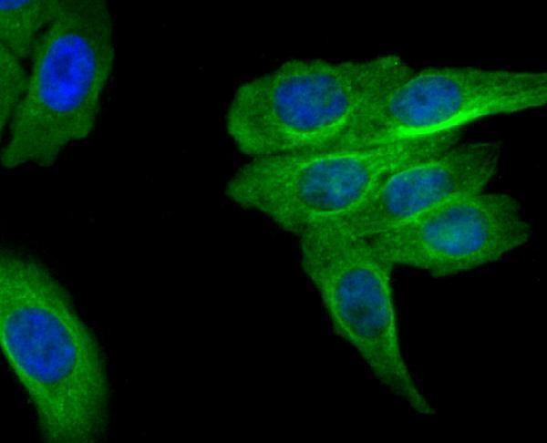
Real IF data ICC staining MRP2 in HepG2 cells (green). The nuclear counter stain is DAPI (blue). Cells were fixed in paraformaldehyde, permeabilised with 0.25% Triton X100/PBS.
Anti-MRP2 ABCC2 Antibody
Cat # A00974

A00974-1 has IHC image data from a paraffin-embedded human liver cancer section (A00974-1 image caption). A00974 lists IHC and ICC applications and has ICC image data from HepG2 cells (A00974 catalog applications; A00974 image caption).

Which to pick: Choose A00974-1 for tissue IHC: its image documents staining in a paraffin-embedded human liver cancer section; the fixative is unreported (A00974-1 image caption). Choose A00974 for IF/ICC: its ICC image documents staining in paraformaldehyde-fixed, permeabilised HepG2 cells (A00974 image caption). Neither is a documented cross-species choice; both list human reactivity only (catalog reactivity).

Each figure is that product's own IHC / IF validation image from its datasheet.

References

  1. UniProt Consortium. UniProt entry Q92887 (MRP2_HUMAN, ATP-binding cassette sub-family C member 2).
  2. Human Protein Atlas. ABCC2 tissue IHC expression (reliability: Enhanced).
  3. Human Protein Atlas. ABCC2 subcellular location (ICC-IF): Membrane.
  4. Human Protein Atlas. ABCC2 antibody validation summary (1 antibodies).
  5. Concurrence of novel mutations causing Gilbert's and Dubin-Johnson syndrome with poor clinical outcomes in a Han Chinese family. Journal of human genetics 2023 — PMC9812767.
  6. Papillary renal neoplasm with reverse polarity is biologically and clinically distinct from eosinophilic papillary renal cell carcinoma. Pathology international 2024 — PMC11551816.
  7. Papillary renal cell carcinoma with high-ABCC2 shows an immune-evasive profile associated with favorable response to immunotherapy. The Journal of pathology 2026 — PMC12805615.
  8. Strain- and sex-dependent circadian changes in abcc2 transporter expression: implications for irinotecan chronotolerance in mouse ileum. PloS one 2011 — PMC3108586.
  9. PubMed PMID:8797578 — UniProt-cited evidence.
  10. PubMed PMID:8662992 — UniProt-cited evidence.
  11. PubMed PMID:10464142 — UniProt-cited evidence.