This website uses cookies to ensure you get the best experience on our website.


- Table of Contents
This IHC-P guide covers membranous and cytoplasmic ABCC3 staining in glandular tissues (HPA tissue IHC). Start with the catalog antibody at 2–5 μg/ml (datasheet A02429-1), and assess membrane staining against the expected basolateral location (UniProt).

Expected localisation, validated protocols, controls and antibodies — the at-a-glance facts below, then the full design guide.
| Expected localisation | Membranous and cytoplasmic tissue staining (HPA tissue IHC) | |
| Staining pattern | Membranous and cytoplasmic staining in glandular cells (HPA tissue IHC) | |
| Antigen retrieval | EDTA pH 8.0 HIER, heat-mediated (datasheet A02429-1) | |
| Positive control | Adrenal gland+4 more · see all | |
| Negative control | Adipose tissue+4 more · see all |
| Fixation | Keep fixation consistent across matched paraffin sections. (selected-SKU IHC image A02429-1) | |
| Caveat | Liver RNA enrichment may not yield cholangiocyte staining (HPA tissue IHC) | |
| Regulation | No staining-linked regulator specified (UniProt) | |
| Isoform / epitope | 5 isoforms; check extracellular versus cytoplasmic epitope (UniProt) |
Compare the catalog antibody’s IHC-P protocol (datasheet A02429-1) with three published ABCC3 IHC methods (PMC8868318; PMC11145118; PMC11450052).
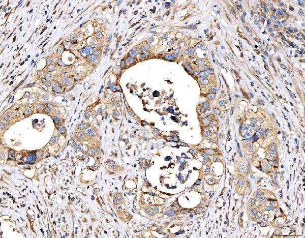
| Sample | Paraffin-embedded human appendiceal adenocarcinoma tissue; fixative not specified (datasheet A02429-1) |
| Fixation | Image fixative and duration unreported (datasheet A02429-1); verify before use. |
| Sectioning | 4–5 µm sections on charged slides (standard) |
| Deparaffinisation | Xylene, graded ethanol series to water (standard) |
| Antigen retrieval | Heat retrieval: EDTA pH 8.0 (datasheet A02429-1); 20 min, 95–100 °C (standard) |
| Peroxidase block | 3% H2O2, 10 min, room temperature (standard) |
| Blocking | 10% goat serum (datasheet A02429-1) |
| Primary antibody | Rabbit anti-ABCC3, 2-5 μg/ml (datasheet A02429-1) |
| Primary incubation | Overnight at 4 °C (datasheet A02429-1) |
| Detection | Streptavidin-biotin complex (SABC), DAB chromogen (datasheet A02429-1) |
| Counterstain | Hematoxylin, blue, dehydrate and mount (standard) |
| Expected result | ABCC3-positive staining in glandular cells of adrenal gland (HPA tissue IHC: High). HPA tissue profile: Membranous and cytoplasmic expression in several tissues including adrenal gland, gastrointestinal tract and gallbladder. No signal in the no-primary control. |
ABCC3 is a 17-transmembrane transporter at the basolateral or basal cell membrane (UniProt O15438 topology and subcellular location). In paraffin-section IHC, expect membrane-enriched staining in appropriate epithelial or glandular cells, with possible cytoplasmic staining (HPA: membranous and cytoplasmic tissue profile). HPA reports high glandular-cell staining in adrenal gland, appendix, colon, gallbladder and rectum (HPA: tissue IHC). Its tissue profile is rated Enhanced, with medium consistency between staining and RNA data (HPA: reliability).
| Membrane-enriched staining in glandular cells of colon or gallbladder, with some cytoplasmic staining (HPA: High glandular cells; membranous and cytoplasmic profile). | This fits the reported tissue pattern. In polarized intestinal epithelium, a basolateral bias is consistent with ABCC3 location (UniProt O15438: basolateral enterocyte membrane). Assess the membrane signal and cell identity together; cytoplasmic staining can coexist with the reported membrane pattern (HPA: tissue IHC profile). |
| Predominantly nuclear staining, with little convincing membrane signal (UniProt O15438: cell-membrane location; HPA: tissue IHC profile). | This is a compartment mismatch and warrants investigation as possible artefact. Review the counterstain, chromogen deposits and secondary-only control before assigning the signal to ABCC3 (standard IHC practice). The supplied sources do not describe nuclear ABCC3 localization (UniProt O15438; HPA: subcellular summary). |
| Prominent staining in adipocytes or liver cholangiocytes (HPA: Not detected in these specified cell types). | This conflicts with the HPA observations for those cells; consider cross-reactivity or endogenous detection activity (HPA: tissue IHC; standard IHC practice). The liver entry concerns cholangiocytes specifically and does not establish that every liver cell should be negative (HPA: liver cholangiocytes Not detected; UniProt O15438: liver expression). |
| Diffuse staining across cells and section background, without a clear cellular boundary (HPA: membrane-enriched tissue profile). | Treat the distribution as uninterpretable until section-wide background is resolved. Check a no-primary or secondary-only control, blocking, washing and detection conditions (standard IHC practice). Cytoplasmic signal alone is not automatically false: HPA reports both membranous and cytoplasmic tissue staining (HPA: tissue IHC profile). |
| No signal in glandular cells of an otherwise suitable colon or gallbladder section (HPA: High in those cell types). | The run may have failed, or the sampled cells may differ from the HPA observations; absence alone does not establish absent ABCC3 protein (HPA: tissue IHC; standard IHC interpretation). Check tissue morphology and the run control, then review retrieval, antibody dilution and detection against the antibody's validated IHC conditions (standard IHC practice). |
| Membrane topology and polarity (UniProt O15438 topology and subcellular location). | ABCC3 has 17 transmembrane segments and is reported at basolateral or basal membranes (UniProt O15438). Use membrane enrichment and the orientation of polarized cells when reading a section; topology alone does not identify the antibody epitope or prescribe retrieval conditions (UniProt O15438 topology; standard IHC interpretation). |
| Choice of tissue and cell compartment (HPA: tissue IHC). | HPA reports High glandular-cell staining in adrenal gland, appendix, colon, fallopian tube, gallbladder, rectum and seminal vesicle (HPA: tissue IHC). Its Not detected calls refer to named cell populations, such as adipocytes and cholangiocytes, rather than every cell in an entire section (HPA: tissue IHC). |
| Strength of antibody-specific evidence (HPA: antibody validation). | HPA048483 has Enhanced IHC validation, while CAB037136 is Approved for IHC (HPA: antibodies). These labels describe antibody-level validation; the overall tissue-profile reliability is separately Enhanced with medium RNA–staining consistency (HPA: antibodies; tissue IHC reliability). Interpret unexpected staining in light of the antibody actually used (standard IHC practice). |
| Isoforms and modifications (UniProt O15438: isoforms and glycosylation). | UniProt lists 5 isoforms and three glycosylation sites for ABCC3 (UniProt O15438). The payload gives no antibody epitope, isoform coverage or modification-specific IHC result; therefore these features cannot explain a particular positive or negative section without further antibody-specific evidence (UniProt O15438; HPA: supplied antibody summaries). |
| IF/ICC Q&A: where should fluorescence localize? (HPA: subcellular ICC-IF). | Mainly at the plasma membrane, a supported HPA ICC-IF location; basal-body localization is additional and uncertain (HPA: subcellular ICC-IF). This answers the localization question only. The supplied ICC-IF summary does not specify an IF protocol or establish that the IHC tissue pattern transfers unchanged to cultured cells (HPA: subcellular ICC-IF; tissue IHC). |
| Fixation and antigen retrieval evidence (HPA: tissue IHC; UniProt O15438). | Neither supplied source establishes ABCC3-specific fixation sensitivity or an optimal retrieval condition (HPA: tissue IHC; UniProt O15438). Retrieval may be checked against the chosen antibody's validated IHC procedure as a general workflow step, without inferring that fixation caused a particular ABCC3 staining result (standard IHC practice). |
| Situation | Likely cause | Next action |
|---|---|---|
| Expected glandular cells lack signal in a colon or gallbladder positive control (HPA: High glandular cells). | A failed staining run or a mismatch between the sampled cells and the reported HPA pattern is possible (HPA: tissue IHC; standard IHC interpretation). | Confirm that the relevant glandular cells are present, inspect the run control, and follow the selected antibody's validated IHC retrieval, dilution and detection settings (standard IHC practice). |
| Nuclei dominate the signal in a tissue expected to show membrane staining (UniProt O15438: membrane location; HPA: tissue IHC profile). | The compartment conflicts with the supplied localization evidence; stain deposits or nonspecific detection are possible (UniProt O15438; standard IHC interpretation). | Inspect a no-primary or secondary-only control and the counterstain; score ABCC3 only where cellular location and tissue context support it (standard IHC practice; HPA: tissue IHC). |
| Adipocytes or liver cholangiocytes stain strongly (HPA: Not detected in those named cell types). | Cross-reactivity or endogenous detection activity could account for unexpected signal (HPA: tissue IHC; standard IHC practice). | Verify cell identity and compare with a no-primary or secondary-only control; assess the unexpected cells separately from any other cell populations in the section (standard IHC practice; HPA: cell-specific calls). |
| A uniform haze obscures membranes and cell boundaries (HPA: membranous and cytoplasmic tissue profile). | Non-specific background from the staining workflow is possible; this appearance cannot be assigned to ABCC3 solely from intensity (standard IHC interpretation). | Check blocking, washes, antibody concentration and detection controls, then reassess whether distinct membrane-enriched cells remain visible (standard IHC practice; UniProt O15438: membrane location). |
| Cytoplasmic staining appears alongside a clear glandular-cell membrane rim (HPA: membranous and cytoplasmic tissue profile). | Both compartments are included in HPA's tissue description; the cytoplasmic component alone does not invalidate the result (HPA: tissue IHC profile). | Record membrane and cytoplasmic patterns separately, including their cell types and relative intensity, and compare them with the selected tissue's HPA observation (standard IHC scoring; HPA: tissue IHC). |
| An IF image shows a basal-body focus without convincing plasma-membrane signal (HPA: subcellular ICC-IF). | HPA calls basal-body localization uncertain and the plasma membrane the supported main location (HPA: subcellular ICC-IF). | Treat the focus as inconclusive for ABCC3 localization; evaluate plasma-membrane signal and appropriate fluorescence controls on the separate IF/ICC workflow (HPA: subcellular ICC-IF; standard IF practice). |


Comprehensive Human Protein Atlas IHC scoring per tissue (reliability: Enhanced — Medium consistency between antibody staining and RNA expression data.). Rows are taken directly from the HPA tissue chart — click any row's HPA link to view the source.
| Tissue | Cell type | Level | Evidence | Source |
|---|---|---|---|---|
| Adrenal gland | Glandular cells | High | Protein (IHC) | HPA → |
| Appendix | Glandular cells | High | Protein (IHC) | HPA → |
| Colon | Glandular cells | High | Protein (IHC) | HPA → |
| Fallopian tube | Glandular cells | High | Protein (IHC) | HPA → |
| Gallbladder | Glandular cells | High | Protein (IHC) | HPA → |
| Tissue | Cell type | Level | Evidence | Source |
|---|---|---|---|---|
| Adipose tissue | Adipocytes | Not detected | Protein (IHC) | HPA → |
| Bone marrow | Hematopoietic cells | Not detected | Protein (IHC) | HPA → |
| Caudate | Glial cells | Not detected | Protein (IHC) | HPA → |
| Cerebellum | Cells in granular layer | Not detected | Protein (IHC) | HPA → |
| Cerebral cortex | Endothelial cells | Not detected | Protein (IHC) | HPA → |
Use the catalog antibody’s documented paraffin-section conditions as a starting point, then judge ABCC3 staining by cell type, membrane pattern and controls.
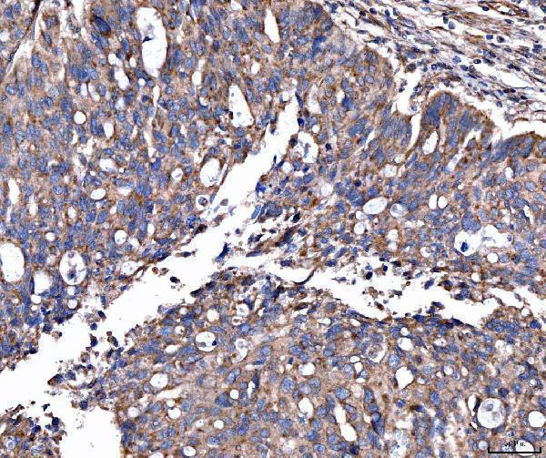
A02429-1 has real IHC data from human paraffin sections of appendiceal and colonic adenocarcinoma, placenta, and stomach cancer (catalog IHC captions); no IF data are supplied (catalog images).
A02429-1 is listed for human IHC and has images from paraffin sections of appendiceal adenocarcinoma and colonic adenocarcinoma (catalog applications, reactivity, and IHC captions). Its other IHC images show human placenta and stomach cancer paraffin sections (catalog IHC captions).
Which to pick: Choose A02429-1 for human paraffin-section IHC: it is a rabbit polyclonal antibody with a listed concentration of 2–5 μg/ml, and its images used EDTA retrieval at pH 8.0 (catalog antibody details and IHC captions). Its listed applications and images provide no IF/ICC validation, so there is no supported IF/ICC pick here (catalog applications and images). A02429-1 lists human reactivity only, so there is no supported cross-species pick; the fixative used for its paraffin sections is unreported (catalog reactivity and IHC captions).