This website uses cookies to ensure you get the best experience on our website.
- Table of Contents
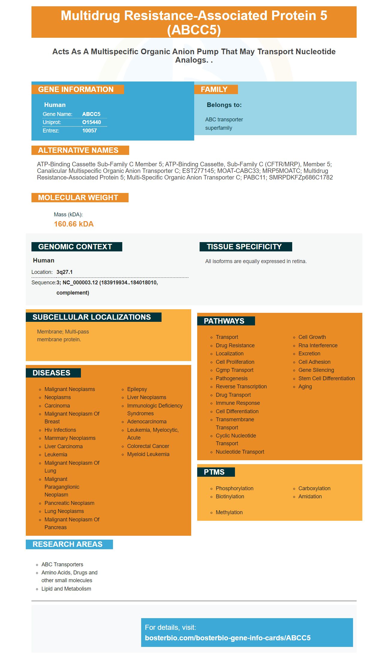
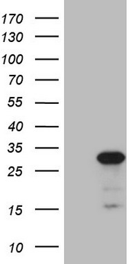
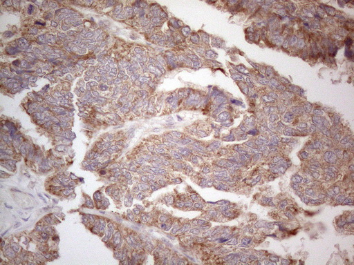
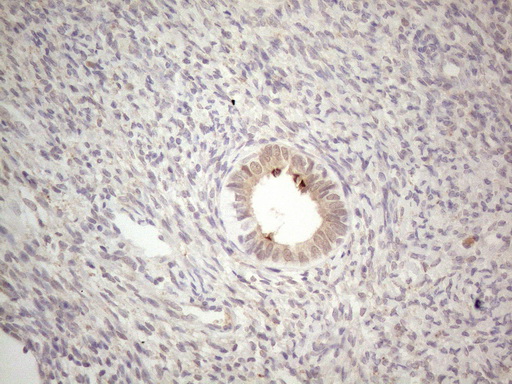
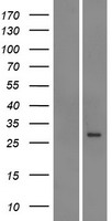
Facts about Multidrug resistance-associated protein 5.
Human | |
---|---|
Gene Name: | ABCC5 |
Uniprot: | O15440 |
Entrez: | 10057 |
Belongs to: |
---|
ABC transporter superfamily |
ATP-binding cassette sub-family C member 5; ATP-binding cassette, sub-family C (CFTR/MRP), member 5; canalicular multispecific organic anion transporter C; EST277145; MOAT-CABC33; MRP5MOATC; multidrug resistance-associated protein 5; Multi-specific organic anion transporter C; pABC11; SMRPDKFZp686C1782
Mass (kDA):
160.66 kDA
Human | |
---|---|
Location: | 3q27.1 |
Sequence: | 3; NC_000003.12 (183919934..184018010, complement) |
All isoforms are equally expressed in retina.
Membrane; Multi-pass membrane protein.
This Boster Bio article provides more information on the ABCC5 mark's benefits. This article will give you valuable information about the ABCC5 markers, including the benefits of protein transefficiency and recording test results. Visit Boster's website for more information. You can also find additional information about Steven Boster, the ABCC5 marker, as well as Steven Boster.
Membrane staining and electrophoretic transfer are two common methods for transferring proteins between cell membranes. Each method differs in the speed and efficiency of the transfer, as well as the equipment required. One thing is clear: each method has both advantages and drawbacks. The most appropriate technique will depend on your experimental requirements and the time available. Here are some tips to help you choose the right transfer method.
First prepare your sample. Prepare a sample comb for the protein transfer process. The sample comb should be gently placed on top of the membrane to prevent bubbles. Incubate at 37 degrees Celsius for 30-60 seconds. After the gel has fully solidified, you can remove the sample. After the gel has cooled, you can apply either the BCIP/NBT substrate or DAB substrate solutions to the membrane. For the best signal to noise ratio you should allow the membrane to incubate for a minimum of 10 minutes.
Protein quantitation assay requires accurate quantification of protein concentrations. The protein concentration must be equal in order to achieve the best results. Boster Bio offers customized Western Blot Services, including total Protein Extraction from Tissues and Premade Blot Construction. There are many options for immunoostaining with any antibody and reagent. After each process, high-quality images are provided by the company.
ELISA provides the best sensitivity. The sensitivity of a commercially-available membrane stain can be improved by preparing a solution with a concentration of 0.5% ferrous sulfate. Transfer your proteins to either a PVDF, or nitrocellulose membrane and allow them to dry at 80 mA. The membranes are then treated with Coomassie Brilliant Blue R-250 dye for reversible detection of protein bands.
The most used method for protein staining is optical oomassie brilliant. It is easy and affordable. The color binds to protein by ionic, hydrophobic, or chemical interactions. It allows for reduced protein extraction. It can stain blood protein and has applications in forensics. It is an excellent choice to sequence proteins.
Researchers are facing many challenges when trying to develop effective treatments for prostate cancer patients with castration-resistant disease. ABCC5 gene expression levels could be linked with drug resistance. Bioinformatics was used to study ABCC5's expression. We conducted cell proliferation assays, migration and invasion assays, as well as western blots. Patients with ABCC5 displayed poor prognosis as well as high levels if drug resistance.
Autoradiography uses photochemical techniques to determine the spatial distribution of radiolabeled chemicals on a sample. Autoradiography is generally divided into two main types: macroautoradiography and microautoradiography. The differences between these two types depend on the type specimen, the emulsion, and the analysis method used. Autoradiography is used most often for nucleic acid mixture and quantitative analysis of samples. The type of test required depends on the emitters used, the label used and the resolution.
This technique was initially developed for medical applications, but has recently been used to analyze environmental samples. Autoradiography labels biological structures using radioactive compounds. These compounds are added to an emulsion that contains silver bromide. As the emulsion forms, the radioactive sample reacts to the silver bromide in the emulsion. This interaction leads to the formation silver grains on top. These images are easily analyzed using microscopy.
Radiographic film is made up of several components. The base, which is made of clear polyester material is 150 um thick. This base is used as a support layer for the film's components. The base can also include a light-blue dye to enhance the image appearance on the viewbox. The emulsion is what makes up the film's main active component. It is composed of small silver halide crystallines suspended in gelatin. The emulsion layer is usually 10 um thick.
There are many different ways to record test results using autoradiography film. You can use a variety of techniques to make the film more reproducible. Among them are apposition and defatting techniques. For example, thawmount autoradiography allows for imaging tissues at the cellular as well as subcellular levels. Another method of recording autoradiography images is incubation receptor autoradiography.
PMID: 9827529 by Belinsky M.G., et al. Characterization of MOAT-C and MOAT-D, new members of the MRP/cMOAT subfamily of transporter proteins.
PMID: 10438534 by McAleer M.A., et al. pABC11 (also known as MOAT-C and MRP5), a member of the ABC family of proteins, has anion transporter activity but does not confer multidrug resistance when overexpressed in human embryonic kidney 293 cells.