ABCC5 / ATP-binding cassette sub-family C member 5 · IHC design guide

Design Immunohistochemistry for ABCC5

Plan paraffin-section ABCC5 IHC with the catalog antibody's validated conditions (datasheet A03040-1). Interpret cytoplasmic tissue staining (HPA tissue IHC) alongside annotated membrane routing (UniProt), while accounting for uncertain staining reliability (HPA tissue IHC).

Evidence assembled Oct 2026 · For research use; verify linked source records and product datasheet before use
Immunohistochemistry protocol sheet for ABCC5 (IHC for ABCC5): expected localisation Cytoplasmic staining in several tissues (HPA tissue IHC), antibody A03040-1, validated IHC image, and IHC protocol steps
Printable ABCC5 IHC protocol sheet — expected localisation Cytoplasmic staining in several tissues (HPA tissue IHC), antibody A03040-1, controls and protocol steps. Open the full ABCC5 IHC guide →

ABCC5 Immunohistochemistry Experimental Design Guide

Expected localisation, validated protocols, controls and antibodies — the at-a-glance facts below, then the full design guide.

Must know before staining
Expected localisation Cytoplasmic staining in several tissues (HPA tissue IHC)
Staining pattern Cytoplasmic signal in kidney tubules and cardiomyocytes (HPA tissue IHC)
Antigen retrieval EDTA pH 8.0 HIER, heat-mediated (datasheet A03040-1)
Positive control ⓘ Appendix+4 more · see all
Negative control ⓘ Adipose tissue+4 more · see all
Important caveats
Reasons your staining may differ from the expected pattern.
Fixation Keep fixation consistent across sections (standard IHC practice; not target-specific); Matched tissue-IHC evidence does not establish the fixation claim. Validate the specimen-specific method before use. (selected-SKU IHC image A03040-1)
Caveat Staining has low concordance with RNA; retesting is pending (HPA tissue IHC)
Regulation Low tissue specificity at the RNA level (HPA tissue RNA)
Isoform / epitope 5 isoforms; check epitope location across membrane and cytoplasmic regions (UniProt)
Section 1

Recommended ABCC5 IHC & IF Protocols

The catalog antibody uses an IHC-P protocol (datasheet A03040-1). The published options below cover paraffin sections (PMC9525794 methods) and frozen sections with chromogenic detection (PMC4053136 methods).

Recommended immunohistochemistry (IHC-P) protocol parameters
SampleParaffin-embedded human gastric cancer tissue; fixative not specified (datasheet A03040-1)
FixationImage fixative and duration unreported (datasheet A03040-1); verify before use.
Sectioning4–5 µm sections on charged slides (standard)
DeparaffinisationXylene, graded ethanol series to water (standard)
Antigen retrievalHeat retrieval: EDTA pH 8.0 (datasheet A03040-1); 20 min, 95–100 °C (standard)
Peroxidase block3% H2O2, 10 min, room temperature (standard)
Blocking10% goat serum (datasheet A03040-1)
Primary antibodyRabbit anti-ABCC5, 2-5μg/ml (datasheet A03040-1)
Primary incubationOvernight at 4 °C (datasheet A03040-1)
DetectionStreptavidin-biotin complex (SABC), DAB chromogen (datasheet A03040-1)
CounterstainHematoxylin, blue, dehydrate and mount (standard)
Expected resultABCC5-positive staining in lymphoid tissue of appendix (HPA tissue IHC: Medium). HPA tissue profile: Cytoplasmic expression in several tissues. No signal in the no-primary control.
💡Decision noteStart with EDTA pH 8.0 heat retrieval for the catalog antibody (datasheet A03040-1); neither published option specifies antigen retrieval (PMC9525794 methods; PMC4053136 methods).
Section 2

What Is the Expected ABCC5 Staining Pattern?

ABCC5 is a 13-transmembrane-segment transporter that usually routes to the basolateral membrane; brain capillary endothelial cells are an apical exception (UniProt O15440 topology and subcellular location). Tissue IHC reports cytoplasmic staining in several tissues, including kidney tubule cells and placental trophoblastic cells at medium levels (HPA tissue IHC). Treat these patterns as provisional: HPA rates the tissue IHC profile Uncertain because antibody staining and RNA expression show low consistency (HPA tissue IHC).

What am I looking at on my slide?
Medium staining in kidney tubule cells or placental trophoblastic cells (HPA tissue IHC).This matches reported positive cell populations and intensity (HPA tissue IHC). Record cytoplasmic and membrane-associated signal separately: HPA describes cytoplasmic tissue staining, while UniProt places ABCC5 at cell membranes (HPA tissue IHC; UniProt O15440 subcellular location). Neither pattern alone establishes antibody specificity because tissue IHC reliability is Uncertain (HPA tissue IHC).
Clear membrane enrichment, with any cytoplasmic staining confined to the same cells.Membrane signal fits ABCC5 topology and routing; intracellular staining can fit its annotated endosome and granule locations (UniProt O15440 topology and subcellular location). HPA tissue IHC describes a broader cytoplasmic pattern, so score both compartments and avoid requiring a sharply outlined membrane in every positive cell (HPA tissue IHC).
Predominantly nuclear staining, without convincing membrane or cytoplasmic signal.Do not call nuclear-only tissue IHC the expected result: HPA tissue IHC describes cytoplasmic staining, and UniProt lists membrane and intracellular locations (HPA tissue IHC; UniProt O15440 subcellular location). HPA also reports nucleoplasm as an additional ICC-IF location, so nuclear signal needs application-specific controls before interpretation (HPA subcellular ICC-IF).
Strong staining in adipocytes or adrenal glandular cells, or widespread color outside cells.These cell populations are listed as not detected in HPA tissue IHC, making strong staining there a specificity or detection-control concern (HPA tissue IHC). Color outside cells may reflect nonspecific background or endogenous chromogenic activity (general IHC practice). The Uncertain HPA tissue profile makes any single negative population an imperfect exclusion control (HPA tissue IHC).
No staining in kidney tubule cells or placental trophoblastic cells.Both are reported at medium staining, so a blank slide prompts a check of the IHC workflow and tissue preservation (HPA tissue IHC; general IHC practice). It does not establish absent ABCC5 expression: the reported tissue pattern has low consistency with RNA expression and is pending retesting (HPA tissue IHC).
💡Expected ABCC5 appearanceCall a provisional positive when discrete cellular signal is seen in reported medium populations such as kidney tubule or placental trophoblastic cells, scoring cytoplasmic and membrane signal separately; diffuse color or strong staining in HPA not-detected populations is suspect (HPA tissue IHC; UniProt O15440 subcellular location; general IHC practice).
How each factor affects the staining
Membrane topology and compartmentABCC5 has 13 transmembrane segments and is annotated at cell membranes, endosomes and cytoplasmic granules (UniProt O15440 topology and subcellular location). The distinction matters when scoring membrane versus cytoplasmic IHC signal; it does not identify which compartment a particular antibody epitope detects.
Tissue distribution and validationHPA reports medium staining in kidney tubules, cardiomyocytes, trophoblastic cells, smooth muscle cells and some lymphoid cells, but not detected in several other listed populations (HPA tissue IHC). Its overall tissue IHC reliability is Uncertain; HPA052295 has an Uncertain IHC rating (HPA tissue IHC; HPA antibodies).
Isoforms and epitope coverageUniProt lists five ABCC5 isoforms (UniProt O15440 isoforms). The supplied record gives no antibody epitope or isoform coverage, so an unstained cell cannot be assigned to an isoform on this evidence. Check antibody documentation before treating variation in staining as a biological isoform difference.
IF/ICC Q&A: where should signal appear?HPA reports plasma membrane and cell junctions as approved main ICC-IF locations, with nucleoplasm as an approved additional location; images are listed for MCF-7 and RT-4 (HPA subcellular ICC-IF). These are IF observations, not an IHC-P protocol or proof that nuclear-only tissue IHC is specific.
Why is my staining missing, weak or wrong?
SituationLikely causeNext action
Reported positive cells are blank, while the section is otherwise interpretable.Detection, antigen retrieval or antibody conditions may need review (general IHC practice); HPA's medium positive calls are themselves Uncertain (HPA tissue IHC).Check the catalog antibody's IHC-P instructions, reagent controls and tissue morphology; compare kidney tubule or placental trophoblastic cells with a negative control on the same run (HPA tissue IHC; general IHC practice).
The whole section has weak, even chromogenic color.Nonspecific antibody binding, incomplete blocking or detection background can produce diffuse signal (general IHC practice).Compare with a primary-antibody-omission control and review blocking, washing and detection steps; score only discrete cellular staining (general IHC practice).
Color persists when the primary antibody is omitted.Endogenous enzyme activity or the detection system can contribute signal in chromogenic IHC (general IHC practice).Review the appropriate endogenous-activity blocking step and detection controls before attributing the color to ABCC5 (general IHC practice).
Strong signal appears in adipocytes or adrenal glandular cells.These cells are not detected in the supplied HPA tissue IHC profile; cross-reactivity or background is possible, although that profile is Uncertain (HPA tissue IHC).Compare positive and negative cell populations under identical staining conditions and inspect primary-omission controls; do not infer specificity from intensity alone (HPA tissue IHC; general IHC practice).
Only nuclei stain in tissue IHC.The pattern differs from HPA's cytoplasmic tissue IHC description and UniProt membrane routing, despite an additional nucleoplasmic ICC-IF location (HPA tissue IHC; UniProt O15440 subcellular location; HPA subcellular ICC-IF).Record the compartment explicitly, inspect detection controls and seek independent antibody or orthogonal support before assigning nuclear tissue signal to ABCC5 (general IHC practice).
Staining varies between reported positive tissues.HPA calls some populations medium and stomach or intestinal glandular cells low; its tissue IHC reliability is Uncertain (HPA tissue IHC).Compare the named cell types rather than whole-section color, and document intensity and compartment for each; avoid converting a weak result into a firm absence call (HPA tissue IHC; general IHC practice).

Sample controls for ABCC5 IHC & IF

🧪Run kidney first: tubular cells should stain (HPA: kidney cells in tubules, Medium); use adipose tissue as the negative tissue, scoring adipocytes (HPA: adipocytes, Not detected). On the kidney slide, cells outside the tubules should lack specific chromogen, but their ABCC5-negative status is unverified by the supplied rows (HPA: kidney cells in tubules, Medium).
Positive control tissue: Appendix (Lymphoid tissue, HPA Medium)
Negative control tissue: Adipose tissue (HPA Not detected)
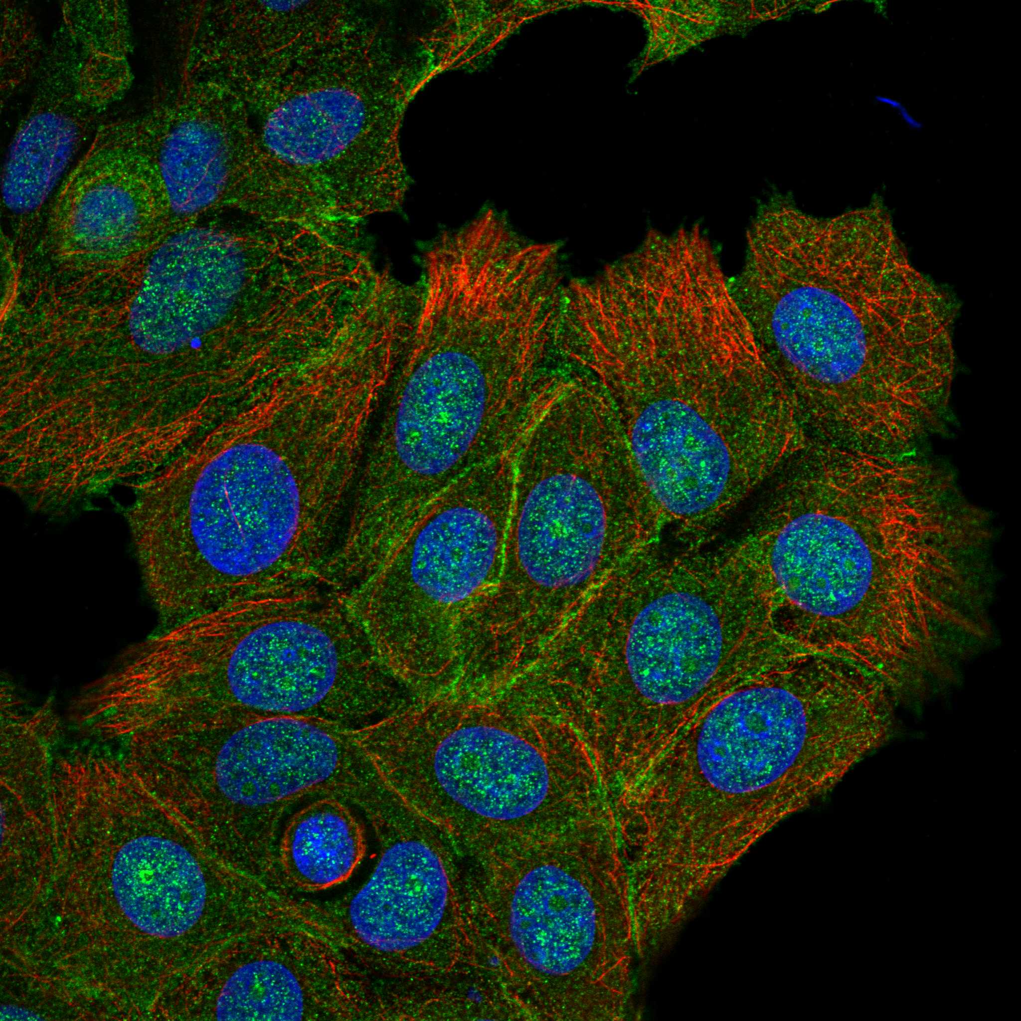
ICC-IF cell lines (HPA subcellular resource): HPA ICC-IF images show ABCC5 in MCF-7, RT-4, with annotated localisation: Plasma membrane (approved), Cell Junctions (approved) (HPA subcellular).
Technical controls: Include no-primary (secondary-only) and rabbit isotype controls matched to the primary antibody’s clonality where available (selected-SKU caption: rabbit primary; standard IHC practice), plus ABCC5 knockout tissue or a validated peptide-block control (standard IHC practice). For kidney, check endogenous peroxidase and biotin background, particularly with the caption’s biotin-based detection (selected-SKU caption: biotinylated secondary and SABC; standard IHC practice).
⚠️Feasibility: The selected-SKU paraffin-section caption reports heat retrieval in EDTA at pH 8.0, but does not establish whether retrieval is required; its fixative is unreported (selected-SKU tissue-IHC caption). A target-specific fixation window or fixation effect is unreported, and the supplied evidence does not establish that frozen sections or IF/ICC are easier (selected-SKU tissue-IHC caption; HPA: ICC-IF images in MCF-7 and RT-4). In kidney, endogenous biotin can complicate interpretation with the caption’s biotin-based detection (selected-SKU tissue-IHC caption; standard IHC practice).

HPA tissue IHC evidence for ABCC5

Comprehensive Human Protein Atlas IHC scoring per tissue (reliability: Uncertain — Low consistency between antibody staining and RNA expression data. Pending retesting.). Rows are taken directly from the HPA tissue chart — click any row's HPA link to view the source.

Positive expression · recommended positive controls

TissueCell typeLevelEvidenceSource
Appendix Lymphoid tissue Medium Protein (IHC) HPA →
Heart muscle Cardiomyocytes Medium Protein (IHC) HPA →
Kidney Cells in tubules Medium Protein (IHC) HPA →
Lymph node Germinal center cells Medium Protein (IHC) HPA →
Placenta Trophoblastic cells Medium Protein (IHC) HPA →

Undetected expression · recommended negative controls

TissueCell typeLevelEvidenceSource
Adipose tissue Adipocytes Not detected Protein (IHC) HPA →
Adrenal gland Glandular cells Not detected Protein (IHC) HPA →
Bone marrow Hematopoietic cells Not detected Protein (IHC) HPA →
Breast Adipocytes Not detected Protein (IHC) HPA →
Bronchus Respiratory epithelial cells Not detected Protein (IHC) HPA →
Section 3

Advanced ABCC5 IHC Tips

Troubleshoot ABCC5 staining in paraffin sections by checking retrieval, compartment, controls and cell-specific scoring before interpreting chromogenic signal.

Which retrieval condition should I start with for ABCC5 paraffin-section IHC?
Start with heat-mediated retrieval in EDTA at pH 8.0 for paraffin sections (datasheet A03040-1). The selected gastric cancer section used this condition with 2 μg/ml primary antibody overnight at 4°C before SABC/DAB detection (datasheet A03040-1). If staining is weak, compare retrieval duration on adjacent sections while keeping antibody concentration and detection constant (standard IHC practice). Assess membrane detail as well as staining intensity, because ABCC5 has 13 transmembrane segments and multiple reported cellular locations (UniProt O15440 topology and subcellular location). Interpret any apparent improvement alongside controls, since tissue staining and RNA expression show low consistency (HPA: reliability Uncertain).
How should I evaluate fixation when ABCC5 staining varies between sections?
The selected paraffin-section caption does not report its fixative, so ABCC5-specific fixation sensitivity is unknown (datasheet A03040-1). Record each specimen’s fixative, time in fixative and processing history before comparing staining between sections (standard IHC practice). Process adjacent sections together with the same EDTA pH 8.0 retrieval and antibody incubation to reduce workflow variation (datasheet A03040-1; standard IHC practice). Examine morphology and the distribution of chromogen, because damaged or unevenly processed tissue can complicate compartment scoring (standard IHC practice). Report fixation differences as a limitation rather than attributing weak staining to a specific ABCC5 epitope without direct evidence (standard IHC practice).
Which compartments should count as plausible ABCC5 staining in tissue?
Prioritise convincing cell-border staining: ABCC5 commonly routes to the basolateral membrane, while brain capillary endothelial cells can show apical localisation (UniProt O15440 subcellular location). Endosome membranes, cytoplasmic granules and the Golgi apparatus are also reported locations, so intracellular staining needs careful morphological review (UniProt O15440 subcellular location). Approved cell-imaging annotations include plasma membrane, cell junctions and an additional nucleoplasmic location (HPA: subcellular). Tissue IHC is described as cytoplasmic across several tissues, but its reliability is Uncertain (HPA: tissue IHC). Score membrane and intracellular patterns separately, and require independent support before treating nuclear-only chromogen as specific ABCC5 staining (standard IHC practice; HPA: subcellular).
Could an unmapped epitope explain different ABCC5 staining patterns?
Check the antibody’s mapped immunogen or epitope before assigning a negative section to absent ABCC5, because the record lists 5 isoforms (UniProt O15440 isoforms). ABCC5 also has 13 transmembrane segments and 8 annotated glycosylation sites, making epitope position relevant to interpretation (UniProt O15440 topology and glycosylation). The supplied caption does not identify the antibody epitope or establish which isoforms it detects (datasheet A03040-1). If sequence information becomes available, compare the epitope with each isoform and its predicted membrane orientation (standard IHC practice). Until then, document the staining compartment without claiming isoform specificity or a glycosylation-dependent staining effect (standard IHC practice).
How can I check the ABCC5 pattern with multiplex IF?
For a separate IF experiment, pair ABCC5 with a validated marker for the cell type being assessed and inspect overlap at individual cell borders (standard IF practice). Choose fluorophores after examining unstained tissue for autofluorescence, and include single-stain controls when setting channels (standard IF practice). Match permeabilisation to the antibody epitope’s side of the membrane: intracellular access may require it, while excessive treatment can obscure membrane outlines (standard IF practice). The supplied IHC caption does not map that epitope or provide an IF permeabilisation condition (datasheet A03040-1). Compare any IF pattern with reported plasma-membrane and junction localisation, without treating IF images as validation of this IHC antibody (HPA: subcellular; standard IF practice).
How do I separate ABCC5 signal from chromogenic background?
The selected section used 10% goat serum blocking, a biotinylated secondary antibody, SABC and DAB (datasheet A03040-1). Run a no-primary control through the same detection steps to reveal secondary, biotin-system or chromogen background (standard IHC practice). Include a peroxidase block for DAB detection, and check whether residual colour persists in the no-primary section (standard chromogenic IHC practice). Review tissue edges, folds and poorly preserved areas separately from intact cells before changing antibody concentration (standard IHC practice). If background remains high, adjust blocking, washing or detection conditions one variable at a time while retaining the documented 2 μg/ml primary condition as a reference (datasheet A03040-1; standard IHC practice).
How should I quantify ABCC5 staining across paraffin sections? ⚠ ANSWER MARKED FOR VERIFICATION
Define the cell population and compartment before scoring, then record membrane and intracellular staining separately (standard IHC practice; UniProt O15440 subcellular location). For cell-based analysis, report percentage positive and an H-score from 0–300 using intensity grades 0–3 (standard IHC practice). Normalise positive-cell counts to the number of eligible cells, or report stained-cell density per mm² of assessed tissue (standard IHC practice). Exclude folds, necrosis and tissue edges by a prespecified rule, and apply identical thresholds across compared slides (standard IHC practice). State the scoring compartment and controls, since the available tissue IHC profile has Uncertain reliability (HPA: tissue IHC).
What makes an ABCC5-positive IHC result convincing?
Look for reproducible staining in intact cells with a plausible membrane or intracellular pattern and little signal in matched no-primary controls (UniProt O15440 subcellular location; standard IHC practice). Check cell identity before comparing tissues: the HPA reports medium staining in kidney tubule cells and low staining in stomach glandular cells (HPA: tissue IHC). Treat uniform nuclear-only colour, edge accentuation and necrotic staining cautiously, while recognising that an additional nucleoplasmic location is reported (HPA: subcellular; standard IHC practice). Investigate chromogen in the no-primary control as possible detection background, including endogenous enzyme activity (standard chromogenic IHC practice). Avoid declaring biological absence or specificity from one section because the tissue staining profile is rated Uncertain (HPA: tissue IHC).
Boster reagents

Best ABCC5 / ATP-binding cassette sub-family C member 5 IHC Antibodies

A03040-1 has IHC images from paraffin-embedded human gastric and mammary cancer sections (A03040-1 image captions). No IF/ICC data are supplied (catalog).

Real IHC data IHC analysis of MRP5/ABCC5 using anti-MRP5/ABCC5 antibody (A03040-1). MRP5/ABCC5 was detected in paraffin-embedded section of human gastric cancer tissue. Heat mediated antigen retrieval was performed in EDTA buffer (pH8.0, epitope retrieval solution). The tissue section was blocked with 10% goat serum. The tissue section was then incubated with 2μg/ml rabbit anti-MRP5/ABCC5 Antibody (A03040-1) overnight at 4°C. Biotinylated goat anti-rabbit IgG was used as secondary antibody and incubated for 30 minutes at 37°C. The tissue section was developed using Strepavidin-Biotin-Complex (SABC) (Catalog # SA1022) with DAB as the chromogen.
Anti-MRP5/ABCC5 Antibody
Cat # A03040-1

A03040-1 is the sole card and is listed for human IHC (catalog: applications and reactivity). Its images show staining in paraffin-embedded human gastric and mammary cancer sections (A03040-1 image captions).

Which to pick: Choose A03040-1 for human paraffin-section chromogenic IHC: it is a rabbit polyclonal antibody with a listed IHC dilution of 2–5 μg/ml (catalog), and its own caption shows EDTA retrieval and DAB detection (A03040-1 image caption). The fixative is unreported (A03040-1 image captions). No listed SKU supports an IF/ICC or cross-species recommendation: A03040-1 lists human reactivity and has no IF/ICC application or image (catalog).

Each figure is that product's own IHC / IF validation image from its datasheet.

References

  1. UniProt Consortium. UniProt entry O15440 (MRP5_HUMAN, ATP-binding cassette sub-family C member 5).
  2. Human Protein Atlas. ABCC5 tissue IHC expression (reliability: Uncertain).
  3. Human Protein Atlas. ABCC5 subcellular location (ICC-IF): Mainly localized to the plasma membrane and cell junctions. In addition localized to the nucleoplasm..
  4. Human Protein Atlas. ABCC5 antibody validation summary (2 antibodies).
  5. Loss of miR-516a-3p mediates upregulation of ABCC5 in prostate cancer and drives its progression. OncoTargets and therapy 2018 — PMC6039062.
  6. Identification and Validation of Three-Gene Signature in Lung Squamous Cell Carcinoma by Integrated Transcriptome and Methylation Analysis. Journal of oncology 2022 — PMC9525794.
  7. Three novel ABCC5 splice variants in human retina and their role as regulators of ABCC5 gene expression. BMC molecular biology 2007 — PMC1890297.
  8. ABCC5 supports osteoclast formation and promotes breast cancer metastasis to bone. Breast cancer research : BCR 2012 — PMC4053136.
  9. PubMed PMID:9827529 — UniProt-cited evidence.
  10. PubMed PMID:10438534 — UniProt-cited evidence.
  11. PubMed PMID:10721709 — UniProt-cited evidence.