This website uses cookies to ensure you get the best experience on our website.
- Table of Contents
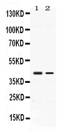
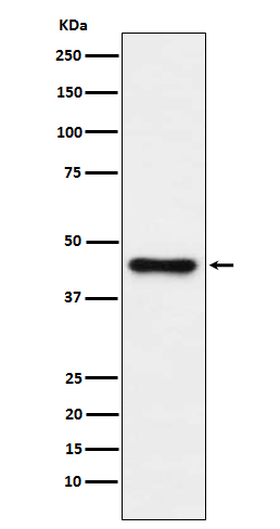
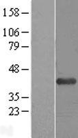
Facts about 1-acylglycerol-3-phosphate O-acyltransferase ABHD5.
Involved in keratinocyte differentiation (PubMed:18832586). Regulates lipid droplet fusion (By similarity).
Human | |
---|---|
Gene Name: | ABHD5 |
Uniprot: | Q8WTS1 |
Entrez: | 51099 |
Belongs to: |
---|
peptidase S33 family |
abhydrolase domain containing 5; Abhydrolase domain-containing protein 5,1-acylglycerol-3-phosphate O-acyltransferase ABHD5; CDS; CGI58; EC 2.3.1.51; IECN2; Lipid droplet-binding protein CGI-58; MGC8731; NCIE2CGI-58
Mass (kDA):
39.096 kDA
Human | |
---|---|
Location: | 3p21.33 |
Sequence: | 3; NC_000003.12 (43690870..43734371) |
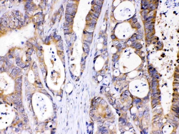
Widely expressed in various tissues, including lymphocytes, liver, skeletal muscle and brain. Expressed by upper epidermal layers and dermal fibroblasts in skin, hepatocytes and neurons (at protein level).
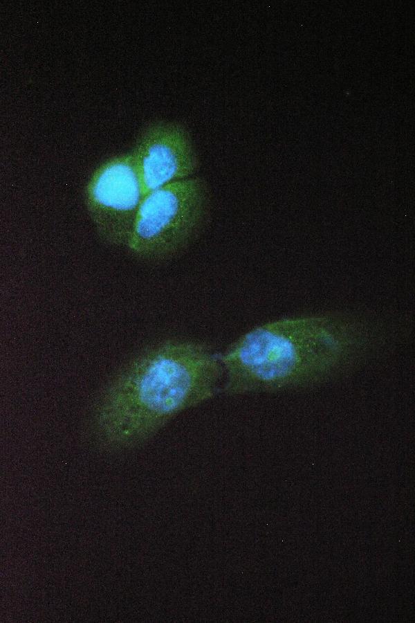
Cytoplasm. Lipid droplet. Cytoplasm, cytosol. Colocalized with PLIN and ADRP on the surface of lipid droplets. The localization is dependent upon the metabolic status of the adipocytes and the activity of PKA (By similarity).
The lysosomal enzyme ABHD5 marks regulates autophagic uracil yeil yield. Its best uses include predicting sensitivity to FU-based chemotherapy, and assessing the level of autophagic uracil in tumors. The ABHD5 is a promising tool for cancer research, as it is widely applicable worldwide.
Lysosomal-RNASET2 is a major player in cancer metabolism. ABHD5 deficiency reduces RNASET2 activities and increases ROS formation, both of which can increase tumor-promoting benefits. ABHD5 insufficient activation activates the ROS–inflammasome pathway, which may be crucial for tumor development. A study in mice showed that ABHD5 deficiency reduced cathepsin activity. Both mRNA expression as well as enzymatic activity were also affected.
Five members of the ABHD group of enzymes have a different amino acids residue. Although some members may be expressed in different tissues or are not yet well understood, they all play a part in many human diseases. Study ABHD5 has been shown to be a useful biomarker in determining CRCs' sensitivity against FU-based chemotherapy. There are many advantages to using ABHD5 as a biomarker for CRC treatment.
The ABHD5 gene is also associated with adipose tissue metabolism. It is encoded by the SIN3B gene, and is a member of the transcription regulator family B. It is thought that decreased expression of SIN3B promotes secondary storage of MPS. The genes ABHD5 (or DGAT1) are involved in the progression of cancer and play a role in regulating N-acyl phospholipid metabolism.
ABHD5 increases RNase activity, which promotes RNA degradation. Consequently, FU-sensitivity can be reduced by knocking out ABHD5 from dMMR cells. The effect of knockdown in ABHD5 and FU sensitivity is small and unremarkable. The correlation coefficient of FU proficiency and response to FU is 0.53.
The ABHD5 gene regulates cytosolic PH and lysosome functions. It is a biomarker that can be used to predict CRC sensitivity to FU chemotherapy. Moreover, it affects cathepsin mRNA expression and lysosomal RNA stability. However, further studies are required to determine the role that ABHD5 plays in CRC.
ABHD5 is expressed in CRCs and is correlated by other markers of autophagy. FU, a powerful cytotoxic agent, inhibits tumor growth and promotes apoptosis in pMMR/ABHD5low cells. However, FU-induced apoptosis only has a small impact on sensitivity.
Interestingly, the ABHD5 gene is required for the biogenesis the lysosome. It is involved with the trafficking of more 50 hydrolytic enzymes from Golgi to the Lysosome. Lysosomal proteins are transported to lysosomes via two distinct pathways. These include receptor-assisted transport and ER mediated transport.
While lysosomes used to be thought of as cellular garbage bins, newer research has shown that they are crucial in controlling cell function. Lysosomes not only recycle macromolecules but also play a key role in energy regulation. They sense nutrient status, and induce starvation adaptations, including translocations of TFEB to a nucleus and lysosomal genesis.
It is not known whether ABHD5 directly influences RNASET2, which, like ABHD5, regulates the lysosome function. Protein-protein docking studies show that ABHD5 & RNASET2 interact through a common domain. The b structure of PDIA5 & ABHD5 also determines the interaction.
ABHD5, a biomarker that can predict sensitivity to FU-based cancer treatment, is a promising candidate. This gene plays a critical role in lysosome function. It competes directly with RNASET2 to interact with PDIA5. RNASET2 is inactive when ABHD5 is not present. This causes RNASET2 to become inactive and promotes exogenous uracil uptake. Moreover, ABHD5+ CRC cells have inherent resistance against FU-based chemotherapy.
The Genomics of Drug Sensitivity in Cancer Project, (GDSP), has collected data from multiple cell lines of CRC. ABHD5 proficiency was found to correlate positively with IC50 to FU in MSI(dMMR), MSS (pMMR), and MSS CRC cells. Further, ABHD5 overexpression and knockdown cells exhibited decreased cell viability under FU challenge.
A new study found that pMMR CRC cell responses to FU-based treatment are not as efficient when they are expressing ABHD5-tagged LC3B plasmid19. FU-induced cellular autophagy signaling is not well-received by dMMRCR cells. However, manipulations in ABHD5-proficient cells had a slight effect on FU sensitivity. The correlation coefficient between FU sensitivity and ABHD5 proficiency in dMMR cells was 0.53.
The ABHD5 genes predict sensitivity to FU based chemotherapy. They also facilitate autophagic uracil generation. This process is prosurvival in cancer cells. Cytotoxicity results from FU-induced FU-induced phagolysis depletion. The increased uracil level of ABHD5-proficient cells could be due to an adaptive mechanism that increases autophagic urail yield.
It targets autophagic Urine Decarboxylases (AUD) and promotes their synthesis and degradation. This is a hallmark feature of autophagy and acts as a prosurvival mechanism for cancer cells. However, FU has an autophagy-inducing effect that inhibits nucleoside metabolism, causing cytotoxicity and death. The increased yield in uracil may serve as a compensatory mechanism to protect cells.
In vitro, the RNASET2 enzyme is required for autophagic uracil yield. Chloroquine, an inhibitor of autophagy flux, rescues intracellular uracil and FU levels in ABHD5 overexpressing SW480 cells. Its mechanism is not yet known but it targets the autophagic uracil production and is therefore an important therapeutic target of URDD.
It inhibits ULK1 (which inhibit autophagy) in vitro. MRT67307 was found to inhibit autophagy in mice. ULK68921 also inhibited this process in a drug-resistant ULK1 cell line. This suggests that ULK1 might be an important target in cancer treatment. It is possible that drugs that inhibit URDD will be developed to target ULK1.
Fluorouracil is a commonly used chemotherapy for colorectal cancer (CRC). It inhibits thymidylate production and suppresses Pyrimidine synthesis. Fluorouracil causes cell death by incorporating into DNA or RNA. This process is essential to cancer survival and treatment resistant. Drugs targeting URDD should target autophagy.
Similarly, TRIM32 regulates the autophagy of muscle cells in response to atrophic stimuli. This complex is used as a scaffold to start autophagy. Here, the phagophore assemblage takes place. Downstream ATG proteins play an important role in autophagic uridine decarboxy, as they recruit URD-MYD63. Autophagy, if inhibited can cause increased DNA damage and ER stress.
ELISA can prove to be a valuable tool, whether you want to detect an enzyme or a specific cellular element. It relies upon antibody-antigen interactions for the visualization of cellular components. Researchers use this technique to optimize sample preparation and staining processes, producing a strong signal. Once you've created an accurate ELISA testing, you can analyze it to determine if the result is a valid biomarker.
Biomarkers are molecules found within bodily fluids. Researchers have long sought biomarkers to help them identify a particular disease. The accessibility of proteins has driven research into identifying new biomarkers. The Genomics and Proteomics Core at BIDMC recently acquired SOMAScan technology, a powerful new application for ultra-sensitive protein biomarker discovery.
New biomarkers have opened the door to more accurate diagnosis of disease. New methods for identifying biomarkers based in antibodies are now available. One tool that can be used to identify biomarkers based on antibodies is the cyclic peptide test. It measures the binding rate of antibodies to synthetic peptides. A peptoid based test does not require prior knowledge about disease.
A single pathogenic organism could signal a disease within a population by using the same data set that the genome. Crohn’s disease, on the other hand, has a decreasing level of complexity. Similarly, bacterial vignanosis has a higher community complexity. This suggests that different microbial biomarkers are associated with different disease phenotypes. Despite this, it is still difficult to find bioinformatics methods that can explain the differences between phenotypes.
PMID: 11590543 by Lefevre C., et al. Mutations in CGI-58, the gene encoding a new protein of the esterase/lipase/thioesterase subfamily, in Chanarin-Dorfman syndrome.
PMID: 16679289 by Lass A., et al. Adipose triglyceride lipase-mediated lipolysis of cellular fat stores is activated by CGI-58 and defective in Chanarin-Dorfman Syndrome.