This website uses cookies to ensure you get the best experience on our website.


- Table of Contents
Start paraffin-section IHC with the catalog antibody at 1:50 (datasheet M08341-1). Compare granular cytoplasmic staining in hepatocytes with adipocytes, while accounting for low agreement between antibody staining and RNA expression (HPA tissue IHC).

Expected localisation, validated protocols, controls and antibodies — the at-a-glance facts below, then the full design guide.
| Expected localisation | Granular cytoplasm observed (HPA tissue IHC); mitochondrion annotated (UniProt) | |
| Staining pattern | Granular cytoplasm in hepatocytes and glandular cells (HPA tissue IHC) | |
| Antigen retrieval | EDTA pH 8.0 HIER, heat-mediated (datasheet M08341-1) | |
| Positive control | Appendix+4 more · see all | |
| Negative control | Adipose tissue+4 more · see all |
| Fixation | Keep fixation consistent across paraffin sections (standard IHC practice; not target-specific) | |
| Caveat | Antibody staining has low concordance with RNA (HPA tissue IHC) | |
| Regulation | No expression regulator annotated (UniProt) | |
| Isoform / epitope | No annotated isoforms; no isoform-dependent epitope map (UniProt) |
The catalog antibody’s IHC-P protocol (datasheet M08341-1) is paired with published ACAA2 IHC methods (PMC12771824; PMC11940541).
| Sample | Paraffin-embedded human colorectal adenocarcinoma tissue; fixative not specified (datasheet M08341-1) |
| Fixation | Image fixative and duration unreported (datasheet M08341-1); verify before use. |
| Sectioning | 4–5 µm sections on charged slides (standard) |
| Deparaffinisation | Xylene, graded ethanol series to water (standard) |
| Antigen retrieval | Heat retrieval: EDTA pH 8.0 (datasheet M08341-1); 20 min, 95–100 °C (standard) |
| Peroxidase block | 3% H2O2, 10 min, room temperature (standard) |
| Blocking | 10% goat serum (datasheet M08341-1) |
| Primary antibody | Rabbit monoclonal (clone 26A90) anti-ACAA2, 1:50 (datasheet M08341-1) |
| Primary incubation | Overnight at 4 °C (datasheet M08341-1) |
| Detection | HRP-conjugated secondary, DAB chromogen (datasheet M08341-1) |
| Counterstain | Hematoxylin, blue, dehydrate and mount (standard) |
| Expected result | ACAA2-positive staining in glandular cells of appendix (HPA tissue IHC: High). HPA tissue profile: Cytoplasmic expression with granular pattern in several tissues. No signal in the no-primary control. |
ACAA2 is mitochondrial and has no annotated transmembrane segment (UniProt P42765 topology). In paraffin sections, expect granular cytoplasmic staining, especially in hepatocytes, kidney tubule cells, and intestinal glandular cells (HPA: tissue IHC). Interpret these examples with care: HPA rates the tissue profile Approved but reports low agreement with RNA data and pending external verification (HPA: tissue reliability).
| Distinct granular cytoplasmic staining in hepatocytes or kidney tubule cells. | This fits the reported tissue pattern and strong staining in those cells (HPA: tissue IHC). Granules are consistent with mitochondrial ACAA2 (UniProt P42765 location); chromogenic IHC alone does not prove organelle identity. |
| Predominantly nuclear, membrane, or extracellular staining. | These compartments conflict with the main mitochondrial assignment (UniProt P42765; HPA: subcellular ICC-IF). Consider nonspecific signal or a detection artefact; assess whether expected cytoplasmic granules are present in the same section. |
| Strong staining in adipocytes or skeletal myocytes. | Both are reported as not detected by HPA tissue IHC. Treat strong signal there as unexpected and investigate cross-reactivity or endogenous detection activity before interpreting it as ACAA2 (HPA: tissue IHC). |
| Broad, smooth colour across cells, stroma, and empty spaces. | This differs from HPA’s granular cytoplasmic pattern (HPA: tissue IHC). General IHC practice: inspect a no-primary control and review blocking, washing, and chromogen development for background signal. |
| No stain in hepatocytes or kidney tubule cells. | Both are reported as High and can serve as positive tissue checks (HPA: tissue IHC). First verify that the run detected its positive control; absent signal alone cannot establish that the test specimen lacks ACAA2. |
| Which cells provide useful tissue contrasts? | Hepatocytes and kidney tubule cells are High; adipocytes and skeletal myocytes are Not detected (HPA: tissue IHC). Compare the named cell populations, since a whole tissue contains more than one cell type. |
| How strong is the tissue-pattern evidence? | HPA labels its tissue IHC profile Approved, while noting low staining–RNA consistency and pending external verification (HPA: tissue reliability). Use the pattern as an expectation, not a specimen-level guarantee. |
| What can the antibody record support? | HPA042303 is listed as IHC Approved and ICC Supported (HPA: antibodies). The supplied record provides no IHC Enhanced designation or second-antibody confirmation; unexpected patterns merit an independent specificity check. |
| What does topology imply for reading the stain? | ACAA2 has no annotated transmembrane segment and is assigned to mitochondria (UniProt P42765 topology/location). A dominant membrane rim is therefore unexpected; topology does not establish which retrieval condition works. |
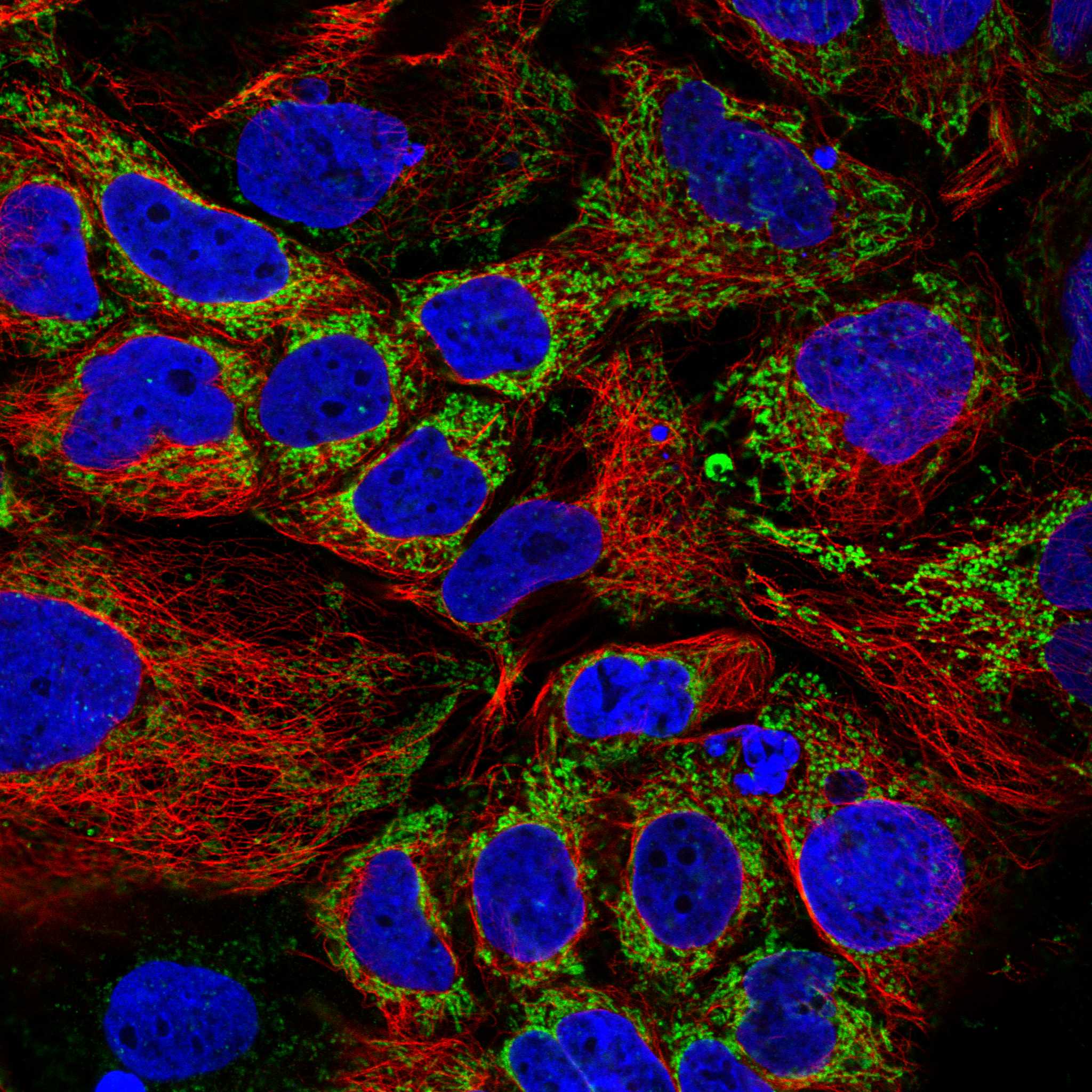
| IF/ICC: where should signal localize? | The main location is mitochondria; cytosol and basal body are additional, uncertain assignments (HPA: subcellular ICC-IF). This is an interpretation cue for the separate IF/ICC guide, not an IHC protocol option. |
| Is ACAA2-specific fixation sensitivity known? | No target-specific fixation effect is supplied by UniProt P42765 or HPA tissue IHC. General IHC practice may assess retrieval and processing when a run fails, without attributing that failure to a proven ACAA2 fixation effect. |
| Situation | Likely cause | Next action |
|---|---|---|
| Expected positive cells are unstained. | A failed detection run or unsuitable staining conditions are possibilities; HPA reports High staining in hepatocytes and kidney tubule cells (HPA: tissue IHC). | Check an on-run positive tissue control and detection reagents. If the run failed, review the catalog antibody’s IHC-P instructions and adjust one general IHC variable at a time. |
| The whole section looks uniformly brown. | General IHC practice: excessive detection signal, residual endogenous activity, or incomplete washing can obscure a cellular pattern. | Compare with a no-primary control, inspect the counterstain and chromogen development, and review blocking and washes. Look again for the granular cytoplasmic pattern reported by HPA (HPA: tissue IHC). |
| Strong colour appears in HPA not-detected cells. | Possible cross-reactivity or endogenous detection activity; adipocytes and skeletal myocytes are Not detected in the reported tissue profile (HPA: tissue IHC). | Identify the stained cell type on the section, compare a no-primary control, and seek an independent specificity check before scoring that signal as ACAA2. |
| Signal is mainly nuclear or outlines cell membranes. | The dominant compartment conflicts with mitochondrial localization (UniProt P42765; HPA: subcellular ICC-IF). | Review staining controls and morphology, then check whether expected positive cells show cytoplasmic granules. Do not score the discordant compartment as a routine positive pattern. |
| Signal is weak in a named positive cell population. | A weak run or biological variation may be involved; HPA’s High category describes its observations, not a guaranteed intensity in every specimen (HPA: tissue IHC). | Compare the same run’s positive control, cell morphology, and background. Review the catalog antibody’s IHC-P conditions before changing interpretation thresholds. |
| Positive control stains, but the test tissue does not. | The tested cell population may differ from HPA’s named positive populations, or the specimen may truly show less detectable signal (HPA: tissue IHC). | Score the relevant cells rather than the tissue as a whole, record the compartment and intensity, and report the result with HPA’s pending external-verification caveat (HPA: tissue reliability). |


Comprehensive Human Protein Atlas IHC scoring per tissue (reliability: Approved — Low consistency between antibody staining and RNA expression data. Pending external verification.). Rows are taken directly from the HPA tissue chart — click any row's HPA link to view the source.
| Tissue | Cell type | Level | Evidence | Source |
|---|---|---|---|---|
| Appendix | Glandular cells | High | Protein (IHC) | HPA → |
| Colon | Glandular cells | High | Protein (IHC) | HPA → |
| Duodenum | Glandular cells | High | Protein (IHC) | HPA → |
| Gallbladder | Glandular cells | High | Protein (IHC) | HPA → |
| Kidney | Cells in tubules | High | Protein (IHC) | HPA → |
| Tissue | Cell type | Level | Evidence | Source |
|---|---|---|---|---|
| Adipose tissue | Adipocytes | Not detected | Protein (IHC) | HPA → |
| Cerebellum | Cells in granular layer | Not detected | Protein (IHC) | HPA → |
| Cervix | Glandular cells | Not detected | Protein (IHC) | HPA → |
| Esophagus | Squamous epithelial cells | Not detected | Protein (IHC) | HPA → |
| Lymph node | Germinal center cells | Not detected | Protein (IHC) | HPA → |
Troubleshoot ACAA2 staining in paraffin sections by checking retrieval, controls, compartment, and scoring before interpreting chromogenic signal.
Both catalog antibodies have IHC figures from human paraffin sections and IF/ICC figures from human cell samples; both list Human, Mouse and Rat reactivity (catalog image captions; catalog reactivity).
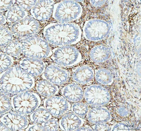
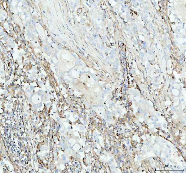
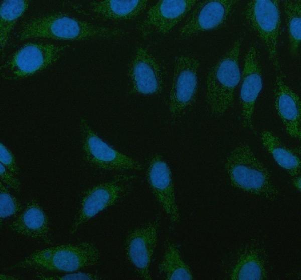
M08341-1 will render with IHC data from a human colorectal adenocarcinoma paraffin section; its IF/ICC figure uses HeLa cells (M08341-1 image captions). PB10022 will render with IHC data from human intestinal cancer paraffin sections; its IF/ICC figure uses U20S cells (PB10022 image captions).
Which to pick: For tissue IHC, choose M08341-1 for the documented 1:50 EDTA pH 8 workflow, or PB10022 for the documented 1 μg/ml citrate pH 6 workflow; both captions show paraffin sections and leave the fixative unreported (respective IHC image captions). For IF/ICC, M08341-1 is a rabbit monoclonal with a HeLa cell figure, while PB10022 has a U20S cell figure and no clone specified (catalog titles and IF image captions). Both list Human, Mouse and Rat reactivity, but their supplied IHC and IF figures show human samples only (catalog reactivity and image captions).