ACACB / Acetyl-CoA carboxylase 2 · IHC design guide

Design Immunohistochemistry for ACACB

Plan ACACB paraffin-section IHC using adipocytes as a positive control (HPA tissue IHC). Compare cytoplasmic and nuclear staining across sections with consistent fixation (HPA tissue IHC; standard IHC practice).

Evidence assembled Oct 2026 · For research use; verify linked source records and product datasheet before use
Immunohistochemistry protocol sheet for ACACB (IHC for ACACB): expected localisation Cytoplasmic and nuclear IHC staining (HPA tissue IHC); mitochondrial protein (UniProt), antibody A03668-2, validated IHC image, and IHC protocol steps
Printable ACACB IHC protocol sheet — expected localisation Cytoplasmic and nuclear IHC staining (HPA tissue IHC); mitochondrial protein (UniProt), antibody A03668-2, controls and protocol steps. Open the full ACACB IHC guide →

ACACB Immunohistochemistry Experimental Design Guide

Expected localisation, validated protocols, controls and antibodies — the at-a-glance facts below, then the full design guide.

Must know before staining
Expected localisation Cytoplasmic and nuclear IHC staining (HPA tissue IHC); mitochondrial protein (UniProt)
Staining pattern Strongest in adipocytes; cytoplasmic and nuclear (HPA tissue IHC)
Antigen retrieval EDTA pH 8.0 HIER, heat-mediated (datasheet A03668-2)
Positive control ⓘ Adipose tissue+3 more · see all
Negative control ⓘ Adrenal gland+4 more · see all
Important caveats
Reasons your staining may differ from the expected pattern.
Fixation Matched tissue-IHC evidence does not establish the fixation claim. Validate the specimen-specific method before use. (selected-SKU IHC image A03668-2)
Caveat Adrenal tissue enriched (UniProt); glandular cells unstained (HPA tissue IHC)
Regulation Adipose-enriched expression (UniProt)
Isoform / epitope 3 isoforms; epitope differences unreported; no propeptide (UniProt)
Section 1

Recommended ACACB IHC & IF Protocols

The catalog antibody’s IHC-P protocol is accompanied by one published ACACB IHC protocol using mouse brain sections (PMC13125043).

Recommended immunohistochemistry (IHC-P) protocol parameters
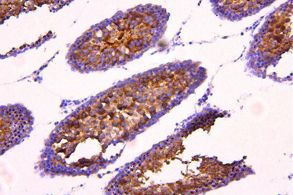
SampleParaffin-embedded rat testis tissue; fixative not specified (datasheet A03668-2)
FixationImage fixative and duration unreported (datasheet A03668-2); verify before use.
Sectioning4–5 µm sections on charged slides (standard)
DeparaffinisationXylene, graded ethanol series to water (standard)
Antigen retrievalHeat retrieval: EDTA pH 8.0 (datasheet A03668-2); 20 min, 95–100 °C (standard)
Peroxidase block3% H2O2, 10 min, room temperature (standard)
Blocking10% goat serum (datasheet A03668-2)
Primary antibodyRabbit anti-ACACB, 0.5-1μg/ml (datasheet A03668-2)
Primary incubationOvernight at 4 °C (datasheet A03668-2)
DetectionHRP-conjugated secondary, DAB chromogen (datasheet A03668-2)
CounterstainHematoxylin, blue, dehydrate and mount (standard)
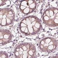
Expected resultACACB-positive staining in adipocytes of adipose tissue (HPA tissue IHC: High). HPA tissue profile: Cytoplasmic and nuclear expression in several tissues, most abundant in adipocytes. No signal in the no-primary control.
💡Decision noteStart with heat-mediated EDTA retrieval at pH 8.0 for the catalog antibody (datasheet A03668-2); the published protocol does not specify retrieval (PMC13125043).
Section 2

What Is the Expected ACACB Staining Pattern?

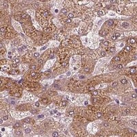
ACACB is a mitochondrial enzyme with no transmembrane segment (UniProt O00763: location, topology). In paraffin-section IHC, expect the strongest staining in adipocytes, with high staining also reported in late spermatids and medium staining in hepatocytes (HPA: tissue IHC). HPA describes cytoplasmic and nuclear staining across several tissues; its tissue profile has Enhanced reliability, with medium consistency between staining and RNA data (HPA: tissue IHC).

What am I looking at on my slide?
Strong cytoplasmic staining in adipocytes, with or without a nuclear component.This fits the most abundant reported IHC pattern (HPA: High in adipocytes; cytoplasmic and nuclear tissue staining). Judge the stained cell, not the overall darkness of adipose tissue (standard IHC practice).
Medium hepatocyte staining or strong staining in elongated or late spermatids.Both are reported positive patterns (HPA: Medium in hepatocytes; High in elongated or late spermatids). Their different intensities should be interpreted by cell type; they need not match the adipocyte control (HPA: tissue IHC).
Signal confined to cell borders, extracellular material, or an unrelated compartment.That distribution does not fit the reported cytoplasmic and nuclear tissue pattern or a mitochondrial enzyme lacking a transmembrane segment (HPA: tissue IHC; UniProt O00763: location, topology). Check for artefact before calling it ACACB.
Strong staining in cells reported as undetected, such as adrenal glandular or bone marrow hematopoietic cells.Treat this as suspect staining and assess cross-reactivity or endogenous detection activity (HPA: Not detected in those cell types; standard IHC practice). UniProt's broad adrenal tissue expression does not identify adrenal glandular cells as an IHC-positive population (UniProt O00763: tissue specificity).
Diffuse, cell-independent color, or no signal in an adipocyte-positive control.Diffuse color favors background from the detection workflow (standard IHC practice). An unstained adipocyte control makes a negative test section uninterpretable until the assay is checked (HPA: High in adipocytes; standard IHC practice).
💡Expected ACACB appearanceCall positive when staining is strongest in adipocytes and chiefly cytoplasmic, allowing the nuclear component reported by HPA; widespread cell-independent color or isolated border staining is suspect (HPA: High in adipocytes; cytoplasmic and nuclear tissue staining; UniProt O00763: topology; standard IHC practice).
How each factor affects the staining
Compartment: how should mitochondrial annotation guide scoring?UniProt places ACACB at mitochondria, while HPA reports cytoplasmic and nuclear tissue IHC (UniProt O00763: location; HPA: tissue IHC). Do not reject nuclear staining solely because of the UniProt annotation; assess whether the stained cells match the tissue pattern.
Isoforms and antibody epitopeThree isoforms are listed, with isoform 3 detected in skeletal muscle, adipose tissue and liver at protein level (UniProt O00763: isoforms, tissue specificity). Epitope coverage is not supplied, so isoform-specific staining cannot be inferred from these tissue results.
Detection chemistryACACB has a biotinyl-binding domain (UniProt O00763: domain 888–962). If using avidin–biotin detection, assess endogenous biotin with a suitable detection control; biotin-related background is a general IHC concern, not an established ACACB tissue pattern (standard IHC practice).
IF/ICC Q&A: is a cellular IF pattern established?HPA provides no main ICC-IF location or cell-line images for ACACB (HPA: subcellular record). UniProt's mitochondrial location suggests a pattern to examine, but this record does not validate an IF/ICC appearance or supply an IF protocol (UniProt O00763: location; HPA: subcellular record).
Strength of tissue-pattern evidenceOne listed rabbit polyclonal antibody, HPA006554, has Enhanced IHC validation; HPA also notes medium consistency between staining and RNA (HPA: antibody record, tissue IHC). Use the cell-specific observations as expectations, with that consistency limit in mind.
Why is my staining missing, weak or wrong?
SituationLikely causeNext action
Adipocyte-positive control has no staining.The run may have failed, or the primary antibody or detection conditions may be unsuitable (standard IHC practice); HPA reports High adipocyte staining (HPA: tissue IHC).Verify control-section quality, antigen retrieval, primary-antibody incubation and detection reagents against the validated IHC workflow before interpreting test negatives (standard IHC practice).
Only hepatocytes look faint beside an adipocyte control.A weaker hepatocyte result can reflect the reported cell-type difference (HPA: Medium in hepatocytes; High in adipocytes), provided the control and staining quality are acceptable.Score hepatocytes against an appropriate liver control and check cellular distribution; do not require adipocyte-level intensity from hepatocytes (HPA: tissue IHC; standard IHC practice).
Color appears throughout tissue, including areas without a clear cellular pattern.Non-specific primary binding or detection background may be present (standard IHC practice); diffuse, cell-independent color does not establish the reported adipocyte or hepatocyte pattern (HPA: tissue IHC).Compare a no-primary control, review blocking and washes, and examine whether chromogen remains confined to cells (standard IHC practice).
Strong color persists in the no-primary control.The detection system or endogenous tissue activity can generate primary-independent signal (standard IHC practice). An avidin–biotin system warrants an endogenous-biotin check because ACACB is a biotin enzyme (UniProt O00763: function).Review detection chemistry and apply the corresponding endogenous-activity control or blocking step before attributing that color to ACACB (standard IHC practice).
Adrenal glandular cells or bone marrow hematopoietic cells stain strongly.Those cells are reported as Not detected, so cross-reactivity or background needs investigation (HPA: tissue IHC; standard IHC practice). UniProt's adrenal tissue-level statement does not resolve the cell-level discrepancy (UniProt O00763: tissue specificity).Check a no-primary control and a positive adipocyte section, then reassess cell identity and staining distribution before scoring the suspect cells positive (HPA: tissue IHC; standard IHC practice).
A nuclear signal is present alongside cytoplasmic staining.Nuclear staining is included in HPA's tissue IHC profile, despite UniProt's mitochondrial annotation (HPA: tissue IHC; UniProt O00763: location). Its presence alone does not establish an artefact.Compare the stained cell types and relative intensities with HPA's tissue pattern; investigate nuclear-only or widespread cell-independent signal with controls (HPA: tissue IHC; standard IHC practice).

Sample controls for ACACB IHC & IF

🧪Run adipose tissue first; adipocytes should stain (HPA: High in adipocytes). Use bone marrow hematopoietic cells as the negative tissue (HPA: Not detected in hematopoietic cells), and assess non-adipocyte cells on the adipose slide for absent chromogenic staining as an internal comparison without assuming they are ACACB-negative (standard IHC practice).
Positive control tissue: Adipose tissue (Adipocytes, HPA High)
Negative control tissue: Adrenal gland (HPA Not detected)
ICC-IF cell lines (HPA subcellular resource): HPA carries no ICC-IF cell line for ACACB; derive a cell-line control from the positive tissue's cell type (Adipocytes) and confirm it by RNA or western blot first.
Technical controls: Include no-primary (secondary-only), host-matched rabbit IgG isotype, and an ACACB-knockout biological negative if available (selected-SKU IHC caption: rabbit primary; standard IHC practice). Quench endogenous peroxidase before DAB detection; block endogenous biotin if using avidin–biotin detection (standard IHC practice).
⚠️Feasibility: A target-specific fixation window or fixation effect is unreported, and the selected-SKU paraffin-section caption does not state a fixative (selected-SKU IHC caption). That rat-testis example uses heat-mediated EDTA retrieval at pH 8.0, but it does not establish a retrieval requirement for adipose tissue (selected-SKU IHC caption). Paraffin IHC has the supplied starting conditions, while matched frozen-section and IF conditions are unreported; adipose lipid vacuoles can complicate interpretation of chromogenic staining (selected-SKU IHC caption; standard IHC practice).

HPA tissue IHC evidence for ACACB

Comprehensive Human Protein Atlas IHC scoring per tissue (reliability: Enhanced — Medium consistency between antibody staining and RNA expression data.). Rows are taken directly from the HPA tissue chart — click any row's HPA link to view the source.

Positive expression · recommended positive controls

TissueCell typeLevelEvidenceSource
Adipose tissue Adipocytes High Protein (IHC) HPA →
Testis Elongated or late spermatids High Protein (IHC) HPA →
Breast Adipocytes Medium Protein (IHC) HPA →
Liver Hepatocytes Medium Protein (IHC) HPA →

Undetected expression · recommended negative controls

TissueCell typeLevelEvidenceSource
Adrenal gland Glandular cells Not detected Protein (IHC) HPA →
Bone marrow Hematopoietic cells Not detected Protein (IHC) HPA →
Caudate Glial cells Not detected Protein (IHC) HPA →
Cerebellum Cells in granular layer Not detected Protein (IHC) HPA →
Cerebral cortex Endothelial cells Not detected Protein (IHC) HPA →
Section 3

Advanced ACACB IHC Tips

Troubleshoot ACACB staining in paraffin section IHC using the catalog antibody’s tissue image, protein annotations, and cell level expression patterns.

What retrieval should I try first when ACACB staining is weak?
Start with heat mediated retrieval in EDTA at pH 8.0 (datasheet A03668-2). That condition accompanies ACACB staining in a paraffin embedded rat testis section, although the caption does not specify heating time or temperature (datasheet A03668-2). Keep retrieval, section thickness, antibody concentration, and development time consistent across a small optimization series so changes in signal can be attributed to one variable (standard IHC practice). The documented primary incubation is 1 μg/ml overnight at 4°C; use it as a starting point when assessing retrieval (datasheet A03668-2). If staining remains weak, compare an alternative buffer or pH on adjacent sections, checking both signal and tissue morphology (standard IHC practice).
How can I troubleshoot fixation related loss of ACACB staining?
The selected paraffin section caption does not state a fixative, so ACACB sensitivity to any fixation method is unknown for this antibody (datasheet A03668-2). Record the actual fixative, fixation duration, processing history, and section age before comparing cases, because these variables can change IHC staining (standard IHC practice). Run sections with known processing histories together using EDTA at pH 8.0 and the documented 1 μg/ml primary incubation overnight at 4°C (datasheet A03668-2). Include a positive control with the same processing history and inspect morphology before attributing a weak signal to fixation (standard IHC practice). HPA tissue staining and UniProt protein annotations do not establish a target specific fixation effect (HPA: tissue IHC; UniProt O00763).
Where should I expect ACACB staining, and how should I assess nuclear signal?
Assess cytoplasmic staining in the expected cell population first: ACACB is annotated as mitochondrial, with no transmembrane segment (UniProt O00763 localisation and topology). HPA reports cytoplasmic and nuclear tissue staining, most abundant in adipocytes, but describes only medium consistency between staining and RNA data (HPA: tissue IHC, Enhanced reliability). A chromogenic paraffin section alone cannot resolve whether a cytoplasmic deposit lies on mitochondria; compare its distribution with morphology and a negative control (standard IHC practice). Record nuclear and cytoplasmic staining separately across at least 2 comparable sections, and check whether nuclear signal persists when nonspecific staining is minimized (standard IHC practice). Treat isolated nuclear staining as unresolved until supported by an independent localization check (UniProt O00763 localisation; HPA: tissue IHC).
Could isoforms or the antibody epitope explain uneven ACACB staining?
ACACB has 3 annotated isoforms, while the supplied antibody caption does not identify its recognized epitope or establish which isoforms it detects (UniProt O00763 isoforms; datasheet A03668-2). Isoform 3 is reported at the protein level in skeletal muscle, adipose tissue, and liver, so tissue comparisons may reflect differing isoform contributions (UniProt O00763 tissue specificity). The protein also has annotated modified residues, including phosphoserines at positions 35 and 91, but no supplied evidence links these modifications to staining with this antibody (UniProt O00763 modified residues; datasheet A03668-2). Review the antibody’s epitope documentation if available, then compare matched sections under the documented EDTA pH 8.0 retrieval condition (datasheet A03668-2; standard IHC practice). Avoid assigning an isoform from staining intensity alone (standard IHC practice).
How should I investigate ACACB localization with multiplex immunofluorescence?
Use IF as a separate localization experiment; the supplied ACACB antibody evidence documents paraffin section chromogenic IHC, while HPA supplies no ICC/IF images or established subcellular IF location (datasheet A03668-2; HPA: subcellular). Pair ACACB with a marker identifying the expected cell population, such as adipocytes in adipose tissue, and assess overlap at the cell level (HPA: high in adipocytes; standard IF practice). Choose fluorophores after checking tissue autofluorescence and include single stain and unstained controls to assess bleed through (standard IF practice). ACACB has no annotated transmembrane segment and is mitochondrial, so optimize permeabilisation for access to its intracellular epitope without assuming which side of a mitochondrial membrane the antibody recognizes (UniProt O00763 localisation and topology; standard IF practice).
What should I change when DAB background obscures ACACB staining?
First compare the affected section with a no primary control and inspect whether color follows tissue edges, damaged areas, or every cell indiscriminately (standard IHC practice). The documented workflow used 10% goat serum, 1 μg/ml primary antibody overnight at 4°C, a peroxidase linked secondary for 30 minutes at 37°C, and DAB (datasheet A03668-2). Use a peroxidase block and titrate development time if the no primary control shows DAB deposition; these are general chromogenic IHC controls (standard IHC practice). ACACB carries biotin as part of its catalytic mechanism, so check for endogenous biotin background if an avidin based detection system is introduced (UniProt O00763 function; standard IHC practice). Compare adjusted staining with expected cell level patterns before calling faint color positive (HPA: tissue IHC; standard IHC practice).
How should I quantify ACACB staining across paraffin sections? ⚠ ANSWER MARKED FOR VERIFICATION
Define the cell compartment and scoring region before reading slides, then report the percentage of positive cells and an H score from staining intensity and positive fraction (standard IHC practice). For spatial questions, report positive cell density per mm² of viable, evaluable tissue and exclude folds, edges, and necrosis by the same rule for every section (standard IHC practice). Normalize comparisons to the relevant cell population or tissue area, since HPA reports high staining in adipocytes and late spermatids but only medium staining in hepatocytes (HPA: tissue IHC). Score nuclear and cytoplasmic deposits separately because HPA describes both, whereas UniProt annotates mitochondrial localization (HPA: tissue IHC; UniProt O00763 localisation). Hold retrieval at EDTA pH 8.0 and detection conditions constant across the quantified set (datasheet A03668-2; standard IHC practice).
How can I distinguish convincing ACACB staining from an artefact?
A credible result should recur in the expected cells and have a cytoplasmic pattern compatible with the mitochondrial annotation, while acknowledging that HPA also reports nuclear tissue staining (HPA: high in adipocytes and late spermatids; UniProt O00763 localisation; HPA: tissue IHC). Interpret isolated nuclear color cautiously, especially if it tracks section edges, damaged tissue, or necrosis (standard IHC practice). Check a no primary control for endogenous peroxidase or nonspecific DAB deposition and compare adjacent sections processed with the same EDTA pH 8.0 retrieval (standard IHC practice; datasheet A03668-2). The catalog image documents staining in rat testis, while HPA’s cell level observations concern human tissues, so do not equate their staining patterns without checking species and cell identity (datasheet A03668-2; HPA: tissue IHC). HPA rates consistency with RNA expression as medium, making independent validation valuable for unexpected patterns (HPA: Enhanced reliability).
Boster reagents

Best ACACB / Acetyl-CoA carboxylase 2 IHC Antibodies

Two anti-ACACB antibodies have IHC images from rat testis and human tissues (catalog IHC captions). Both list Human, Mouse and Rat reactivity; one also lists IF validation (catalog applications and reactivity).

Real IHC data IHC analysis of ACACB using anti-ACACB antibody (A03668-2). ACACB was detected in paraffin-embedded section of rat testis tissue. Heat mediated antigen retrieval was performed in EDTA buffer (pH8.0, epitope retrieval solution). The tissue section was blocked with 10% goat serum. The tissue section was then incubated with 1 μg/ml rabbit anti-ACACB Antibody (A03668-2) overnight at 4°C. Peroxidase Conjugated Goat Anti-rabbit IgG was used as secondary antibody and incubated for 30 minutes at 37°C. The tissue section was developed using HRP Conjugated Rabbit IgG Super Vision Assay Kit (Catalog # SV0002) with DAB as the chromogen.
Anti-Acetyl Coenzyme A Carboxylase/ACACB Antibody ®
Cat # A03668-2
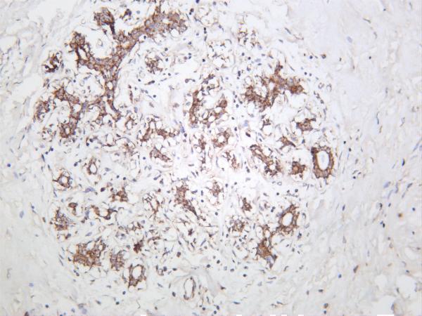
Real IHC data Human breast carcinoma was stained with anti-ACACB rabbit antibody
Anti-ACACB Rabbit Monoclonal Antibody
Cat # M03668-1

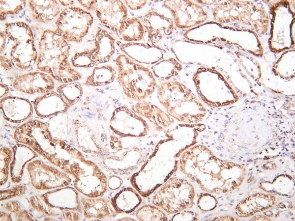
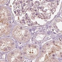
A03668-2 shows ACACB staining in a paraffin-embedded rat testis section (A03668-2 IHC caption). M03668-1 shows staining in human breast carcinoma and kidney and lists IF as an application, although no IF image is supplied (M03668-1 IHC captions; catalog applications and images).

Which to pick: Choose A03668-2 for paraffin-section IHC when a documented retrieval and detection example is useful: its rat testis caption specifies EDTA retrieval and DAB detection but does not report the fixative (A03668-2 IHC caption). Choose M03668-1 for IF/ICC planning because it is the monoclonal antibody with listed IF validation and a 1:200–1:1000 IF range; its human IHC captions do not report tissue processing or fixation (M03668-1 catalog applications and dilution; M03668-1 IHC captions). Both list Human, Mouse and Rat reactivity, but the supplied IHC images document rat tissue for A03668-2 and human tissues for M03668-1 (catalog reactivity; IHC captions).

Each figure is that product's own IHC / IF validation image from its datasheet.

References

  1. UniProt Consortium. UniProt entry O00763 (ACACB_HUMAN, Acetyl-CoA carboxylase 2).
  2. Human Protein Atlas. ACACB tissue IHC expression (reliability: Enhanced).
  3. Human Protein Atlas. ACACB subcellular location (ICC-IF): Highest expression in MCF-7: 16.2 nTPM.
  4. Human Protein Atlas. ACACB antibody validation summary (1 antibodies).
  5. ACACB is a novel metabolism-related biomarker in the prediction of response to cetuximab therapy inmetastatic colorectal cancer. Acta biochimica et biophysica Sinica 2022 — PMC9828296.
  6. Oleic acid improves pathological changes in Aβ1-42-induced astrocytes and Alzheimer's disease mouse models through PKA/ACACB/CPT1A. Frontiers in neuroscience 2026 — PMC13125043.
  7. Acetyl Co-A Carboxylase Inhibition Halts Hyperglycemia Induced Upregulation of De Novo Lipogenesis in Podocytes and Proximal Tubular Cells. Metabolites 2022 — PMC9610518.
  8. PubMed PMID:9099716 — UniProt-cited evidence.
  9. PubMed PMID:16854592 — UniProt-cited evidence.
  10. PubMed PMID:16541075 — UniProt-cited evidence.