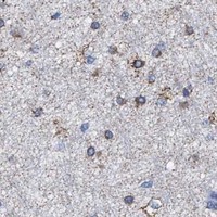
ACADS / Short-chain specific acyl-CoA dehydrogenase, mitochondrial · IHC design guide

Design Immunohistochemistry for ACADS

Plan ACADS IHC in paraffin sections using granular cytoplasmic staining as the expected tissue pattern (HPA tissue IHC). The catalog antibody is listed at 2–5 μg/ml (datasheet A05028-1); liver hepatocytes provide a high-staining reference (HPA tissue IHC).

Evidence assembled Oct 2026 · For research use; verify linked source records and product datasheet before use
Immunohistochemistry protocol sheet for ACADS (IHC for ACADS): expected localisation Granular cytoplasm (HPA tissue IHC); mitochondrial matrix expected (UniProt), antibody A05028-1, validated IHC image, and IHC protocol steps
Printable ACADS IHC protocol sheet — expected localisation Granular cytoplasm (HPA tissue IHC); mitochondrial matrix expected (UniProt), antibody A05028-1, controls and protocol steps. Open the full ACADS IHC guide →

ACADS Immunohistochemistry Experimental Design Guide

Expected localisation, validated protocols, controls and antibodies — the at-a-glance facts below, then the full design guide.

Must know before staining
Expected localisation Granular cytoplasm (HPA tissue IHC); mitochondrial matrix expected (UniProt)
Staining pattern Granular cytoplasm in hepatocytes and glandular cells (HPA tissue IHC)
Antigen retrieval EDTA pH 8.0 HIER, heat-mediated (datasheet A05028-1)
Positive control ⓘ Colon+4 more · see all
Negative control ⓘ None in HPA (detected in all 45 tissues); use no-primary + isotype controls
Important caveats
Reasons your staining may differ from the expected pattern.
Fixation Matched tissue-IHC evidence does not establish the fixation claim. Validate the specimen-specific method before use. (selected-SKU IHC image A05028-1)
Caveat Staining and RNA show only medium consistency (HPA tissue IHC)
Regulation No expression regulator annotated (UniProt)
Isoform / epitope No isoforms annotated; mature chain starts at residue 25 (UniProt)
Section 1

Recommended ACADS IHC & IF Protocols

The catalog antibody has an IHC-P protocol (datasheet: A05028-1). Three published ACADS IHC methods add tissue-specific examples (PMC13022253; PMC9631217; PMC11351355).

Recommended immunohistochemistry (IHC-P) protocol parameters
SampleParaffin-embedded human colon cancer tissue; fixative not specified (datasheet A05028-1)
FixationImage fixative and duration unreported (datasheet A05028-1); verify before use.
Sectioning4–5 µm sections on charged slides (standard)
DeparaffinisationXylene, graded ethanol series to water (standard)
Antigen retrievalHeat retrieval: EDTA pH 8.0 (datasheet A05028-1); 20 min, 95–100 °C (standard)
Peroxidase block3% H2O2, 10 min, room temperature (standard)
Blocking10% goat serum (datasheet A05028-1)
Primary antibodyRabbit anti-ACADS, 2-5μg/ml (datasheet A05028-1)
Primary incubationOvernight at 4 °C (datasheet A05028-1)
DetectionHRP-conjugated secondary, DAB chromogen (datasheet A05028-1)
CounterstainHematoxylin, blue, dehydrate and mount (standard)
Expected resultACADS-positive staining in glandular cells of colon (HPA tissue IHC: High). HPA tissue profile: General cytoplasmic expression with a granular, mitochondrial pattern. No signal in the no-primary control.
💡Decision noteStart with heat-mediated EDTA, pH 8.0, for the catalog antibody (datasheet: A05028-1). Citrate, pH 6.0, is a published alternative (PMC13022253).
Section 2

What Is the Expected ACADS Staining Pattern?

ACADS is a mitochondrial matrix enzyme without a transmembrane segment (UniProt P16219 topology). In paraffin-section IHC, expect granular cytoplasmic staining, particularly in hepatocytes, intestinal glandular cells, kidney collecting ducts, lung type II alveolar cells, and Leydig cells (HPA: High in these cell types). HPA rates the tissue staining Enhanced, while reporting medium consistency between antibody staining and RNA expression (HPA: reliability).

What am I looking at on my slide?
Granular cytoplasmic signal in hepatocytes and intestinal glandular cells, with little nuclear emphasis.This matches the mitochondrial matrix location (UniProt P16219) and HPA's reported pattern. Both cell types stain at a High level in HPA tissue IHC; judge granularity and cell identity alongside intensity.
Predominantly nuclear or cell-surface staining, without a granular cytoplasmic pattern.Treat this IHC result as discordant with the supported mitochondrial location (HPA: subcellular; UniProt P16219 topology). HPA lists nucleoplasm only as an uncertain additional ICC-IF location; it does not establish nuclear-dominant tissue IHC.
Strong signal appears mainly in cells outside the expected tissue pattern, while known-positive cells remain weak.Consider antibody cross-reactivity or endogenous detection activity (general IHC practice). Compare cell identity with HPA tissue IHC: collecting ducts and hepatocytes are High, whereas soft-tissue fibroblasts are Low, not confirmed negative.
Brown chromogen spreads across many compartments or the surrounding tissue, obscuring individual cells.Diffuse background cannot establish ACADS localisation (general IHC practice). Reassess blocking, chromogen development, and a no-primary control; the expected tissue pattern is granular cytoplasmic staining (HPA: tissue IHC).
No convincing signal appears in an otherwise interpretable liver section.Hepatocytes are High in HPA tissue IHC, so first examine assay performance (HPA: liver). A failed run, inadequate retrieval, or overly weak detection can produce a false negative (general IHC practice); absence alone does not establish absent ACADS.
💡Expected ACADS appearanceCall a result positive when expected cells show clear granular cytoplasmic staining, often strong in HPA High cell types; dominant nuclear or diffuse extracellular color is suspect (HPA: tissue IHC; UniProt P16219 topology).
How each factor affects the staining
Compartment and topologyACADS belongs to the mitochondrial matrix and has no transmembrane segment (UniProt P16219). Expect intracellular granules, not a membrane outline; use morphology to locate the stained cells.
Processing and isoformsUniProt annotates a mature chain spanning residues 25–412 and no isoforms (UniProt P16219). The supplied record does not locate the antibody epitope, so it cannot predict how processing changes IHC recognition.
Tissue comparisonHPA reports High staining in several specified cell types but Low staining in soft-tissue fibroblasts; it lists no negative tissue (HPA: tissue IHC). Use fibroblasts only as a lower-signal comparison.
Strength of evidenceTwo listed antibodies have Enhanced IHC status (HPA: HPA022271, CAB019284). HPA also reports medium consistency with RNA expression; neither statement guarantees specificity in a new specimen.
IF/ICC Q: What localisation should appear?A: Mainly mitochondrial staining (HPA: supported ICC-IF location). HPA also lists uncertain nucleoplasm and centrosome locations; consult the separate IF/ICC guide for that application's workflow.
Why is my staining missing, weak or wrong?
SituationLikely causeNext action
A liver positive-control section has little or no hepatocyte signal.The assay may have insufficient accessible signal or an ineffective detection step (general IHC practice); hepatocytes are High in HPA tissue IHC.Check the IHC-validated antibody's stated IHC-P conditions, retrieval and detection controls, then repeat with an interpretable liver section (general IHC practice; HPA: High in hepatocytes).
Most cells look uniformly brown, and mitochondrial granules cannot be resolved.Excess chromogen development, antibody concentration, or nonspecific binding may obscure localisation (general IHC practice).Review a no-primary control, shorten development or titrate the antibody, and score only interpretable cellular signal (general IHC practice; HPA: granular cytoplasmic pattern).
The stain is concentrated in nuclei.A nuclear-dominant IHC pattern conflicts with the supported mitochondrial assignment (HPA: subcellular; UniProt P16219).Check the control and cellular morphology, then confirm with an independently validated IHC antibody if needed (general IHC practice; HPA: two Enhanced IHC antibodies).
Unexpected cells stain strongly while the expected cells do not.Cross-reactivity or endogenous detection activity is possible (general IHC practice); HPA tissue levels describe observed cell types, not universal exclusions.Verify cell identity against the HPA tissue pattern and examine no-primary and detection controls before assigning ACADS positivity (HPA: tissue IHC; general IHC practice).
Soft-tissue fibroblasts stain faintly.Low fibroblast staining is reported and should not automatically be classified as a failed negative control (HPA: Low in fibroblasts).Compare their signal with HPA High cell types on interpretable sections; use a no-primary control to assess background (HPA: tissue IHC; general IHC practice).
A plausible granular signal varies greatly between specimens.Biological expression or assay conditions may differ (general IHC practice); HPA reports medium antibody-to-RNA consistency rather than uniform staining.Keep retrieval, detection, and scoring consistent, and interpret each specimen against cell identity and run controls (general IHC practice; HPA: reliability and tissue IHC).

Sample controls for ACADS IHC & IF

🧪Run colon first; its glandular cells should stain (HPA: High in colon glandular cells). HPA detects ACADS in all 45 scored tissues, so there is no HPA-negative tissue; use no-primary and isotype controls for the negative, and expect only background in any unstained cells within the positive section without assuming those cells lack ACADS (HPA: detected in all 45 scored tissues).
Positive control tissue: Colon (Glandular cells, HPA High)
Negative control tissue: None in HPA: ACADS is detected in all 45 scored tissues. Use a no-primary (secondary-only) and an isotype control instead.
ICC-IF cell lines (HPA subcellular resource): HPA ICC-IF images show ACADS in Hep-G2, RT-4, SK-MEL-30, JURKAT, Sperm, with annotated localisation: Mitochondria (supported) (HPA subcellular).
Technical controls: Include a secondary-only section, an isotype control matched to the primary antibody’s rabbit host and clonality, and ACADS-knockout tissue or cells as a biological negative (A05028-1 caption: rabbit primary). Quench endogenous peroxidase in colon sections before HRP/DAB detection to limit false staining (A05028-1 caption: colon section with HRP/DAB detection).
⚠️Feasibility: No target-specific fixation window or fixation effect is reported, and the selected paraffin-section caption does not state a fixative (A05028-1 caption: fixative unreported). Heat retrieval with EDTA at pH 8.0 is documented for that example, but whether ACADS staining depends on retrieval is unreported (A05028-1 caption: EDTA retrieval). The supplied evidence does not establish whether frozen sections or IF are easier; endogenous peroxidase is a potential artefact in the documented colon HRP/DAB assay (A05028-1 caption: colon HRP/DAB detection).

HPA tissue IHC evidence for ACADS

Comprehensive Human Protein Atlas IHC scoring per tissue (reliability: Enhanced — Medium consistency between antibody staining and RNA expression data.). Rows are taken directly from the HPA tissue chart — click any row's HPA link to view the source.

Positive expression · recommended positive controls

TissueCell typeLevelEvidenceSource
Colon Glandular cells High Protein (IHC) HPA →
Duodenum Glandular cells High Protein (IHC) HPA →
Kidney Collecting ducts High Protein (IHC) HPA →
Liver Hepatocytes High Protein (IHC) HPA →
Lung Alveolar cells type II High Protein (IHC) HPA →

Undetected expression · recommended negative controls

TissueCell typeLevelEvidenceSource
None in HPA: ACADS is detected in all 45 scored tissues. Use a no-primary (secondary-only) and an isotype control instead.
Section 3

Advanced ACADS IHC Tips

ACADS staining should be evaluated against its mitochondrial matrix location and the cell patterns reported in tissue IHC (UniProt P16219; HPA tissue IHC).

What retrieval should I start with if ACADS staining is weak?
Start with heat-mediated retrieval in EDTA at pH 8.0 for paraffin sections (datasheet A05028-1). The selected tissue-IHC image used this condition, followed by 2 μg/ml primary antibody overnight at 4°C; those settings provide a documented starting point for troubleshooting (caption A05028-1). If staining remains weak, compare retrieval duration on adjacent sections while holding antibody concentration and detection constant (standard IHC practice). Check for granular cytoplasmic staining in an expected positive compartment, such as colon glandular cells, before increasing retrieval intensity (HPA tissue IHC: High in colon glandular cells; HPA tissue IHC: granular mitochondrial pattern). Include a no-primary control to distinguish stronger signal from retrieval-associated background (standard IHC practice).
Could fixation explain weak or patchy ACADS staining?
Target-specific fixation sensitivity is unknown: the selected paraffin-section caption does not state a fixative (caption A05028-1). Record the actual fixative, fixation duration, tissue thickness, and processing history for each specimen before comparing staining across batches (standard IHC practice). Compare similarly processed sections using the documented EDTA retrieval at pH 8.0, with the same detection conditions, to isolate processing effects (datasheet A05028-1; standard IHC practice). Evaluate morphology alongside signal because poor preservation can make granular cytoplasmic staining difficult to judge (HPA tissue IHC: granular mitochondrial pattern; standard IHC practice). Do not assign a target-specific fixation effect from tissue-expression patterns or protein modifications alone (HPA tissue IHC; UniProt P16219).
Where should ACADS appear in a positive tissue section?
Expect a granular cytoplasmic pattern consistent with mitochondrial staining, because ACADS is assigned to the mitochondrial matrix (HPA tissue IHC: granular mitochondrial pattern; UniProt P16219). Review signal within identifiable cells rather than treating diffuse staining across the section as equivalent evidence (standard IHC practice). Colon glandular cells, liver hepatocytes, and kidney collecting ducts have reported High tissue-IHC staining and can guide selection of comparison regions (HPA tissue IHC). The protein has no annotated transmembrane segment, so a crisp plasma-membrane outline would warrant scrutiny rather than automatic acceptance as ACADS (UniProt P16219 topology). Compare the distribution with a no-primary control and preserved tissue morphology before calling an unusual compartment positive (standard IHC practice).
How should epitope uncertainty affect ACADS staining decisions?
The supplied record lists 0 isoforms and identifies the annotated mature chain as residues 25–412 (UniProt P16219). It also lists several modified residues, including phosphothreonine at position 27 and acetylation or succinylation annotations at other positions (UniProt P16219). Without a stated antibody epitope, these annotations cannot establish whether processing or modification changes this antibody’s staining (record P16219; standard IHC interpretation). Keep retrieval, primary concentration, and detection matched when comparing specimens, beginning with EDTA at pH 8.0 and 2 μg/ml as documented (caption A05028-1; standard IHC practice). Resolve discordant patterns with an independent antibody or orthogonal expression evidence where available (standard IHC practice).
How can I adapt the ACADS question to multiplex IF?
Treat IF as a separate assay requiring its own antibody and condition validation; the supplied catalog example documents chromogenic paraffin-section IHC (caption A05028-1; standard IF practice). Pair ACADS with a marker identifying the cell population under study, then assess whether punctate cytoplasmic signal lies within those cells (HPA tissue IHC: granular mitochondrial pattern; standard IF practice). Choose fluorophores and imaging channels after measuring tissue autofluorescence in an unstained control (standard IF practice). Because ACADS is a mitochondrial matrix protein, optimise permeabilisation for access across mitochondrial membranes while checking that cell structure remains intact (UniProt P16219; standard IF practice). Use single-stain and no-primary controls to assess bleed-through and nonspecific fluorescence (standard IF practice).
What should I adjust when DAB obscures granular ACADS staining?
First inspect a no-primary section and a section lacking the detection reagent to locate background introduced by the detection workflow (standard IHC practice). The selected example used 10% goat serum blocking, a peroxidase-conjugated secondary, and DAB development (caption A05028-1). Block endogenous peroxidase as part of the general chromogenic workflow, then review whether DAB development time or primary concentration is saturating the cytoplasm (standard IHC practice). Retain the documented 2 μg/ml primary condition as a reference when testing a concentration series (caption A05028-1; standard IHC practice). Count staining as convincing only when granular cytoplasmic signal exceeds control background in intact cells (HPA tissue IHC: granular mitochondrial pattern; standard IHC practice).
How should I score ACADS across heterogeneous tissue regions? ⚠ ANSWER MARKED FOR VERIFICATION
Define the cell population and tissue region before scoring, using morphology and a consistent counterstain to identify eligible cells (standard IHC practice). Record both the percentage of positive cells and intensity, then calculate an H-score on the same intensity scale across slides (standard IHC practice). Report the scoring denominator as eligible cells within each region, and normalise positive-cell counts to that denominator rather than total section area (standard IHC practice). If spatial density is the endpoint, report positive cells per mm² of viable, analysable tissue and exclude folds or necrosis (standard IHC practice). Review granular cytoplasmic signal against matched controls, since ACADS staining is described as mitochondrial in pattern (HPA tissue IHC).
Which patterns would make an apparent ACADS positive questionable?
A convincing result shows granular cytoplasmic staining in intact cells, consistent with the mitochondrial matrix assignment (HPA tissue IHC: granular mitochondrial pattern; UniProt P16219). High staining is reported in colon glandular cells and liver hepatocytes, whereas soft-tissue fibroblasts are listed as Low; interpret each cell type against its own context (HPA tissue IHC). Flag isolated nuclear or sharp membrane staining for review before treating it as the expected tissue-IHC pattern (HPA tissue IHC; UniProt P16219 topology). Check whether signal concentrates at section edges, folds, or necrotic regions, which can generate staining artefacts (standard IHC practice). Compare suspicious DAB signal with a no-primary control and an endogenous-peroxidase-blocked section (standard IHC practice).
Boster reagents

Best ACADS / Short-chain specific acyl-CoA dehydrogenase, mitochondrial IHC Antibodies

The catalog antibody A05028-1 has documented paraffin-section IHC in human colon and liver cancer tissue and IF in A549 cells (catalog image captions).

Real IHC data IHC analysis of ACADS/SCAD using anti-ACADS/SCAD antibody (A05028-1). ACADS/SCAD was detected in a paraffin-embedded section of human colon cancer tissue. Heat mediated antigen retrieval was performed in EDTA buffer (pH 8.0, epitope retrieval solution). The tissue section was blocked with 10% goat serum. The tissue section was then incubated with 2 μg/ml rabbit anti-ACADS/SCAD Antibody (A05028-1) overnight at 4°C. Peroxidase Conjugated Goat Anti-rabbit IgG was used as secondary antibody and incubated for 30 minutes at 37°C. The tissue section was developed using HRP Conjugated Rabbit IgG Super Vision Assay Kit (Catalog # SV0002) with DAB as the chromogen.
Anti-ACADS/SCAD Antibody ®
Cat # A05028-1

A05028-1 is the SKU with a rendered IHC figure, showing human colon cancer paraffin sections (A05028-1 IHC caption). Its additional captions show human liver cancer paraffin sections by IHC and A549 cells by IF (A05028-1 IHC and IF captions).

Which to pick: Choose A05028-1 for tissue IHC when a documented paraffin-section example matters; its IHC captions do not report the fixative (A05028-1 IHC captions). Choose A05028-1 for IF/ICC because it lists both applications and has an A549 IF example (catalog applications; A05028-1 IF caption). For cross-species work, both A05028-1 and monoclonal M05028-1 list Human, Mouse and Rat reactivity, but the supplied IHC images document human tissue only (catalog reactivity; A05028-1 IHC captions).

Each figure is that product's own IHC / IF validation image from its datasheet.

References

  1. UniProt Consortium. UniProt entry P16219 (ACADS_HUMAN, Short-chain specific acyl-CoA dehydrogenase, mitochondrial).
  2. Human Protein Atlas. ACADS tissue IHC expression (reliability: Enhanced).
  3. Human Protein Atlas. ACADS subcellular location (ICC-IF): Mainly localized to the mitochondria. In addition localized to the nucleoplasm, centrosome, mid piece, principal piece and end piece..
  4. Human Protein Atlas. ACADS antibody validation summary (3 antibodies).
  5. Short-chain acyl-CoA dehydrogenase initiates mtDNA demethylation and leakage to fuel antitumor immunity in colorectal cancer. Signal transduction and targeted therapy 2026 — PMC13022253.
  6. Sodium butyrate mediates histone crotonylation and alleviated neonatal rats hypoxic-ischemic brain injury through gut-brain axis. Frontiers in microbiology 2022 — PMC9631217.
  7. Acetylome Analyses Provide New Insights into the Effect of Chronic Intermittent Hypoxia on Hypothalamus-Dependent Endocrine Metabolism Impairment. Biology 2024 — PMC11351355.
  8. DNA methylation of ACADS promotes immunogenic cell death in hepatocellular carcinoma. Cell & bioscience 2025 — PMC11727568.
  9. PubMed PMID:2565344 — UniProt-cited evidence.
  10. PubMed PMID:9383286 — UniProt-cited evidence.
  11. PubMed PMID:15489334 — UniProt-cited evidence.