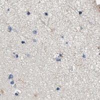
ACADVL / Very long-chain acyl-CoA dehydrogenase, mitochondrial · IHC design guide

Design Immunohistochemistry for ACADVL

Plan ACADVL chromogenic IHC on paraffin sections using the granular cytoplasmic pattern seen in cardiomyocytes (HPA tissue IHC). Catalog antibody PB10023 lists 2–5 μg/ml for IHC (datasheet PB10023); score staining by cell type and intensity.

Evidence assembled Oct 2026 · For research use; verify linked source records and product datasheet before use
Immunohistochemistry protocol sheet for ACADVL (IHC for ACADVL): expected localisation Granular cytoplasm (HPA tissue IHC); mitochondrial inner membrane (UniProt), antibody PB10023, validated IHC image, and IHC protocol steps
Printable ACADVL IHC protocol sheet — expected localisation Granular cytoplasm (HPA tissue IHC); mitochondrial inner membrane (UniProt), antibody PB10023, controls and protocol steps. Open the full ACADVL IHC guide →

ACADVL Immunohistochemistry Experimental Design Guide

Expected localisation, validated protocols, controls and antibodies — the at-a-glance facts below, then the full design guide.

Must know before staining
Expected localisation Granular cytoplasm (HPA tissue IHC); mitochondrial inner membrane (UniProt)
Staining pattern Granular cytoplasmic staining in cardiomyocytes (HPA tissue IHC)
Antigen retrieval EDTA pH 8.0 HIER, heat-mediated (datasheet PB10023)
Positive control ⓘ Adrenal gland+4 more · see all
Negative control ⓘ Soft tissue
Important caveats
Reasons your staining may differ from the expected pattern.
Fixation Keep section fixation consistent. (standard IHC practice; not target-specific)
Caveat Cell-type variation: chondrocytes were not detected (HPA tissue IHC)
Regulation Predominant in heart and skeletal muscle (UniProt)
Isoform / epitope 3 isoforms; mature chain spans 41–655; epitope coverage unknown (UniProt)
Section 1

Recommended ACADVL IHC & IF Protocols

Compare the catalog antibody’s IHC-P protocol (datasheet: PB10023) with two published ACADVL IHC protocols (PMC11308078; PMC9877169).

Recommended immunohistochemistry (IHC-P) protocol parameters
SampleParaffin-embedded human liver cancer tissue; fixative not specified (datasheet PB10023)
FixationImage fixative and duration unreported (datasheet PB10023); verify before use.
Sectioning4–5 µm sections on charged slides (standard)
DeparaffinisationXylene, graded ethanol series to water (standard)
Antigen retrievalHeat retrieval: EDTA pH 8.0 (datasheet PB10023); 20 min, 95–100 °C (standard)
Peroxidase block3% H2O2, 10 min, room temperature (standard)
Blocking10% goat serum (datasheet PB10023)
Primary antibodyRabbit anti-ACADVL, 2-5 μg/ml (datasheet PB10023)
Primary incubationOvernight at 4 °C (datasheet PB10023)
DetectionHRP-conjugated secondary, DAB chromogen (datasheet PB10023)
CounterstainHematoxylin, blue, dehydrate and mount (standard)
Expected resultACADVL-positive staining in glandular cells of adrenal gland (HPA tissue IHC: High). HPA tissue profile: General cytoplasmic expression with a granular pattern. No signal in the no-primary control.
💡Decision noteStart with heat-mediated EDTA retrieval at pH 8.0 (datasheet: PB10023); the published excerpts do not specify a retrieval method (PMC11308078; PMC9877169).
Section 2

What Is the Expected ACADVL Staining Pattern?

ACADVL is associated with the mitochondrial inner membrane and has no annotated transmembrane segment (UniProt P49748 topology). In paraffin sections, expect granular cytoplasmic staining rather than a membrane outline (HPA: general granular cytoplasmic IHC profile). HPA reports high staining in heart cardiomyocytes, lung macrophages, and several glandular or ciliated cell populations (HPA: tissue IHC). Its tissue IHC reliability is Enhanced, with medium consistency between antibody staining and RNA data (HPA: reliability).

What am I looking at on my slide?
High, granular cytoplasmic staining in heart cardiomyocytes.This fits the reported cell-level IHC result (HPA: High in cardiomyocytes) and the mitochondrial location (UniProt P49748 subcellular location). Judge staining in identified cardiomyocytes; do not treat a uniformly dark section as confirmation (general IHC interpretation).
A crisp cell-surface outline or exclusively nuclear chromogenic signal dominates.Neither is the expected tissue IHC profile (HPA: granular cytoplasmic expression; UniProt P49748 topology). Recheck compartment assignment and controls before calling it ACADVL; HPA's additional nuclear locations are uncertain ICC-IF observations (HPA: subcellular).
Strong staining appears in chondrocytes, or staining ignores the expected cell populations.HPA reports ACADVL as not detected in soft-tissue chondrocytes (HPA: tissue IHC). Unexpected signal can reflect cross-reactivity or endogenous chromogenic activity; compare a negative reagent control and the expected granular pattern (general IHC practice).
Color spreads across extracellular spaces or obscures cell boundaries.This cannot be scored as the reported granular cytoplasmic pattern (HPA: tissue IHC). Diffuse deposit can arise from nonspecific reagent binding, endogenous detection activity, or excessive chromogen development (general chromogenic IHC practice).
No granular signal is visible in heart cardiomyocytes.That conflicts with the High HPA cardiomyocyte result and UniProt's predominant heart expression (HPA: tissue IHC; UniProt P49748 tissue specificity). Check the run controls and assay steps before interpreting the specimen as biologically negative (general IHC practice).
💡Expected ACADVL appearanceCall a positive result when identified cardiomyocytes show High, granular cytoplasmic chromogenic signal (HPA: High in cardiomyocytes; granular IHC profile); an isolated nuclear stain or cell-surface outline does not match the expected pattern (HPA: tissue IHC; UniProt P49748 topology).
How each factor affects the staining
Compartment and topologyUniProt places ACADVL at the mitochondrial inner membrane but annotates no transmembrane segment (UniProt P49748 topology); at light-microscope resolution, use HPA's granular cytoplasmic IHC pattern as the practical readout (HPA: tissue IHC).
Cell and tissue contextHigh staining is reported in cardiomyocytes, lung macrophages, and specified glandular and ciliated cells (HPA: tissue IHC). UniProt describes predominant expression in heart and skeletal muscle, with protein also detected in kidney and liver (UniProt P49748 tissue specificity).
Low and undetected comparatorsHPA lists low staining in adipocytes and smooth-muscle cells, and no detected staining in soft-tissue chondrocytes (HPA: tissue IHC). Use these as pattern comparisons, allowing for tissue and cell identification during scoring (general IHC practice).
Strength of the staining evidenceThe tissue profile is rated Enhanced with medium antibody-staining versus RNA consistency (HPA: reliability). Two listed antibodies have Enhanced IHC validation (HPA: HPA019006 and HPA020595); that supports the pattern, but does not validate every stain in an individual run.
Protein formsUniProt lists three isoforms and a mature chain spanning residues 41–655 (UniProt P49748 isoforms and processing). Without a supplied antibody epitope, these facts cannot predict which forms the catalog antibody detects or justify a retrieval setting.
Why is my staining missing, weak or wrong?
SituationLikely causeNext action
Heart cardiomyocytes stain weakly or remain blank.A missed assay step or failed detection may explain a result that differs from HPA's High cardiomyocyte staining (HPA: tissue IHC; general IHC practice).Review the catalog antibody's IHC-P instructions and run controls; check retrieval, reagent application, detection, and chromogen development against the laboratory's validated workflow (general IHC practice).
The section shows diffuse brown background.Nonspecific reagent binding or endogenous detection activity can create diffuse color (general chromogenic IHC practice); HPA describes granular cytoplasmic staining (HPA: tissue IHC).Compare a negative reagent control, review blocking and washes, and adjust detection or chromogen development only within a validated workflow (general IHC practice).
Nuclear staining is stronger than cytoplasmic granules.The reported tissue pattern is cytoplasmic (HPA: tissue IHC); HPA labels additional nucleoplasm and nucleoli ICC-IF locations uncertain (HPA: subcellular).Check the negative control and reassess cell boundaries and granular cytoplasmic signal before scoring nuclear staining as ACADVL (general IHC interpretation).
Chondrocytes show strong staining.This differs from HPA's not detected chondrocyte result (HPA: soft-tissue IHC); cross-reactivity or background is possible (general IHC practice).Confirm cell identity and compare negative reagent controls and an expected positive cell population before accepting the signal (general IHC practice; HPA: tissue IHC).
Every cell looks equally intense.An indiscriminate stain conflicts with HPA's cell-specific High, Low, and not-detected observations (HPA: tissue IHC); excessive background is possible (general IHC practice).Assess staining at the cell level, inspect the negative control, and review antibody concentration and detection development under the validated IHC workflow (general IHC practice).
IF/ICC shows mitochondrial granules plus nuclear signal—is the nuclear signal expected?Mitochondria are the supported main ICC-IF location; nucleoplasm and nucleoli are additional uncertain locations (HPA: subcellular).Interpret mitochondrial localization as the supported finding and treat nuclear signal as uncertain; consult the separate IF/ICC guide for its assay design (HPA: subcellular).

Sample controls for ACADVL IHC & IF

🧪Run colon first and expect staining in its glandular cells (HPA: High in colon glandular cells). Use soft tissue chondrocytes as the negative tissue (HPA: Not detected); on the colon slide, use any unstained stromal cells as internal background comparators without assuming that all stromal cells lack ACADVL (standard IHC interpretation).
Positive control tissue: Adrenal gland (Glandular cells, HPA High)
Negative control tissue: Soft tissue (HPA Not detected)
ICC-IF cell lines (HPA subcellular resource): HPA ICC-IF images show ACADVL in A-431, A-549, U2OS, Sperm, KOLF2.1J, NIH 3T3, with annotated localisation: Mitochondria (supported) (HPA subcellular).
Technical controls: Include no-primary (secondary-only), host-matched rabbit IgG isotype, and a confirmed ACADVL knockout specimen as controls (selected-SKU caption: rabbit primary; standard IHC controls). For HRP/DAB staining, quench endogenous peroxidase and inspect the colon no-primary slide for mucus or luminal debris that could resemble staining (selected-SKU caption: HRP/DAB; standard IHC practice).
⚠️Feasibility: The selected-SKU paraffin-section caption reports heat retrieval in EDTA at pH 8.0, but does not establish that retrieval is required; its fixative is unreported (PB10023 tissue-IHC caption). A target-specific fixation window or fixation effect is unreported in the supplied evidence (PB10023 tissue-IHC caption; HPA). Frozen sections are not shown to be easier, while ICC-IF offers a mitochondrial localization readout that needs separate optimization; in colon, distinguish cellular signal from luminal mucus or debris (HPA: mitochondrial location supported; standard IHC practice).

HPA tissue IHC evidence for ACADVL

Comprehensive Human Protein Atlas IHC scoring per tissue (reliability: Enhanced — Medium consistency between antibody staining and RNA expression data.). Rows are taken directly from the HPA tissue chart — click any row's HPA link to view the source.

Positive expression · recommended positive controls

TissueCell typeLevelEvidenceSource
Adrenal gland Glandular cells High Protein (IHC) HPA →
Bronchus Ciliated cells (cell body) High Protein (IHC) HPA →
Colon Glandular cells High Protein (IHC) HPA →
Duodenum Glandular cells High Protein (IHC) HPA →
Gallbladder Glandular cells High Protein (IHC) HPA →

Undetected expression · recommended negative controls

TissueCell typeLevelEvidenceSource
Soft tissue Chondrocytes Not detected Protein (IHC) HPA →
Section 3

Advanced ACADVL IHC Tips

Troubleshoot ACADVL staining in paraffin sections by checking retrieval, compartment, cell type and controls before comparing chromogenic signal (UniProt P49748; HPA tissue IHC).

Which retrieval conditions should I try first when ACADVL staining is weak?
Start with heat-mediated antigen retrieval in EDTA at pH 8.0 for paraffin sections (datasheet PB10023). The catalog antibody stained a human liver cancer section after that retrieval, followed by 2 μg/ml primary antibody overnight at 4°C (datasheet PB10023). Keep section thickness, heating and cooling conditions consistent while comparing runs, since those variables affect retrieval and tissue integrity (standard IHC practice). If staining remains weak, compare a different retrieval buffer only as a documented fallback, alongside the EDTA condition and a positive control (standard IHC practice). Evaluate whether any gain preserves granular cytoplasmic staining rather than increasing diffuse background (HPA tissue IHC; standard IHC practice).
How should I troubleshoot fixation when ACADVL staining varies between sections?
Target-specific fixation sensitivity for ACADVL is unknown from the supplied evidence; the PB10023 caption identifies a paraffin section but does not state its fixative (datasheet PB10023). Record the fixative, fixation duration and processing history for each specimen before attributing a weak result to the antibody (standard IHC practice). Compare similarly processed positive-control tissue within the same staining run, then vary retrieval or primary incubation one condition at a time (standard IHC practice). Avoid claiming that mitochondrial location or annotated acetylation and succinylation predict a particular fixation response (UniProt P49748 topology and modified residues). Check morphology and section retention when assessing harsh processing, because damaged tissue can make chromogenic signal difficult to interpret (standard IHC practice).
What ACADVL staining pattern should I expect in paraffin tissue sections?
Expect granular cytoplasmic staining at light-microscope resolution, consistent with the tissue IHC profile and the protein’s mitochondrial location (HPA tissue IHC; UniProt P49748). ACADVL is assigned to the mitochondrial inner membrane despite having no annotated transmembrane segment, so IHC cannot establish membrane sidedness from a DAB pattern (UniProt P49748 topology; standard IHC practice). Heart cardiomyocytes are reported as highly stained, whereas soft-tissue chondrocytes are reported as undetected, making cell identity important during review (HPA tissue IHC). If signal appears predominantly nuclear or uniformly cytosolic, inspect controls, counterstain and tissue preservation before accepting it as ACADVL (UniProt P49748; standard IHC practice). Additional nuclear and nucleolar IF locations carry uncertain support and should not override the tissue IHC pattern (HPA subcellular).
Can isoforms or epitope accessibility explain inconsistent ACADVL IHC staining?
ACADVL has 3 annotated isoforms, but the supplied antibody caption does not identify its epitope or establish isoform coverage (UniProt P49748; datasheet PB10023). The annotated mature chain spans residues 41–655; that annotation alone cannot show which isoforms or processed forms the antibody detects (UniProt P49748 processing). Check the antibody’s documented immunogen or epitope information, if available, before assigning an isoform-specific explanation to staining differences (standard IHC practice). ACADVL also has annotated modified residues, including acetylated, succinylated and nitrosylated sites, but their effect on this antibody’s IHC signal is unreported (UniProt P49748 modified residues). Compare retrieval conditions and matched controls before interpreting altered intensity as altered isoform abundance (standard IHC practice).
How can I use IF to investigate an ambiguous ACADVL IHC pattern?
Use IF as a separate validation experiment while interpreting the chromogenic section against its established granular cytoplasmic pattern (HPA tissue IHC; standard IF practice). Multiplex ACADVL with a marker for the expected cell type, such as a cardiomyocyte marker in heart, and a mitochondrial marker to assess the claimed compartment (HPA tissue IHC; UniProt P49748; standard IF practice). Select fluorophores after checking tissue autofluorescence and include single-label controls for channel bleed-through (standard IF practice). For an intracellular epitope, optimise permeabilisation to provide access to the relevant side of the mitochondrial inner membrane; the antibody’s epitope side is not supplied (UniProt P49748 topology; standard IF practice). Treat apparent nuclear overlap cautiously because the additional nuclear IF locations have uncertain support (HPA subcellular).
How do I distinguish weak ACADVL staining from chromogenic background?
Run a no-primary control and inspect whether DAB develops in the same structures as the test section; block endogenous peroxidase before HRP detection (standard IHC practice). The catalog example used 10% goat serum, a peroxidase-conjugated secondary antibody and DAB, so compare background under those documented conditions when following that example (datasheet PB10023). Reassess blocking, washes and primary concentration if color appears broadly distributed rather than granular within cells (HPA tissue IHC; standard IHC practice). Include a tissue region with reliably stained cell types where available; HPA reports high staining in heart cardiomyocytes and lung macrophages (HPA tissue IHC). Score the test section only after the no-primary control shows acceptably low signal (standard IHC practice).
What should I measure when comparing ACADVL IHC across tissue samples? ⚠ ANSWER MARKED FOR VERIFICATION
Define the cell population and region of interest before scoring, because ACADVL staining differs among reported cell types (HPA tissue IHC; standard IHC practice). Within comparable regions, report the percentage of positive cells and an intensity-weighted H-score, with a fixed threshold and blinded review where feasible (standard IHC practice). For sparse populations, positive-cell density per mm² can supplement those measures, provided the sampled area and exclusion rules are recorded (standard IHC practice). Normalise comparisons to the relevant cell population or viable tissue area, and account for differences in cell composition rather than treating whole-section DAB area as protein abundance (standard IHC practice). Use the same staining run and controls when comparing intensity between specimens (standard IHC practice).
When is an apparent ACADVL-positive area likely to be an artefact?
A credible result places granular cytoplasmic signal in intact cells, consistent with mitochondrial ACADVL and the tissue IHC profile (UniProt P49748; HPA tissue IHC). Examine cell identity before calling a region positive: HPA reports high staining in heart cardiomyocytes and lung macrophages, but no detected signal in soft-tissue chondrocytes (HPA tissue IHC). Predominantly nuclear staining needs independent verification because reported nuclear and nucleolar IF locations are uncertain (HPA subcellular). Signal confined to section edges or necrotic areas should be checked against morphology and a no-primary control (standard IHC practice). Persistent DAB in that control suggests detection background, including possible endogenous peroxidase activity, rather than demonstrated ACADVL staining (standard IHC practice).
Boster reagents

Best ACADVL / Very long-chain acyl-CoA dehydrogenase, mitochondrial IHC Antibodies

PB10023 has paraffin-section IHC images from human liver cancer, human ovarian cancer and rat heart, plus IF data from U2OS cells (PB10023 image captions).

Real IHC data IHC analysis of ACADVL using anti-ACADVL antibody (PB10023). ACADVL was detected in a paraffin-embedded section of human liver cancer tissue. Heat mediated antigen retrieval was performed in EDTA buffer (pH 8.0, epitope retrieval solution). The tissue section was blocked with 10% goat serum. The tissue section was then incubated with 2 μg/ml rabbit anti-ACADVL Antibody (PB10023) overnight at 4°C. Peroxidase Conjugated Goat Anti-rabbit IgG was used as secondary antibody and incubated for 30 minutes at 37°C. The tissue section was developed using HRP Conjugated Rabbit IgG Super Vision Assay Kit (Catalog # SV0002) with DAB as the chromogen.
Anti-ACADVL Antibody ®
Cat # PB10023

PB10023 will render with its own IHC images from human liver cancer, human ovarian cancer and rat heart, and an IF image from U2OS cells (PB10023 image captions). M02607 lists human IHC reactivity but has no IHC or IF image in the payload, so it has no figure-backed card (M02607 catalog).

Which to pick: For tissue IHC, choose PB10023: its captions document paraffin sections, with the fixative unreported (PB10023 IHC image captions). For IF/ICC and cross-species work, PB10023 lists both applications and Human, Mouse and Rat reactivity; its images document IF in U2OS cells and tissue IHC in Human and Rat samples (PB10023 catalog and image captions). M02607 is a rabbit monoclonal option for human IHC, though its payload supplies no IHC or IF image (M02607 catalog).

Each figure is that product's own IHC / IF validation image from its datasheet.

References

  1. UniProt Consortium. UniProt entry P49748 (ACADV_HUMAN, Very long-chain acyl-CoA dehydrogenase, mitochondrial).
  2. Human Protein Atlas. ACADVL tissue IHC expression (reliability: Enhanced).
  3. Human Protein Atlas. ACADVL subcellular location (ICC-IF): Mainly localized to the mitochondria. In addition localized to the nucleoplasm and nucleoli..
  4. Human Protein Atlas. ACADVL antibody validation summary (2 antibodies).
  5. Proteomics Analysis Provides Insights into the Role of Lipid Metabolism in T2DM-Related Sarcopenia. ACS omega 2024 — PMC11308078.
  6. AAV9 gene replacement therapy for respiratory insufficiency in very-long chain acyl-CoA dehydrogenase deficiency. Journal of inherited metabolic disease 2019 — PMC6739149.
  7. Synthetic mRNA rescues very long-chain acyl-CoA dehydrogenase deficiency in patient fibroblasts and a murine model. Molecular genetics and metabolism 2023 — PMC9877169.
  8. Unveiling the aging-immune axis in irritable bowel syndrome: a multi-omics and machine learning approach to biomarker discovery and validation. Frontiers in immunology 2026 — PMC13437292.
  9. PubMed PMID:7668252 — UniProt-cited evidence.
  10. PubMed PMID:8845838 — UniProt-cited evidence.
  11. PubMed PMID:8554625 — UniProt-cited evidence.