This website uses cookies to ensure you get the best experience on our website.


- Table of Contents
Plan ACLY staining in paraffin sections using the observed cytoplasmic and nuclear tissue pattern (HPA tissue IHC). Compare defined positive and negative cell populations while interpreting intensity cautiously because antibody staining and RNA levels show low consistency (HPA tissue IHC).

Expected localisation, validated protocols, controls and antibodies — the at-a-glance facts below, then the full design guide.
| Expected localisation | Cytosol expected (UniProt); cytoplasmic and nuclear tissue staining (HPA tissue IHC) | |
| Staining pattern | Cytoplasmic and nuclear staining in several tissues (HPA tissue IHC) | |
| Antigen retrieval | EDTA pH 8.0 HIER, heat-mediated (datasheet PB10024) | |
| Positive control | Pancreas+4 more · see all | |
| Negative control | Esophagus+4 more · see all |
| Fixation | Keep fixation consistent across sections. (standard IHC practice; not target-specific) | |
| Caveat | Antibody staining and RNA levels show low consistency (HPA tissue IHC) | |
| Regulation | Staining regulation is not annotated (UniProt) | |
| Isoform / epitope | 3 isoforms; epitope coverage is unknown (UniProt) |
The catalog antibody uses EDTA pH 8.0 heat retrieval (datasheet PB10024). The four published ACLY IHC protocols below provide additional staining conditions (PMC7855479; PMC4427316; PMC12806462; PMC6774899).
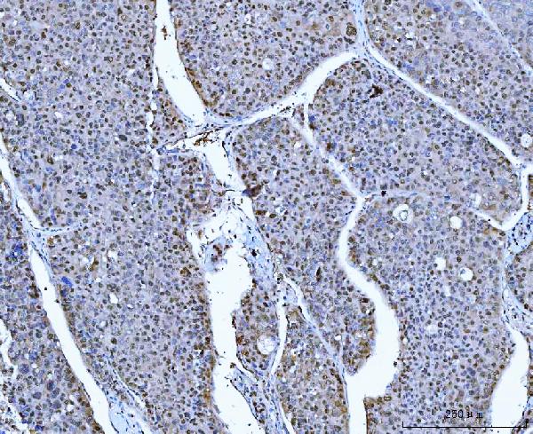
| Sample | Paraffin-embedded human colonic adenocarcinoma tissue; fixative not specified (datasheet PB10024) |
| Fixation | Image fixative and duration unreported (datasheet PB10024); verify before use. |
| Sectioning | 4–5 µm sections on charged slides (standard) |
| Deparaffinisation | Xylene, graded ethanol series to water (standard) |
| Antigen retrieval | Heat retrieval: EDTA pH 8.0 (datasheet PB10024); 20 min, 95–100 °C (standard) |
| Peroxidase block | 3% H2O2, 10 min, room temperature (standard) |
| Blocking | 10% goat serum (datasheet PB10024) |
| Primary antibody | Rabbit anti-ACLY, 1-2μg/ml (datasheet PB10024) |
| Primary incubation | Overnight at 4 °C (datasheet PB10024) |
| Detection | HRP-conjugated secondary, DAB chromogen (datasheet PB10024) |
| Counterstain | Hematoxylin, blue, dehydrate and mount (standard) |
| Expected result | ACLY-positive staining in pancreatic endocrine cells of pancreas (HPA tissue IHC: High). HPA tissue profile: Cytoplasmic and nuclear expression in several tissues. No signal in the no-primary control. |
ACLY is a cytosolic enzyme with no transmembrane segment (UniProt P53396). In paraffin sections, expect mainly cytoplasmic staining, with possible nuclear staining in some tissues (HPA: tissue IHC profile). High staining is reported in pancreatic endocrine cells, prostate glandular cells, and testicular Leydig cells (HPA: tissue IHC). The HPA IHC profile is rated Enhanced, but its antibody staining has low consistency with RNA expression data (HPA: reliability description).
| Strong cytoplasmic staining in pancreatic endocrine cells, prostate glandular cells, or Leyig cells. | These are reported high-staining cell populations (HPA: tissue IHC). Compare staining within the identified cell population; a positive result does not require every cell in the section to stain (general IHC practice). |
| Cytoplasmic staining accompanied by nuclear staining in some cells. | HPA describes cytoplasmic and nuclear staining across several tissues (HPA: tissue IHC profile). UniProt places ACLY in the cytosol (UniProt P53396); assess nuclear signal alongside the tissue and antibody controls rather than automatically rejecting it. |
| Predominantly membranous or extracellular staining, with little cytoplasmic signal. | This does not match the reported cytosolic location or the absence of a transmembrane segment (UniProt P53396). Check morphology and detection controls before attributing the pattern to ACLY (general IHC practice). |
| Strong staining in a cell population reported as not detected by HPA. | For example, HPA reports no staining in esophageal squamous epithelial cells (HPA: tissue IHC). Unexpected staining raises a specificity or endogenous detection concern; the HPA reliability caveat means it is not conclusive evidence of cross-reactivity (HPA: reliability description; general IHC practice). |
| Uniform color over cells and tissue spaces, without a clear cellular pattern. | Diffuse color makes compartment and cell-type assessment unreliable (general IHC practice). Evaluate background with a no-primary control and compare a reported high-staining tissue in the same run (general IHC practice; HPA: tissue IHC). |
| Tissue and cell selection | HPA reports high staining in pancreatic endocrine, prostate glandular, and Leyig cells, but no detected staining in esophageal squamous epithelial cells (HPA: tissue IHC). Use the specified cell populations when comparing controls; HPA also reports low tissue RNA specificity and low staining–RNA consistency (HPA: RNA specificity and reliability description). |
| Antibody evidence | Four listed rabbit polyclonal antibodies have Enhanced IHC status; the listed rabbit monoclonal has Approved IHC status (HPA: antibody validation). Enhanced indicates reproduction by independent antibodies or orthogonal data (HPA: antibody validation definition). This supports the observed pattern, subject to HPA's staining–RNA consistency caveat (HPA: reliability description). |
| Isoforms and epitope coverage | UniProt lists three ACLY isoforms and an ATP-grasp domain at residues 4–265 (UniProt P53396). The supplied records do not map an IHC antibody epitope or establish isoform-specific staining; check the chosen antibody's documented target region before interpreting a missing cell signal as isoform absence (general IHC practice). |
| Intracellular target and modifications | ACLY has no signal peptide, propeptide, or transmembrane segment; UniProt lists multiple modified residues, including phosphorylation sites (UniProt P53396). These facts support evaluating intracellular staining, but do not establish how processing, phosphorylation, or fixation affects this antibody's IHC signal. |
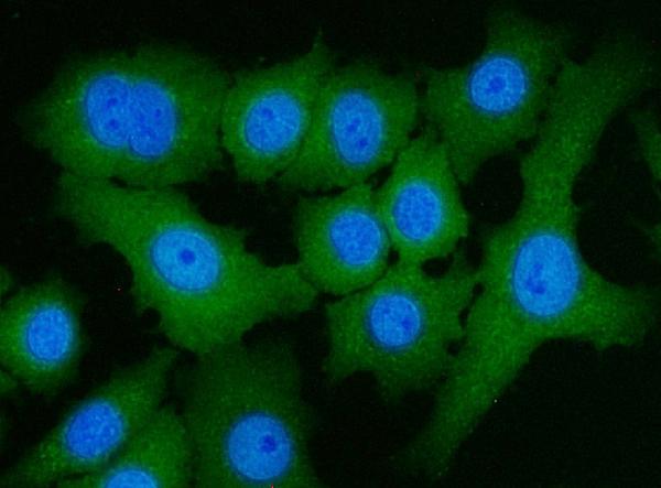
| IF/ICC Q&A: where should signal appear? | Mainly in the cytosol (HPA: ICC-IF, supported). HPA lists nucleoplasm, primary cilium transition zone, and basal body as uncertain additional locations, and principal piece as approved (HPA: ICC-IF). Treat those location labels as ICC-IF evidence; they do not establish an IHC staining requirement. |
| Situation | Likely cause | Next action |
|---|---|---|
| No staining in pancreatic endocrine cells, prostate glandular cells, or Leyig cells. | These are reported high-staining populations, so a blank result may reflect a run or reagent problem (HPA: tissue IHC; general IHC practice). HPA staining–RNA inconsistency also limits certainty about any single specimen (HPA: reliability description). | Confirm the expected cells are present, inspect the positive control and detection reagents, and review the antibody's validated IHC-P instructions (general IHC practice). If needed, optimize retrieval as a general IHC step; no ACLY-specific fixation effect is established here. |
| High background obscures the cytoplasm. | Nonspecific binding or detection background can hide a cellular pattern (general IHC practice). The HPA tissue profile cannot identify which workflow step caused background in a particular section. | Run a no-primary control; review blocking, wash, detection, and counterstain conditions, then compare the cellular pattern with a reported positive population (general IHC practice; HPA: tissue IHC). |
| Color appears chiefly on cell borders or outside cells. | A border or extracellular pattern conflicts with ACLY's cytosolic annotation and lack of a transmembrane segment (UniProt P53396). Section morphology or background may complicate the apparent compartment (general IHC practice). | Inspect morphology at higher magnification, compare a no-primary control, and reassess compartment assignment before scoring the section (general IHC practice). |
| Unexpected strong staining appears in esophageal squamous epithelium. | HPA reports this cell population as not detected (HPA: tissue IHC). Cross-reactivity or endogenous detection activity is possible, but HPA's staining–RNA consistency caveat limits a categorical call (HPA: reliability description; general IHC practice). | Check a no-primary control and compare staining in a reported high-staining cell population; seek independent antibody or orthogonal evidence if the unexpected result drives the conclusion (general IHC practice; HPA: tissue IHC and antibody validation). |
| Nuclear staining seems stronger than cytoplasmic staining. | HPA reports nuclear expression in several tissues, while UniProt annotates ACLY as cytosolic (HPA: tissue IHC profile; UniProt P53396). The supplied evidence does not set a nuclear-to-cytoplasmic intensity cutoff. | Score compartments separately, confirm the cell population, and compare with the no-primary control and another validated antibody where available (general IHC practice; HPA: antibody validation). |
| An IF/ICC image is being used to judge an IHC section. | HPA supports cytosol as the main ICC-IF location, with other locations carrying distinct confidence labels (HPA: ICC-IF). ICC-IF localization alone does not define the appearance of a chromogenic tissue section. | Judge the IHC section against HPA tissue IHC cell patterns and the section's controls; use the ICC-IF location labels only to inform compartment interpretation (HPA: tissue IHC and ICC-IF; general IHC practice). |





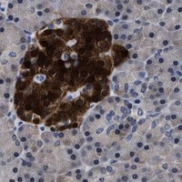


Comprehensive Human Protein Atlas IHC scoring per tissue (reliability: Enhanced — Low consistency between antibody staining and RNA expression data.). Rows are taken directly from the HPA tissue chart — click any row's HPA link to view the source.
| Tissue | Cell type | Level | Evidence | Source |
|---|---|---|---|---|
| Pancreas | Pancreatic endocrine cells | High | Protein (IHC) | HPA → |
| Prostate | Glandular cells | High | Protein (IHC) | HPA → |
| Testis | Leydig cells | High | Protein (IHC) | HPA → |
| Adrenal gland | Glandular cells | Medium | Protein (IHC) | HPA → |
| Cerebellum | Cells in molecular layer | Medium | Protein (IHC) | HPA → |
| Tissue | Cell type | Level | Evidence | Source |
|---|---|---|---|---|
| Esophagus | Squamous epithelial cells | Not detected | Protein (IHC) | HPA → |
| Fallopian tube | Ciliated cells (cell body) | Not detected | Protein (IHC) | HPA → |
| Oral mucosa | Squamous epithelial cells | Not detected | Protein (IHC) | HPA → |
| Ovary | Ovarian stroma cells | Not detected | Protein (IHC) | HPA → |
| Parathyroid gland | Glandular cells | Not detected | Protein (IHC) | HPA → |
Troubleshoot ACLY staining in paraffin sections by checking retrieval, staining controls, cellular location, and how signal is scored.
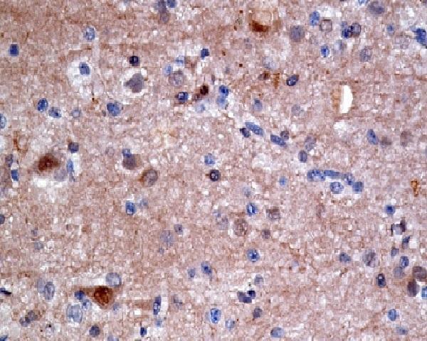
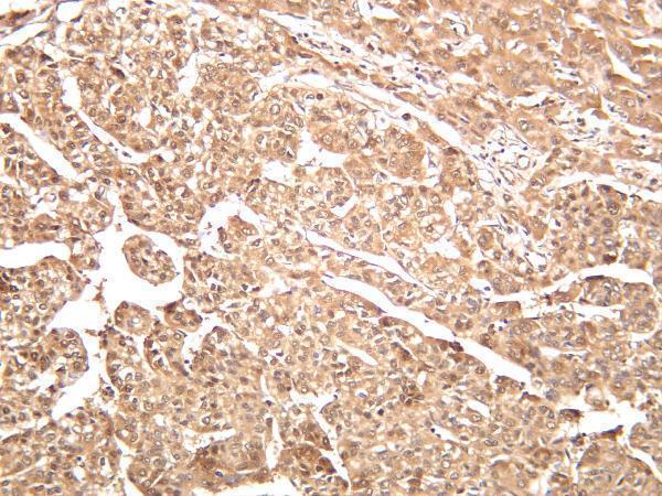
The catalog includes IHC images from human, mouse, and rat paraffin sections and IF/ICC images from A594 and MCF7 cells; all four antibodies list human, mouse, and rat reactivity (catalog image captions; catalog reactivity).
PB10024 has human colonic adenocarcinoma paraffin IHC and A594 IF/ICC images; M02372-1 has human pancreatic cancer and mouse and rat pancreas paraffin IHC images, plus an MCF7 IF/ICC image (catalog image captions). M02372 has a human brain carcinoma paraffin IHC image, while M02372-3 has a human hepatocellular carcinoma IHC image with section processing unreported (catalog image captions).
Which to pick: For tissue IHC, PB10024 offers a documented paraffin-section example with EDTA pH 8.0 retrieval and DAB detection; the fixative is unreported (PB10024 IHC image caption). For IF/ICC, M02372-1 is a mouse monoclonal with an MCF7 IF/ICC image; it also has mouse and rat pancreas paraffin IHC images, making it the strongest pictured option for cross-species tissue work (M02372-1 catalog applications, clone, and image captions).