Click image to see more details
-

-
-
-
-
+4
Product Info Summary
| SKU: | M02372-1 |
|---|---|
| Size: | 100 μg/vial |
| Reactive Species: | Human, Mouse, Rat |
| Host: | Mouse |
| Application: | Flow Cytometry, IF, IHC, ICC, WB |
Customers Who Bought This Also Bought

Product info
Product Name
Anti-ATP citrate lyase ACLY Antibody Picoband® (monoclonal, 5I2)
SKU/Catalog Number
M02372-1
Size
100 μg/vial
Form
Lyophilized
Description
Boster Bio Anti-ATP citrate lyase ACLY Antibody Picoband® (monoclonal, 5I2) catalog # M02372-1. Tested in Flow Cytometry, IF, IHC, ICC, WB applications. This antibody reacts with Human, Mouse, Rat. The brand Picoband indicates this is a premium antibody that guarantees superior quality, high affinity, and strong signals with minimal background in Western blot applications. Only our best-performing antibodies are designated as Picoband, ensuring unmatched performance.
Storage & Handling
Store at -20˚C for one year from date of receipt. After reconstitution, at 4˚C for one month. It can also be aliquotted and stored frozen at -20˚C for six months. Avoid repeated freeze-thaw cycles.
Cite This Product
Anti-ATP citrate lyase ACLY Antibody Picoband® (monoclonal, 5I2) (Boster Biological Technology, Pleasanton CA, USA, Catalog # M02372-1)
Host
Mouse
Contents
Each vial contains 4mg Trehalose, 0.9mg NaCl, 0.2mg Na2HPO4, 0.05mg NaN3.
Clonality
Monoclonal
Clone Number
5I2
Isotype
Mouse IgG2b
Immunogen
E. coli-derived human ATP citrate lyase recombinant protein (Position: M1-I180). Human ATP citrate lyase shares 95% amino acid (aa) sequence identity with both mouse and rat ATP citrate lyase.
Cross-reactivity
No cross-reactivity with other proteins.
Reactive Species
M02372-1 is reactive to ACLY in Human, Mouse, Rat
Observed Molecular Weight
121 kDa
Calculated molecular weight
120.8 kDa
Background of ACLY
ATP citrate lyase, aslo known as ACLY, is an enzyme that in animals represents an important step in fatty acid biosynthesis. ATP citrate lyase is the primary enzyme responsible for the synthesis of cytosolic acetyl-CoA in many tissues. The enzyme is a tetramer of apparently identical subunits. The product, acetyl-CoA, in animals serves several important biosynthetic pathways, including lipogenesis and cholesterogenesis. It is activated by insulin. In nervous tissue, ATP citrate-lyase may be involved in the biosynthesis of acetylcholine.In plants, ATP citrate lyase generates the acetyl-CoA for cytosolically-synthesized metabolites.
Antibody Validation
Boster validates all antibodies on WB, IHC, ICC, Immunofluorescence, and ELISA with known positive control and negative samples to ensure specificity and high affinity, including thorough antibody incubations.
Application & Images
Applications
M02372-1 is guaranteed for Flow Cytometry, IF, IHC, ICC, WB Boster Guarantee
Recommend Dilution
| Application | Dilution | Species |
|---|---|---|
| Western blot | 0.1-0.5μg/ml | |
| Immunohistochemistry (Paraffin-embedded Section) | 0.5-1μg/ml | |
| Immunocytochemistry/Immunofluorescence | 2μg/ml | Human |
| Flow Cytometry (Fixed) | 1-3μg/1x106 cells |
Tested application
Suggested blocking solution with 5% non-fat milk or BSA; (*)Recommended protein loading: 20-40 µg per lane
Use TE buffer pH 9.0 for antigen retrieval; (*) citrate buffer pH 6.0 is an alternative.
Validation Images & Assay Conditions
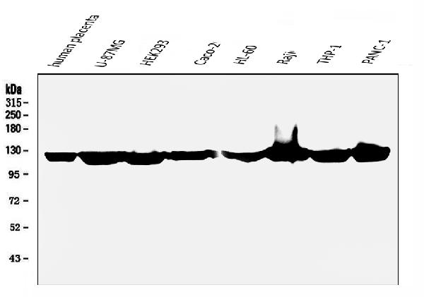
Click image to see more details
Western blot analysis of ATP citrate lyase using anti-ATP citrate lyase antibody (M02372-1).
Electrophoresis was performed on a 5-20% SDS-PAGE gel at 70V (Stacking gel) / 90V (Resolving gel) for 2-3 hours. The sample well of each lane was loaded with 50ug of sample under reducing conditions.
Lane 1: human placenta tissue lysates,
Lane 2: U-87MG whole cell lysates,
Lane 3: HEK293 whole cell lysates,
Lane 4: Caco-2 whole cell lysates,
Lane 5: HL-60 whole cell lysates,
Lane 6: Raji whole cell lysates,
Lane 7: THP-1 whole cell lysates,
Lane 8: PANC-1 whole cell lysates.
After Electrophoresis, proteins were transferred to a Nitrocellulose membrane at 150mA for 50-90 minutes. Blocked the membrane with 5% Non-fat Milk/ TBS for 1.5 hour at RT. The membrane was incubated with mouse anti-ATP citrate lyase antigen affinity purified monoclonal antibody (Catalog # M02372-1) at 0.5 μg/mL overnight at 4°C, then washed with TBS-0.1%Tween 3 times with 5 minutes each and probed with a goat anti-mouse IgG-HRP secondary antibody at a dilution of 1:5000 for 1.5 hour at RT. The signal is developed using an Enhanced Chemiluminescent detection (ECL) kit (Catalog # EK1001) with Tanon 5200 system. A specific band was detected for ATP citrate lyase at approximately 121KD. The expected band size for ATP citrate lyase is at 121KD.

Click image to see more details
Western blot analysis of ATP citrate lyase using anti-ATP citrate lyase antibody (M02372-1).
Electrophoresis was performed on a 5-20% SDS-PAGE gel at 70V (Stacking gel) / 90V (Resolving gel) for 2-3 hours. The sample well of each lane was loaded with 50ug of sample under reducing conditions.
Lane 1: rat lung tissue lysates,
Lane 2: rat testicular tissue lysates,
Lane 3: rat kidney tissue lysates,
Lane 4: rat brain tissue lysates,
Lane 5: mouse lung tissue lysates,
Lane 6: mouse testicular tissue lysates,
Lane 7: mouse kidney tissue lysates.
After Electrophoresis, proteins were transferred to a Nitrocellulose membrane at 150mA for 50-90 minutes. Blocked the membrane with 5% Non-fat Milk/ TBS for 1.5 hour at RT. The membrane was incubated with mouse anti-ATP citrate lyase antigen affinity purified monoclonal antibody (Catalog # M02372-1) at 0.5 μg/mL overnight at 4°C, then washed with TBS-0.1%Tween 3 times with 5 minutes each and probed with a goat anti-mouse IgG-HRP secondary antibody at a dilution of 1:5000 for 1.5 hour at RT. The signal is developed using an Enhanced Chemiluminescent detection (ECL) kit (Catalog # EK1001) with Tanon 5200 system. A specific band was detected for ATP citrate lyase at approximately 121KD. The expected band size for ATP citrate lyase is at 121KD.

Click image to see more details
IHC analysis of ATP citrate lyase using anti-ATP citrate lyase antibody (M02372-1).
ATP citrate lyase was detected in paraffin-embedded section of human pancreatic cancer tissue. Heat mediated antigen retrieval was performed in EDTA buffer (pH8.0, epitope retrieval solution). The tissue section was blocked with 10% goat serum. The tissue section was then incubated with 1μg/ml mouse anti-ATP citrate lyase Antibody (M02372-1) overnight at 4°C. Biotinylated goat anti-mouse IgG was used as secondary antibody and incubated for 30 minutes at 37°C. The tissue section was developed using Strepavidin-Biotin-Complex (SABC) (Catalog # SA1021) with DAB as the chromogen.

Click image to see more details
IHC analysis of ATP citrate lyase using anti-ATP citrate lyase antibody (M02372-1).
ATP citrate lyase was detected in paraffin-embedded section of human testis cancer tissue. Heat mediated antigen retrieval was performed in EDTA buffer (pH8.0, epitope retrieval solution). The tissue section was blocked with 10% goat serum. The tissue section was then incubated with 1μg/ml mouse anti-ATP citrate lyase Antibody (M02372-1) overnight at 4°C. Biotinylated goat anti-mouse IgG was used as secondary antibody and incubated for 30 minutes at 37°C. The tissue section was developed using Strepavidin-Biotin-Complex (SABC) (Catalog # SA1021) with DAB as the chromogen.

Click image to see more details
IHC analysis of ATP citrate lyase using anti-ATP citrate lyase antibody (M02372-1).
ATP citrate lyase was detected in paraffin-embedded section of mouse pancreas tissue. Heat mediated antigen retrieval was performed in EDTA buffer (pH8.0, epitope retrieval solution). The tissue section was blocked with 10% goat serum. The tissue section was then incubated with 1μg/ml mouse anti-ATP citrate lyase Antibody (M02372-1) overnight at 4°C. Biotinylated goat anti-mouse IgG was used as secondary antibody and incubated for 30 minutes at 37°C. The tissue section was developed using Strepavidin-Biotin-Complex (SABC) (Catalog # SA1021) with DAB as the chromogen.

Click image to see more details
IHC analysis of ATP citrate lyase using anti-ATP citrate lyase antibody (M02372-1).
ATP citrate lyase was detected in paraffin-embedded section of rat pancreas tissue. Heat mediated antigen retrieval was performed in EDTA buffer (pH8.0, epitope retrieval solution). The tissue section was blocked with 10% goat serum. The tissue section was then incubated with 1μg/ml mouse anti-ATP citrate lyase Antibody (M02372-1) overnight at 4°C. Biotinylated goat anti-mouse IgG was used as secondary antibody and incubated for 30 minutes at 37°C. The tissue section was developed using Strepavidin-Biotin-Complex (SABC) (Catalog # SA1021) with DAB as the chromogen.

Click image to see more details
Flow Cytometry analysis of A549 cells using anti-ATP citrate lyase antibody (M02372-1).
Overlay histogram showing A549 cells stained with M02372-1 (Blue line). To facilitate intracellular staining, cells were fixed with 4% paraformaldehyde and permeabilized with permeabilization buffer. The cells were blocked with 10% normal goat serum. And then incubated with mouse anti-ATP citrate lyase Antibody (M02372-1,1μg/1x106 cells) for 30 min at 20°C. DyLight®488 conjugated goat anti-mouse IgG (BA1126, 5-10μg/1x106 cells) was used as secondary antibody for 30 minutes at 20°C. Isotype control antibody (Green line) was mouse IgG (1μg/1x106) used under the same conditions. Unlabelled sample (Red line) was also used as a control.

Click image to see more details
IF analysis of ATP citrate lyase using anti-ATP citrate lyase antibody (M02372-1).
ATP citrate lyase was detected in immunocytochemical section of MCF7 cells. Enzyme antigen retrieval was performed using IHC enzyme antigen retrieval reagent (AR0022) for 15 mins. The cells were blocked with 10% goat serum. And then incubated with 2μg/mL mouse anti-ATP citrate lyase Antibody (M02372-1) overnight at 4°C. DyLight®488 Conjugated Goat Anti-Mouse IgG (BA1126) was used as secondary antibody at 1:100 dilution and incubated for 30 minutes at 37°C. The section was counterstained with DAPI. Visualize using a fluorescence microscope and filter sets appropriate for the label used.
Specific Publications For Anti-ATP citrate lyase ACLY Antibody Picoband® (monoclonal, 5I2) (M02372-1)
Loading publications
Recommended Resources
Here are featured tools and databases that you might find useful.
- Boster's Pathways Library
- Protein Databases
- Bioscience Research Protocol Resources
- Data Processing & Analysis Software
- Photo Editing Software
- Scientific Literature Resources
- Research Paper Management Tools
- Molecular Biology Software
- Primer Design Tools
- Bioinformatics Tools
- Phylogenetic Tree Analysis
Customer Reviews
Have you used Anti-ATP citrate lyase ACLY Antibody Picoband® (monoclonal, 5I2)?
Share your experimental results or join a short interview to earn up to $1,000 in product credits or other rewards.
0 Reviews For Anti-ATP citrate lyase ACLY Antibody Picoband® (monoclonal, 5I2)
Customer Q&As
Have a question?
Find answers in Q&As, reviews.
Can't find your answer?
Submit your question
6 Customer Q&As for Anti-ATP citrate lyase ACLY Antibody Picoband® (monoclonal, 5I2)
Question
Is there a BSA free version of anti-ATP citrate lyase antibody (monoclonal, 5I2) M02372-1 available?
Verified Customer
Verified customer
Asked: 2020-05-01
Answer
I appreciate your recent telephone inquiry. I can confirm that some lots of this anti-ATP citrate lyase antibody (monoclonal, 5I2) M02372-1 are BSA free. For now, these lots are available and we can make a BSA free formula for you free of charge. It will take 3 extra days to prepare. If you require this antibody BSA free again in future, please do not hesitate to contact me and I will be pleased to check which lots we have in stock that are BSA free.
Boster Scientific Support
Answered: 2020-05-01
Question
See below the WB image, lot number and protocol we used for cervix carcinoma erythroleukemia using anti-ATP citrate lyase antibody (monoclonal, 5I2) M02372-1. Please let me know if you require anything else.
Verified Customer
Verified customer
Asked: 2019-08-14
Answer
Thank you very much for the data. Our lab team are working to resolve this as quickly as possible, and we appreciate your patience and understanding! You have provided everything we needed. Please let me know if there is anything you need in the meantime.
Boster Scientific Support
Answered: 2019-08-14
Question
I was wanting to use your anti-ATP citrate lyase antibody (monoclonal, 5I2) for IHC-P for mouse cervix carcinoma erythroleukemia on frozen tissues, but I want to know if it has been tested for this particular application. Has this antibody been tested and is this antibody a good choice for mouse cervix carcinoma erythroleukemia identification?
Verified Customer
Verified customer
Asked: 2019-05-10
Answer
You can see on the product datasheet, M02372-1 anti-ATP citrate lyase antibody (monoclonal, 5I2) has been validated for Flow Cytometry, IHC-P, WB on human, mouse, rat tissues. We have an innovator award program that if you test this antibody and show it works in mouse cervix carcinoma erythroleukemia in IHC-frozen, you can get your next antibody for free.
Boster Scientific Support
Answered: 2019-05-10
Question
I see that the anti-ATP citrate lyase antibody (monoclonal, 5I2) M02372-1 works with IHC-P, what is the protocol used to produce the result images on the product page?
Verified Customer
Verified customer
Asked: 2018-04-24
Answer
You can find protocols for IHC-P on the "support/technical resources" section of our navigation menu. If you have any further questions, please send an email to support@bosterbio.com
Boster Scientific Support
Answered: 2018-04-24
Question
Would anti-ATP citrate lyase antibody (monoclonal, 5I2) M02372-1 work for IHC-P with cervix carcinoma erythroleukemia?
L. Williams
Verified customer
Asked: 2017-12-08
Answer
According to the expression profile of cervix carcinoma erythroleukemia, ACLY is highly expressed in cervix carcinoma erythroleukemia. So, it is likely that anti-ATP citrate lyase antibody (monoclonal, 5I2) M02372-1 will work for IHC-P with cervix carcinoma erythroleukemia.
Boster Scientific Support
Answered: 2017-12-08
Question
I am interested in to test anti-ATP citrate lyase antibody (monoclonal, 5I2) M02372-1 on mouse cervix carcinoma erythroleukemia for research purposes, then I may be interested in using anti-ATP citrate lyase antibody (monoclonal, 5I2) M02372-1 for diagnostic purposes as well. Is the antibody suitable for diagnostic purposes?
E. Banerjee
Verified customer
Asked: 2016-10-17
Answer
The products we sell, including anti-ATP citrate lyase antibody (monoclonal, 5I2) M02372-1, are only intended for research use. They would not be suitable for use in diagnostic work. If you have the means to develop a product into diagnostic use, and are interested in collaborating with us and develop our product into an IVD product, please contact us for more discussions.
Boster Scientific Support
Answered: 2016-10-17
