This website uses cookies to ensure you get the best experience on our website.


- Table of Contents
Plan chromogenic ACOX1 IHC-P with catalog antibody M03054 at 1:10–1:50 (datasheet: M03054). Use high-staining hepatocytes as a positive tissue reference and assess granular cytoplasmic staining (HPA tissue IHC).

Expected localisation, validated protocols, controls and antibodies — the at-a-glance facts below, then the full design guide.
| Expected localisation | Granular cytoplasm (observed); peroxisome (molecular) (HPA tissue IHC; UniProt) | |
| Staining pattern | Granular cytoplasmic staining across tissues (HPA tissue IHC) | |
| Antigen retrieval | Citrate pH 6.0 HIER, 95–98 °C, 20 min (rule: cytoplasmic / membrane antigen) | |
| Positive control | Duodenum+4 more · see all | |
| Negative control | None in HPA (detected in all 45 tissues); use no-primary + isotype controls |
| Fixation | Keep formalin fixation consistent across paraffin samples. (standard IHC practice; not target-specific) | |
| Caveat | Antibody staining has low consistency with RNA expression (HPA tissue IHC) | |
| Regulation | Isoform abundance varies by tissue (UniProt) | |
| Isoform / epitope | 3 isoforms; B/C chains split at 468/469—check epitope (UniProt) |
The catalog antibody protocol is accompanied by published ACOX1 IHC methods for colorectal cancer sections, rat kidney, rat brown adipose tissue, and tumor xenografts (PMC11757735; PMC9453879; PMC10154930; PMC10601207).
| Sample | FFPE human colon carcinoma tissue (datasheet M03054) |
| Fixation | Image formalin-fixed; duration unreported (datasheet M03054); verify before use. |
| Sectioning | 4–5 µm sections on charged slides (standard) |
| Deparaffinisation | Xylene, graded ethanol series to water (standard) |
| Antigen retrieval | Heat-induced epitope retrieval in citrate buffer, pH 6.0, 20 min at 95–98 °C (standard rule: cytoplasmic / membrane antigen) |
| Peroxidase block | 3% H2O2, 10 min, room temperature (standard) |
| Blocking | 10% normal serum of the secondary host, 30 min, room temperature (standard) |
| Primary antibody | Mouse monoclonal (clone 153CT43.1.1) anti-ACOX1, 1:10-1:50 (datasheet M03054) |
| Primary incubation | Overnight at 4 °C (standard) |
| Detection | HRP-polymer secondary, DAB chromogen 5–10 min (standard) |
| Counterstain | Hematoxylin, blue, dehydrate and mount (standard) |
| Expected result | ACOX1-positive staining in glandular cells of duodenum (HPA tissue IHC: High). HPA tissue profile: General cytoplasmic expression with a granular pattern. No signal in the no-primary control. |
ACOX1 is a peroxisomal protein with no transmembrane segment (UniProt Q15067 topology and subcellular location). In paraffin-section IHC, expect granular cytoplasmic staining, especially in liver hepatocytes and duodenal and small-intestinal glandular cells (HPA: tissue IHC profile; High in these cells). HPA rates its tissue staining Approved but reports low consistency between antibody staining and RNA expression, so interpret intensity with that limitation (HPA: reliability description).
| Granular cytoplasmic staining in hepatocytes or intestinal glandular cells. | This fits the reported tissue pattern and High staining in those cells (HPA: tissue IHC). Granules are consistent with peroxisomal localization (UniProt Q15067; HPA: subcellular). Brightfield IHC does not resolve every granule as an individual peroxisome (general IHC practice). |
| Predominantly membranous or nuclear staining, with little granular cytoplasmic signal. | Question target assignment: ACOX1 has no transmembrane segment and is peroxisomal (UniProt Q15067 topology and location). Nucleolar localization is additionally reported by ICC-IF, so nucleolar signal alone cannot be declared an artefact, but it does not replace the expected tissue IHC pattern (HPA: subcellular; tissue IHC). |
| Strong staining in unexpected cells while nearby expected cells show little signal. | Consider cross-reactivity or endogenous detection activity, then compare morphology and controls (general IHC practice). HPA reports widespread cytoplasmic expression and lists no negative tissues; an unlisted cell type is not automatically ACOX1-negative (HPA: tissue IHC). |
| Uniform haze across tissue, or color in tissue-free areas. | A field-wide signal lacks the reported granular cytoplasmic pattern (HPA: tissue IHC). Background from detection reagents, nonspecific binding, or incomplete washing can obscure cell-level interpretation; assess reagent controls and washing before scoring (general IHC practice). |
| No convincing signal in liver hepatocytes or intestinal glandular cells. | These are reported High-staining cells, so first check section quality, retrieval and detection controls (HPA: tissue IHC; general IHC practice). A failed run is possible, but the HPA reliability caveat means one unstained specimen does not by itself settle antibody specificity or biological absence (HPA: reliability description). |
| Compartment and optical resolution | ACOX1 is peroxisomal (UniProt Q15067; HPA: subcellular). The tissue IHC readout is granular cytoplasm (HPA: tissue IHC); do not require every granule to be separately resolved in chromogenic sections (general IHC practice). |
| Tissue and cell selection | HPA reports High staining in liver hepatocytes and duodenal and small-intestinal glandular cells, and Low staining in several listed cell types (HPA: tissue IHC). Use the High group as practical positive references; Low is not a validated absence control (HPA: tissue IHC). |
| Isoforms and processing | UniProt lists 3 isoforms and full-length, B-chain and C-chain annotations; isoform balance varies by tissue (UniProt Q15067). Without an antibody epitope in the payload, no isoform or chain-specific IHC interpretation is justified (UniProt Q15067; general antibody interpretation). |
| Antibody evidence | HPA lists four antibodies with Approved IHC status, while its tissue profile reports low staining-to-RNA consistency (HPA: antibody statuses; reliability description). Approved supports using the observations as a reference, but does not establish that every granular signal in a new run is specific (general IHC practice). |
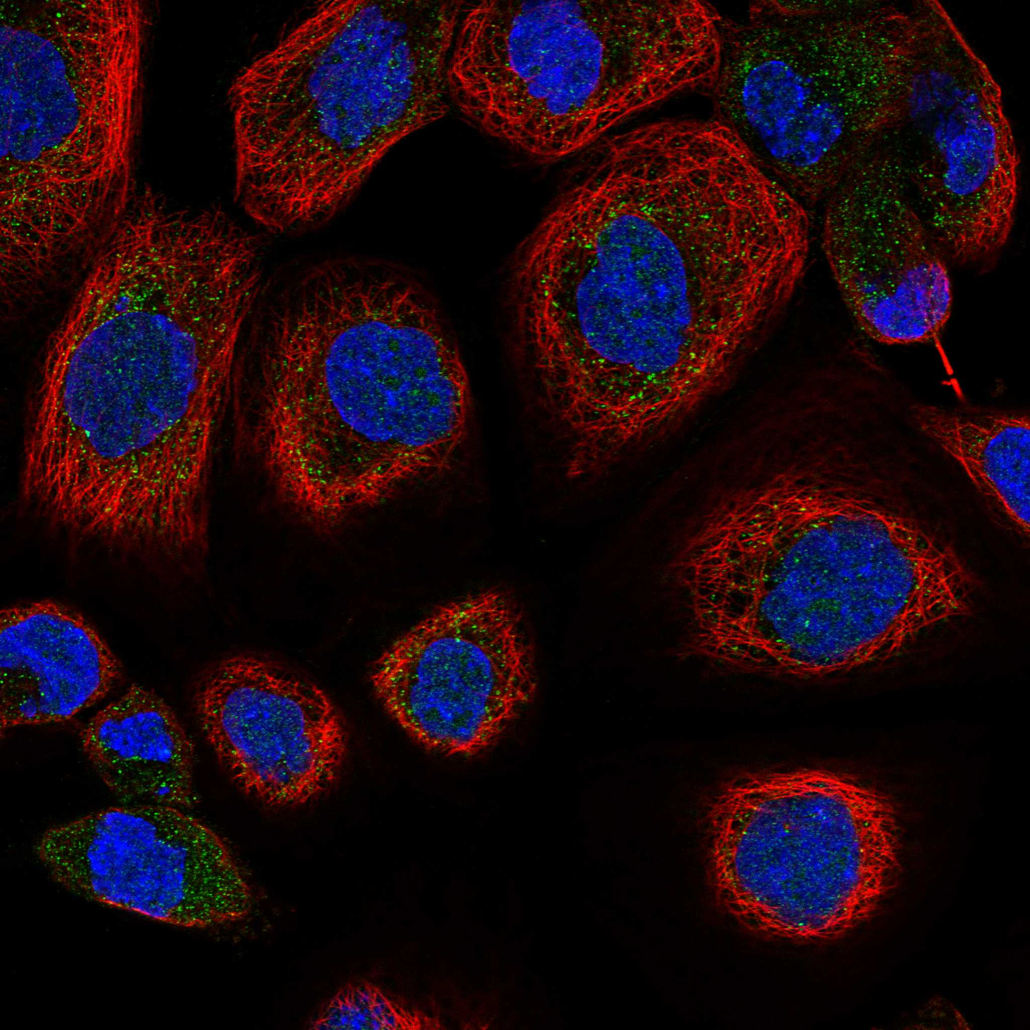
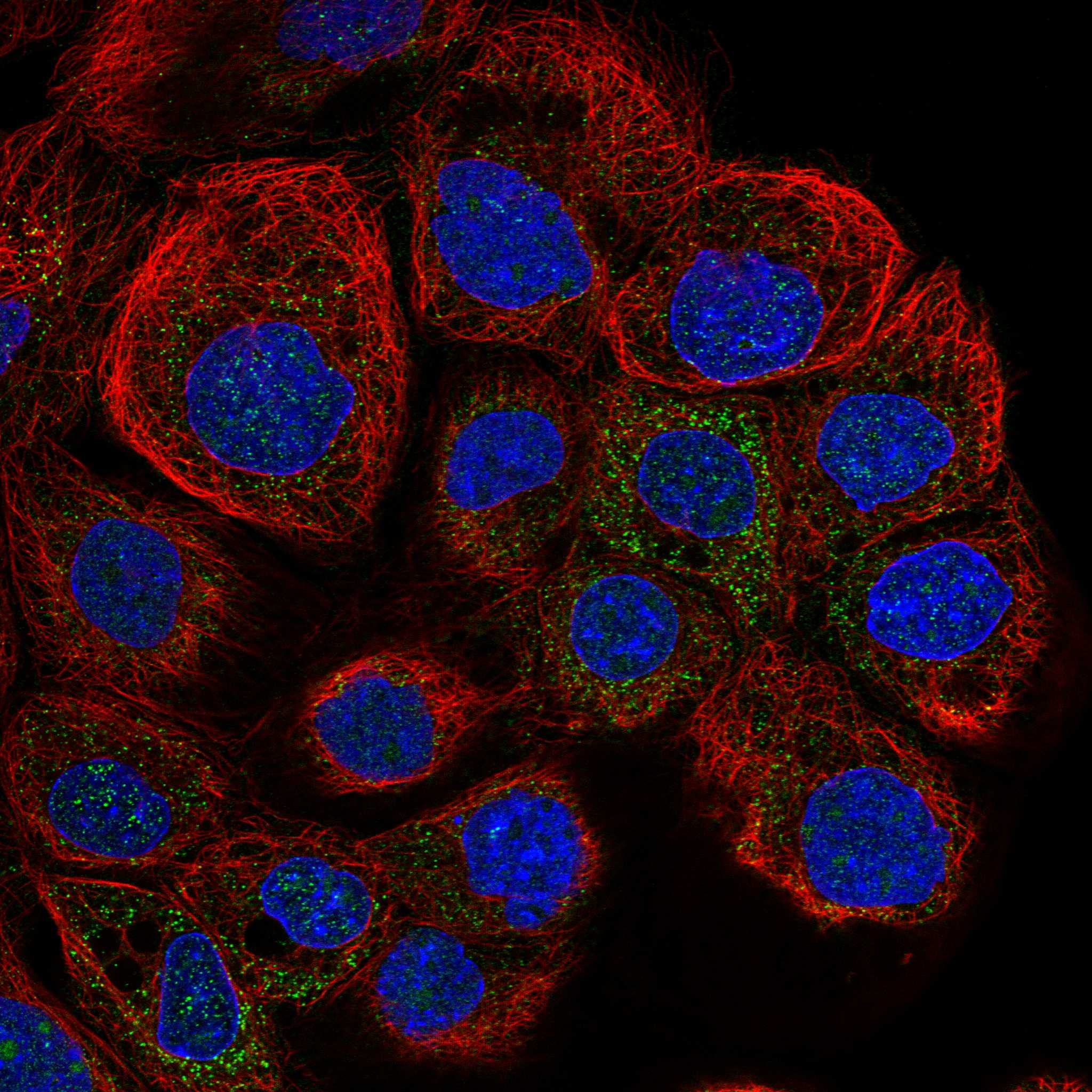
| IF/ICC Q&A: should the same pattern appear? | In ICC-IF, HPA reports peroxisomes as the main location and nucleoli as an additional location, with images from A-431, U-251MG and U2OS (HPA: subcellular). This supports a localization comparison, not an IF protocol or an expectation that nucleolar tissue IHC must be visible (HPA: subcellular; general microscopy practice). |
| Detection chemistry | Endogenous enzyme activity or nonspecific secondary-reagent binding can produce color unrelated to ACOX1 (general IHC practice). A reagent control helps identify that contribution; the supplied HPA and UniProt records do not quantify its size in any listed tissue. |
| Situation | Likely cause | Next action |
|---|---|---|
| Positive-reference cells show no signal. | The run may have insufficient retrieval or a detection failure (general IHC practice); target-specific fixation sensitivity is unreported in the supplied records. | Check a contemporaneous positive section and detection controls, then adjust retrieval within the chosen IHC workflow (general IHC practice). Use hepatocytes or intestinal glandular cells as reported High references (HPA: tissue IHC). |
| Granules are faint but tissue-wide haze is strong. | Background may mask the granular cytoplasmic pattern reported by HPA (HPA: tissue IHC; general IHC practice). | Review reagent-only controls, blocking, primary-antibody concentration and wash steps; compare cells against adjacent background before scoring (general IHC practice). No ACOX1-specific dilution is supplied. |
| Signal outlines cell membranes. | A predominantly membrane pattern conflicts with peroxisomal localization and the absence of a transmembrane segment (UniProt Q15067 topology; HPA: subcellular). | Check morphology and control sections, then repeat with the IHC-validated antibody and its documented conditions if needed (general IHC practice). Do not score membrane-only color as the expected ACOX1 pattern. |
| A surprising cell population stains strongly. | Cross-reactivity or endogenous detection activity is possible (general IHC practice), but ACOX1 tissue expression is broad and HPA supplies no negative-tissue list (HPA: tissue IHC). | Compare the compartment and staining pattern with nearby expected cells; inspect reagent controls before assigning specificity or declaring the population biologically negative (HPA: tissue IHC; general IHC practice). |
| Nucleolar signal appears in an IF comparison. | HPA reports additional nucleolar localization in ICC-IF alongside the main peroxisomal location (HPA: subcellular). | Assess whether peroxisomal signal is also present and keep the ICC-IF observation separate from tissue IHC scoring (HPA: subcellular; tissue IHC). Follow the separate IF/ICC guide for experimental conditions. |
| Two specimens have different staining intensities. | HPA reports tissue-dependent staining levels, and UniProt reports tissue-dependent isoform balance; HPA also notes low consistency with RNA expression (HPA: tissue IHC; UniProt Q15067 tissue specificity). | Compare matched cell types and run controls under the same IHC conditions before calling a biological change; do not infer a particular isoform or chain from intensity alone (general IHC practice; UniProt Q15067). |


Comprehensive Human Protein Atlas IHC scoring per tissue (reliability: Approved — Low consistency between antibody staining and RNA expression data.). Rows are taken directly from the HPA tissue chart — click any row's HPA link to view the source.
| Tissue | Cell type | Level | Evidence | Source |
|---|---|---|---|---|
| Duodenum | Glandular cells | High | Protein (IHC) | HPA → |
| Liver | Hepatocytes | High | Protein (IHC) | HPA → |
| Small intestine | Glandular cells | High | Protein (IHC) | HPA → |
| Adrenal gland | Glandular cells | Medium | Protein (IHC) | HPA → |
| Appendix | Glandular cells | Medium | Protein (IHC) | HPA → |
| Tissue | Cell type | Level | Evidence | Source |
|---|---|---|---|---|
| None in HPA: ACOX1 is detected in all 45 scored tissues. Use a no-primary (secondary-only) and an isotype control instead. | ||||
Troubleshoot ACOX1 staining by checking retrieval, tissue context and peroxisomal localisation before comparing chromogenic IHC results across sections.
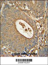
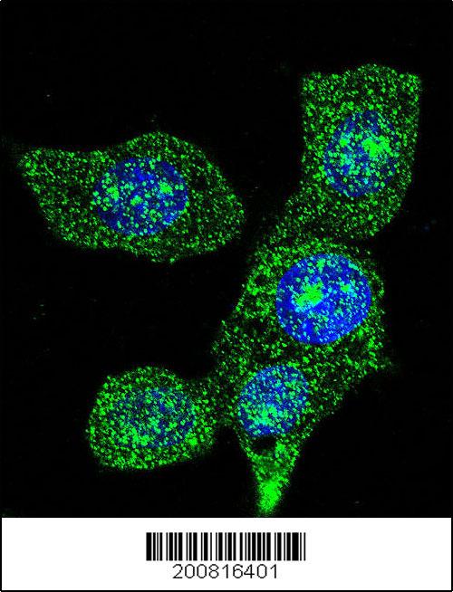
M03054 has IHC data from formalin-fixed, paraffin-embedded human colon carcinoma and IF data from HeLa cells (catalog IHC and IF captions).
M03054 is the card that will render; its IHC image shows peroxidase/DAB staining of formalin-fixed, paraffin-embedded human colon carcinoma (catalog IHC caption). Its IF image shows staining of HeLa cells, and its listed reactivity is human (catalog IF caption; catalog reactivity).
Which to pick: Choose mouse monoclonal M03054 for human tissue IHC-P because its own image documents formalin-fixed, paraffin-embedded colon carcinoma stained with peroxidase/DAB (catalog host/clone; catalog IHC caption). M03054 also has an IF image in HeLa cells; rabbit polyclonal A03054-1 lists both IF and ICC (catalog IF caption; catalog host/applications). For mouse or rat samples, consider A03054-1 because those species are listed as reactive, but its stated paraffin-section IHC condition is for human samples (catalog reactivity; datasheet: IHC dilution).