ACP5 / Tartrate-resistant acid phosphatase type 5 · IHC design guide

Design Immunohistochemistry for ACP5

Use lung macrophages as a high-staining reference for ACP5 chromogenic IHC in paraffin sections (HPA tissue IHC). Start with the IHC-validated antibody at 2–5 μg/mL (datasheet A03277-1), and assess cytoplasmic staining (HPA tissue IHC).

Evidence assembled Oct 2026 · For research use; verify linked source records and product datasheet before use
Immunohistochemistry protocol sheet for ACP5 (IHC for ACP5): expected localisation Cytoplasmic in lung macrophages (HPA tissue IHC), antibody A03277-1, validated IHC image, and IHC protocol steps
Printable ACP5 IHC protocol sheet — expected localisation Cytoplasmic in lung macrophages (HPA tissue IHC), antibody A03277-1, controls and protocol steps. Open the full ACP5 IHC guide →

ACP5 Immunohistochemistry Experimental Design Guide

Expected localisation, validated protocols, controls and antibodies — the at-a-glance facts below, then the full design guide.

Must know before staining
Expected localisation Cytoplasmic in lung macrophages (HPA tissue IHC)
Staining pattern Cytoplasmic staining in lung macrophages (HPA tissue IHC)
Antigen retrieval EDTA pH 8.0 HIER, heat-mediated (datasheet A03277-1)
Positive control ⓘ Lung
Negative control ⓘ Adipose tissue+4 more · see all
Important caveats
Reasons your staining may differ from the expected pattern.
Fixation Keep fixation consistent across sections. (standard IHC practice; not target-specific)
Caveat Secreted ACP5 may produce extracellular signal (UniProt)
Regulation Macrophage-rich areas may stain more (HPA tissue IHC)
Isoform / epitope 5a is uncleaved; 5b is cleaved, so epitope choice matters (UniProt)
Section 1

Recommended ACP5 IHC & IF Protocols

Compare the catalog antibody’s IHC-P protocol (datasheet: A03277-1) with two published chromogenic IHC protocols for ACP5 (PMC7066063; PMC12639494).

Recommended immunohistochemistry (IHC-P) protocol parameters
SampleParaffin-embedded human spleen tissue; fixative not specified (datasheet A03277-1)
FixationImage fixative and duration unreported (datasheet A03277-1); verify before use.
Sectioning4–5 µm sections on charged slides (standard)
DeparaffinisationXylene, graded ethanol series to water (standard)
Antigen retrievalHeat retrieval: EDTA pH 8.0 (datasheet A03277-1); 20 min, 95–100 °C (standard)
Peroxidase block3% H2O2, 10 min, room temperature (standard)
Blocking10% goat serum (datasheet A03277-1)
Primary antibodyRabbit anti-ACP5, 2-5 μg/ml (datasheet A03277-1)
Primary incubationOvernight at 4 °C (datasheet A03277-1)
DetectionStreptavidin-biotin complex (SABC), DAB chromogen (datasheet A03277-1)
CounterstainHematoxylin, blue, dehydrate and mount (standard)
Expected resultACP5-positive staining in macrophages of lung (HPA tissue IHC: High). HPA tissue profile: Cytoplasmic expression in macrophages in lung. No signal in the no-primary control.
💡Decision noteStart with heat-mediated EDTA retrieval at pH 8.0 for the catalog antibody (datasheet: A03277-1); one published protocol specifies a different retrieval solution (PMC12639494).
Section 2

What Is the Expected ACP5 Staining Pattern?

ACP5 should give predominantly cytoplasmic staining in lung macrophages, where HPA reports High expression (HPA: lung macrophages). Lysosomal localisation and secretion are both annotated, with no transmembrane segment (UniProt P13686: subcellular location and topology). HPA rates the tissue IHC evidence Enhanced while noting low consistency between antibody staining and RNA expression (HPA: tissue IHC reliability); interpret an isolated positive signal in that context.

What am I looking at on my slide?
Strong cytoplasmic signal is concentrated in lung macrophages, while nearby respiratory epithelial cells lack convincing signal.This matches the reported High macrophage staining in lung and absence of detected staining in bronchial respiratory epithelium (HPA: tissue IHC). Score the identified cells and their cytoplasm rather than calling the whole section positive.
Nuclei dominate the staining, with little or no cytoplasmic signal in identifiable macrophages.A predominantly nuclear result conflicts with the cytoplasmic lung pattern (HPA: tissue IHC) and the lysosomal annotation (UniProt P13686: subcellular location). Treat it as suspect; inspect counterstain overlap and staining controls before assigning an ACP5 result.
Widespread strong staining appears in bronchial respiratory epithelium or other cells expected to be negative.HPA reports respiratory epithelial cells in bronchus as Not detected (HPA: tissue IHC). Cross-reactivity or endogenous detection activity is a possible explanation (standard IHC practice); verify the cell identity and compare an appropriate detection-only control.
A diffuse haze covers cells, extracellular spaces, or much of the section without clear macrophage enrichment.That distribution is less persuasive than macrophage-centred cytoplasmic staining (HPA: tissue IHC). Secreted ACP5 is annotated (UniProt P13686: subcellular location), so extracellular signal alone does not identify its source; assess background controls and morphology before scoring.
No signal appears in lung macrophages selected as the positive reference.This conflicts with the reported High level in lung macrophages (HPA: tissue IHC). First confirm that macrophages are present; then examine primary-antibody performance, retrieval and detection with controls (standard IHC practice) before interpreting other fields as negative.
💡Expected ACP5 appearanceCall a result positive when identifiable lung macrophages show clear, strong cytoplasmic staining (HPA: High in lung macrophages); predominant nuclear or widespread epithelial colour is suspect (HPA: tissue IHC; standard IHC practice).
How each factor affects the staining
Cell identity in the sectionThe expected positive population is lung macrophages, not every lung cell (HPA: tissue IHC). HPA lists bronchial respiratory epithelium as Not detected (HPA: tissue IHC), making cell identification essential when comparing fields.
Intracellular and secreted poolsACP5 is annotated in lysosomes and as secreted, without a transmembrane segment (UniProt P13686: subcellular location and topology). For tissue IHC, the observed reference remains cytoplasmic macrophage staining (HPA: tissue IHC); extracellular colour needs cautious interpretation.
Proteolytic formsThe uncleaved 5a form is secreted by macrophages and dendritic cells; cleaved 5b is secreted by osteoclasts (UniProt P13686: processing and subcellular location). The supplied IHC record does not show which form an antibody detects, so staining cannot identify an isoform.
Evidence and antibody choiceHPA lists IHC as Enhanced for HPA057655 and HPA059463, and Approved for CAB002584 (HPA: antibody validation). Its tissue summary also reports low staining–RNA consistency (HPA: tissue IHC reliability); retain the tissue pattern as a reference, not a guarantee for every section.
Why is my staining missing, weak or wrong?
SituationLikely causeNext action
The lung reference has no stained cells.The examined field may lack identifiable macrophages, or the IHC run may have failed (HPA: High in lung macrophages; standard IHC practice).Locate macrophage-rich fields by morphology, then review the run controls, retrieval conditions and detection steps (standard IHC practice). Do not call a tissue negative until the positive reference works.
Nuclei stain more strongly than macrophage cytoplasm.This disagrees with HPA's cytoplasmic lung pattern (HPA: tissue IHC); counterstain or nonspecific staining may be contributing (standard IHC practice).Compare the chromogen with the nuclear counterstain, check a detection-only control and rescore identifiable macrophage cytoplasm (standard IHC practice).
Strong colour appears across respiratory epithelium.Bronchial respiratory epithelial cells are reported as Not detected (HPA: tissue IHC); cross-reactivity or endogenous detection activity is possible (standard IHC practice).Check epithelial versus macrophage morphology and a detection-only control; interpret broad epithelial colour cautiously against the HPA reference (HPA: tissue IHC; standard IHC practice).
The section shows diffuse colour with poor cell boundaries.Nonspecific background is possible (standard IHC practice). ACP5 can also be secreted (UniProt P13686: subcellular location), so a diffuse signal alone cannot establish its cellular source.Inspect background controls and tissue morphology, then score only convincing cellular staining in macrophages (standard IHC practice; HPA: tissue IHC).
Two antibodies give different staining patterns.HPA assigns distinct validation statuses: Enhanced for HPA057655 and HPA059463, Approved for CAB002584 (HPA: antibody validation). That summary does not establish the cause of a particular disagreement.Compare matched sections and controls, prioritise reproducible cytoplasmic staining in lung macrophages, and record the disagreement (HPA: tissue IHC; standard IHC practice).
Can ICC/IF localisation settle an ambiguous IHC result?HPA's ICC/IF summary calls the cytosol location uncertain, while tissue IHC reports cytoplasmic staining in lung macrophages (HPA: subcellular ICC/IF; HPA: tissue IHC).Use ICC/IF as supporting context, not as a substitute for identifying stained cells and checking IHC controls (HPA: subcellular ICC/IF; standard IHC practice).

Sample controls for ACP5 IHC & IF

🧪Run lung first and look for ACP5 staining in macrophages (High; HPA: lung macrophages). Run adipose tissue and score adipocytes as the negative population (Not detected; HPA: adipose adipocytes); on the lung slide, use neighboring nonmacrophage cells with only background staining as an internal negative reference, while confirming their identity by morphology (HPA: lung macrophages High).
Positive control tissue: Lung (Macrophages, HPA High)
Negative control tissue: Adipose tissue (HPA Not detected)
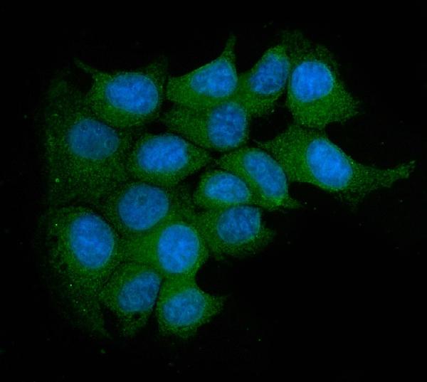
ICC-IF cell lines (HPA subcellular resource): HPA ICC-IF images show ACP5 in A-431, U-251MG, with annotated localisation: Cytosol (uncertain) (HPA subcellular).
Technical controls: Include no-primary (secondary-only) and matched rabbit IgG isotype controls, plus an ACP5 knockout biological negative if available (caption: rabbit primary antibody; standard IHC practice). For chromogenic detection, control endogenous peroxidase and, if using the caption’s biotin-based detection, endogenous biotin; assess lung macrophage autofluorescence if performing IF (caption: biotin-based detection; standard IHC/IF practice).
⚠️Feasibility: A target-specific fixation window and fixation effect are unreported, and the selected A03277-1 tissue-IHC caption does not report a fixative (caption: fixative not stated). The paraffin-section example uses heat retrieval in EDTA at pH 8.0, but does not establish that retrieval is required; whether frozen sections or IF are easier is unreported (caption: EDTA retrieval; HPA: ICC-IF images in A-431 and U-251MG). In lung, macrophage pigment or endogenous enzyme activity can complicate chromogenic scoring, so compare signal with the control slides (HPA: lung macrophages High; standard IHC practice).

HPA tissue IHC evidence for ACP5

Comprehensive Human Protein Atlas IHC scoring per tissue (reliability: Enhanced — Low consistency between antibody staining and RNA expression data.). Rows are taken directly from the HPA tissue chart — click any row's HPA link to view the source.

Positive expression · recommended positive controls

TissueCell typeLevelEvidenceSource
Lung Macrophages High Protein (IHC) HPA →

Undetected expression · recommended negative controls

TissueCell typeLevelEvidenceSource
Adipose tissue Adipocytes Not detected Protein (IHC) HPA →
Adrenal gland Glandular cells Not detected Protein (IHC) HPA →
Appendix Glandular cells Not detected Protein (IHC) HPA →
Bone marrow Hematopoietic cells Not detected Protein (IHC) HPA →
Breast Adipocytes Not detected Protein (IHC) HPA →
Section 3

Advanced ACP5 IHC Tips

Use the catalog antibody’s spleen IHC conditions as a starting point, then judge ACP5 staining by cell type, compartment and controls.

Which antigen retrieval should I start with for ACP5 paraffin IHC?
Start with heat mediated retrieval in EDTA pH 8.0 for paraffin sections (datasheet A03277-1). The demonstrated spleen workflow then used 10% goat serum and 2 μg/ml primary antibody overnight at 4°C (datasheet A03277-1). If staining is weak, adjust heating duration on matched sections while holding antibody concentration and detection conditions constant, and check tissue preservation after retrieval (standard IHC practice). Compare each condition against the same positive tissue and a primary omission control, because a stronger DAB signal alone cannot establish improved ACP5 specificity (standard IHC practice).
Could fixation explain weak ACP5 staining in my paraffin sections?
Target specific fixation sensitivity is unknown: the selected paraffin spleen caption does not state its fixative (datasheet A03277-1). Record the fixative, fixation duration and tissue thickness for each specimen, then compare matched sections processed with the same EDTA pH 8.0 retrieval (standard IHC practice; datasheet A03277-1). Check whether weak staining tracks processing batches while tissue morphology and the assay’s positive control remain interpretable (standard IHC practice). Do not attribute a fixation effect to ACP5’s glycosylation sites at residues 116 and 147; those annotations do not establish fixation sensitivity (UniProt P13686 glycosylation; standard IHC practice).
What ACP5 staining pattern should I expect in tissue?
Evaluate cytoplasmic staining in the relevant cells: HPA reports high cytoplasmic ACP5 expression in lung macrophages (HPA tissue IHC). A granular intracellular pattern can be consistent with lysosomal ACP5, while extracellular signal is biologically plausible because ACP5 is also secreted (UniProt P13686 subcellular annotation). HPA separately lists an uncertain cytosolic location from cell imaging, so diffuse staining should be assessed alongside morphology and controls (HPA subcellular; standard IHC practice). ACP5 has no annotated transmembrane segment; a crisp, isolated plasma membrane rim should therefore prompt review for nonspecific edge or detection staining (UniProt P13686 topology; standard IHC practice).
Can this antibody distinguish ACP5 5a from 5b in IHC?
Do not score the IHC signal as isoform specific without epitope mapping or separate validation for the catalog antibody (standard IHC practice). ACP5 can occur as an uncleaved monomer called 5a or a proteolytically processed, disulfide linked two chain form called 5b (UniProt P13686 subunit annotation). Macrophages and dendritic cells secrete 5a, whereas osteoclasts secrete 5b, but cell context alone does not identify which form an antibody detected (UniProt P13686 subcellular annotation; standard IHC practice). Interpret a change in staining as a change in detectable ACP5 antigen until the antibody’s epitope and its recognition of both processed forms are established (standard IHC practice).
How should I check ACP5 localisation by multiplex IF?
For a separate IF experiment, pair ACP5 with a validated macrophage marker and inspect whether signals occur in the same cells, since HPA reports high ACP5 staining in lung macrophages (HPA tissue IHC; standard IF practice). Select a red or far red fluorophore after checking unstained tissue for autofluorescence in each channel (standard IF practice). For an intracellular epitope, use a controlled permeabilisation step because ACP5 can localise to lysosomes or cytosol; avoid assuming permeabilisation is needed to examine an extracellular pool (UniProt P13686 subcellular annotation; HPA subcellular; standard IF practice). The catalog antibody’s supplied tissue demonstration uses chromogenic IHC, so establish its IF performance with appropriate controls (datasheet A03277-1; standard IF practice).
How can I distinguish ACP5 signal from chromogenic background?
Inspect a primary omission section for signal from the biotin based detection workflow used in the demonstrated spleen IHC (datasheet A03277-1; standard IHC practice). Include an endogenous peroxidase block before DAB development, and assess whether residual pigment or precipitate persists in control sections (standard chromogenic IHC practice). The demonstrated workflow used 10% goat serum, a biotinylated secondary for 30 minutes at 37°C, and DAB; keep these conditions documented when comparing runs (datasheet A03277-1). If background is widespread, check blocking, washing and detection exposure before interpreting faint staining as ACP5, especially outside the expected cellular pattern (standard IHC practice; HPA tissue IHC).
How should I quantify ACP5 positive cells across sections? ⚠ ANSWER MARKED FOR VERIFICATION
Define the tissue compartment and cell population before scoring, since HPA identifies lung macrophages as a high staining population (HPA tissue IHC; standard IHC practice). Within comparable regions, report the percentage of positive macrophages and staining intensity, or calculate an H-score on its conventional 0–300 scale (standard IHC practice). If counting cells, report ACP5 positive macrophages per mm² and normalise that count to total macrophages in the same sampled area (standard IHC practice). Use identical retrieval, DAB development, counterstain and image thresholds across groups, and exclude folds, necrosis and section edges by a prespecified rule (standard IHC practice).
When is apparent ACP5 positivity convincing rather than artefactual?
Give most weight to staining in morphologically plausible cells with a cytoplasmic pattern, consistent with the high signal reported in lung macrophages (HPA tissue IHC; standard IHC practice). Review isolated membrane rims, section edges and necrotic areas as possible artefacts; ACP5 has no annotated transmembrane segment (UniProt P13686 topology; standard IHC practice). Separate antibody staining from endogenous enzyme or detection signal with primary omission and detection controls, particularly when assessing weak DAB deposits (standard chromogenic IHC practice). HPA reports low consistency between antibody staining and RNA expression, so corroborate surprising tissue patterns with an independent assay before assigning biological meaning (HPA tissue IHC; standard IHC practice).
Boster reagents

Best ACP5 / Tartrate-resistant acid phosphatase type 5 IHC Antibodies

Antibodies with IHC and IF images cover human and mouse paraffin sections, human liver tissue IF, and MCF-7 cell IF (catalog image captions).

Real IHC data IHC analysis of Acid Phosphatase/ACP5 using anti-Acid Phosphatase/ACP5 antibody (A03277-1). Acid Phosphatase/ACP5 was detected in a paraffin-embedded section of human spleen tissue. Heat mediated antigen retrieval was performed in EDTA buffer (pH 8.0, epitope retrieval solution). The tissue section was blocked with 10% goat serum. The tissue section was then incubated with 2 μg/ml rabbit anti-Acid Phosphatase/ACP5 Antibody (A03277-1) overnight at 4°C. Biotinylated goat anti-rabbit IgG was used as secondary antibody and incubated for 30 minutes at 37°C. The tissue section was developed using Strepavidin-Biotin-Complex (SABC) (Catalog # SA1022) with DAB as the chromogen.
Anti-Acid phosphatase/ACP5 Antibody ®
Cat # A03277-1
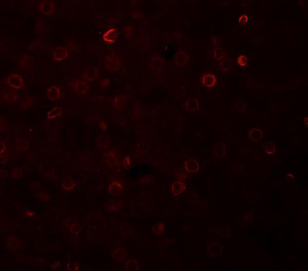
Real IF data Immunofluorescence of TRAP in human liver tissue with TRAP antibody at 20 μg/mL.
Anti-TRAP ACP5 Antibody
Cat # A03277

A03277-1 has IHC images from paraffin sections of human spleen, human lung, human renal cancer, and mouse lung, plus IF in MCF-7 cells (A03277-1 image captions). A03277 has IF data from human liver tissue at 20 μg/mL (A03277 image caption).

Which to pick: For tissue IHC, choose A03277-1: its own caption documents paraffin sections, EDTA retrieval at pH 8.0, and 2 μg/mL primary antibody; the fixative is unreported (A03277-1 IHC image caption). For IF/ICC, A03277-1 has an MCF-7 cell image and lists both applications, while A03277 has a human liver IF image and lists IF; clonality is unreported for both (catalog applications and image captions). For work across species, A03277-1 lists human, mouse, and rat reactivity, with IHC images from human and mouse sections; rat IHC evidence is unreported (A03277-1 catalog reactivity and IHC image captions).

Each figure is that product's own IHC / IF validation image from its datasheet.

References

  1. UniProt Consortium. UniProt entry P13686 (PPA5_HUMAN, Tartrate-resistant acid phosphatase type 5).
  2. Human Protein Atlas. ACP5 tissue IHC expression (reliability: Enhanced).
  3. Human Protein Atlas. ACP5 subcellular location (ICC-IF): Localized to the cytosol..
  4. Human Protein Atlas. ACP5 antibody validation summary (3 antibodies).
  5. Extracellular vesicles derived from dental follicle stem cells regulate tooth eruption by inhibiting osteoclast differentiation. Frontiers in cell and developmental biology 2024 — PMC11744031.
  6. ACP5-positive macrophages contribute to cerebral oedema and neuroinflammation after traumatic brain injury. Clinical and translational medicine 2026 — PMC13613102.
  7. Tartrate-Resistant Acid Phosphatase 5/ACP5 Interacts with p53 to Control the Expression of SMAD3 in Lung Adenocarcinoma. Molecular therapy oncolytics 2020 — PMC7066063.
  8. Ultra-small coordination dihydromyricetin nanodots attenuate alveolar bone resorption in periodontitis through inhibition of ITGB3-Driven osteoclast differentiation. Materials today. Bio 2025 — PMC12639494.
  9. PubMed PMID:2909539 — UniProt-cited evidence.
  10. PubMed PMID:2338077 — UniProt-cited evidence.
  11. PubMed PMID:14702039 — UniProt-cited evidence.