This website uses cookies to ensure you get the best experience on our website.
- Table of Contents
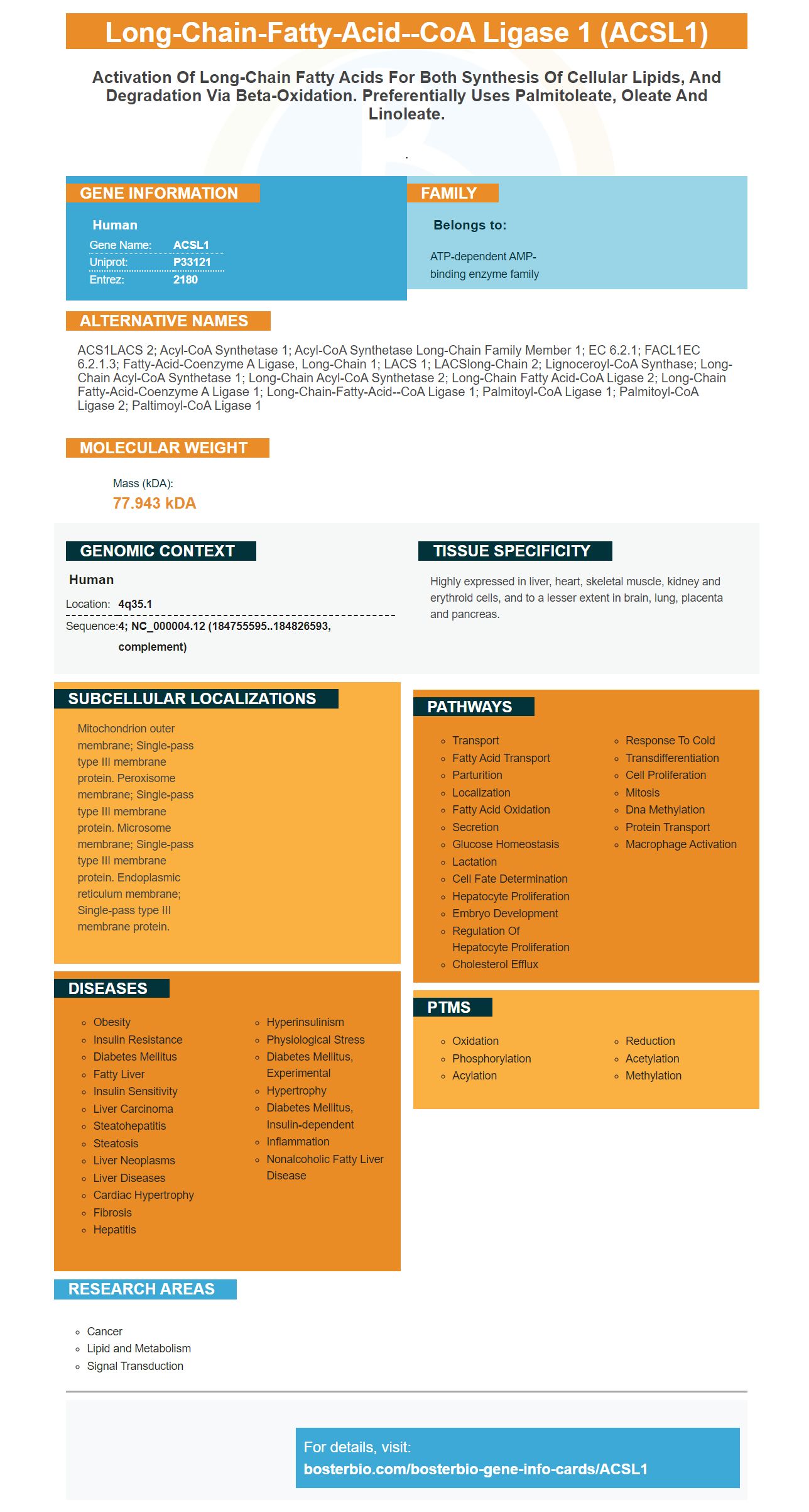
Facts about Long-chain-fatty-acid--CoA ligase 1.
.
Human | |
---|---|
Gene Name: | ACSL1 |
Uniprot: | P33121 |
Entrez: | 2180 |
Belongs to: |
---|
ATP-dependent AMP-binding enzyme family |
ACS1LACS 2; Acyl-CoA synthetase 1; acyl-CoA synthetase long-chain family member 1; EC 6.2.1; FACL1EC 6.2.1.3; fatty-acid-Coenzyme A ligase, long-chain 1; LACS 1; LACSlong-chain 2; lignoceroyl-CoA synthase; Long-chain acyl-CoA synthetase 1; Long-chain acyl-CoA synthetase 2; Long-chain fatty acid-CoA ligase 2; long-chain fatty-acid-coenzyme A ligase 1; long-chain-fatty-acid--CoA ligase 1; Palmitoyl-CoA ligase 1; Palmitoyl-CoA ligase 2; paltimoyl-CoA ligase 1
Mass (kDA):
77.943 kDA
Human | |
---|---|
Location: | 4q35.1 |
Sequence: | 4; NC_000004.12 (184755595..184826593, complement) |
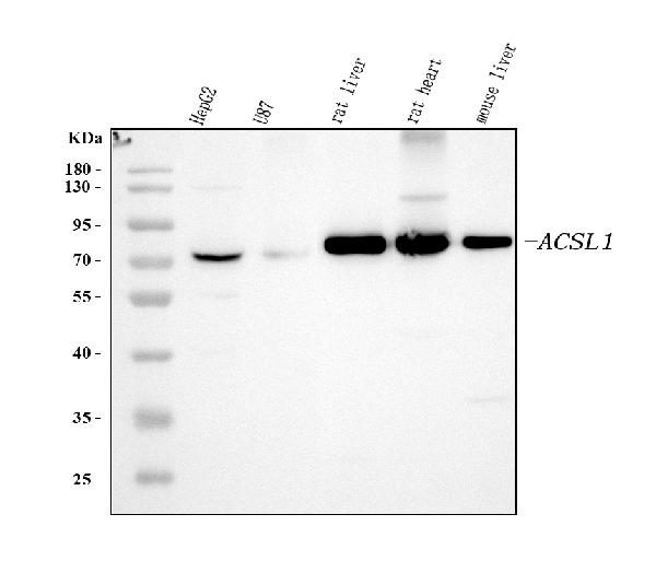
Highly expressed in liver, heart, skeletal muscle, kidney and erythroid cells, and to a lesser extent in brain, lung, placenta and pancreas.
Mitochondrion outer membrane; Single-pass type III membrane protein. Peroxisome membrane; Single-pass type III membrane protein. Microsome membrane; Single-pass type III membrane protein. Endoplasmic reticulum membrane; Single-pass type III membrane protein.
In this article, we'll go over the top uses for the ACSL1 Marker, which includes Western Blot, Insulin sensitivities and cellular lipid content. This article is ideal for researchers. It will discuss the most important functions of the ACSL1 marker as well as the anti-ACSL1 antibody. However, before we get into the details let's take a look at what these markers can do for you.
The Anti-ACSL1 antibody Picoband is available from Boster Bio. It is able to react with mouse, human and rat cells. This antibody has been validated to be used in a variety of biological tests. It can be diluted between 10 to 15 uL for each data point. It is made up of Trehalose. Antibodies for the ACSL1 protein are produced in rabbits and mice.
The ACSL1 gene encodes a long-chain fatty-acid-CoA ligase. Members of the ACSL1 family differ in substrate specificity, subcellular localization, and tissue distribution. They play an important role in the biosynthesis of lipids and are found most often in peroxisomes. Boster Bio has validated antibodies for ACSL1. They were tested on various types of cells to evaluate their effectiveness.
ACSL1 expression in the peripheral blood of patients suffering from acute myocardial injuries (AMI) was higher than in healthy subjects. This study confirms previous research that have shown that ACSL1 triggers an inflammation response in the peripheral blood of patients with AMI. This study has a few limitations. These limitations are discussed below. ACSL1 expression is significantly higher levels in patients suffering from AMI than in healthy controls.
Activation ACSL1 results in the incorporation of aESA into DAGs. This suggests that ACSL1 is involved in the conversion of cholesterol esters to fat acids. This could also suggest the role of lipid drops in ferroptosis. In addition, ACSL1 is a part of the family of sterol regulatory element binding protein (SREBP1) that controls the incorporation of fatty acids into cholesterol esters.
The liver cells are responsible for activating ACSL1 in order to regulate metabolism. It has been suggested that a reduced expression of ACSL1 can cause an increase in the risk of AMI. This is significant because this gene is actively involved in the production of fats in liver cells. Thus, metabolic disorders can be caused by malfunctions in metabolic pathways. ACSL1 expression levels in the PBL were associated with AMI however the connection between this gene and the risk of AMI is not certain. However, the increased expression of ACSL1 is associated with the number of lesions found in the main branches of the coronary artery, which suggests a more severe type of atherosclerosis.
To measure the level of ACSL1 in cells We used human hepatocytes that were transfected with an ACSL1 vector overexpression or RNAi. ACSL1 knockdown and overexpression vectors induced green fluorescence in liver cells. We also tested cells that were transfected by the ACSL1 knockdown or overexpression vectors and determined the viability of each cell. The p-values are listed above the bars of comparison.
In addition to regulating levels of triglycerides ACSL1 also regulates oxidation of fat acids. Additionally, overexpression of ACSL1 reduced the expression of the PPARg gene, which is involved in fatty acid synthesis. This finding is in opposition to other studies that show ACSL1 overexpression increases the levels of triglycerides in blood. The results of this study, although not conclusive are consistent with other studies that have proven that ACSL1 can affect the oxidation and boxidation processes of fatty acids. pathways.
It is possible to aid in treatment decisions by combining the ACSL1 marker and insulin sensitivity. While longitudinal data aren't easy to collect and are not readily available, the gene expression profiles of ACSL1 as well as insulin sensitivity may be valuable to this end. ACSL1 and ACSL4, both candidate genes, were chosen. They were also found to be upregulated across different datasets. The data were gathered from different continents as well as Affymetrix and Illumina array platforms.
The association between ACSL1 and AMI remains unclear. The presence of atherosclerotic plaques inside the coronary arteries has been linked with the expression of ACSL1 in PBL. A higher level of ACSL1 expression is linked to more severe atherosclerosis. Insulin sensitivity is closely linked to ACSL1 expression, which suggests that they are both regulated. Although ACSL1 and insulin sensitivity expression may be related however, the connection between them isn't known.
The ACSL1 expression is a diagnostic tool with high accuracy. ACSL1 expression at 0.93+0.02 (area below the curve) is a reliable predictor of AMI. Both its specificity and sensitivity were high. A high level of ACSL1 implies the risk of AMI. The positive and negative predictive values were 84 85 percent and 85% respectively. These results are encouraging for the development of new tests that accurately assess the role of ACSL1 in predicting the risk of AMI.
ACSL1 expression was significantly greater in patients suffering from AMI in Northern China. The researchers speculated that increased ACSL1 levels could increase the risk of AMI. AMI and other cardiovascular disorders can also be detected early due to ACSL1 overexpression. They are currently trying to determine a marker for the gene as well as to study the relationship between ACSL1 and AMI. If they can establish a precise connection, this study will be beneficial in gaining a better understanding of how ACSL1 and AMI could work together.
It is unclear what role ACSL1 plays in regulation of lipid metabolism. It has been linked to macrophage activation and proinflammatory mediator release. This activation occurs through intracellular fatty acids crystals, which induce lysosomal injury and promote the release IL1B. It has been related to other biomarkers.
This publication presents the findings of a review of the literature on the ACSL1 marker that measures cholesterol content of cells. ACSL1 expression was significantly elevated in neutrophil cultures that were exposed to plasma from septic patients according to researchers. Participants of the workshop looked up the literature about each ACSL gene. Keywords like cell type, biomolecules, biological processes, as well as diseases were noted and identified by hand. The results were compiled by identifying common themes in the literature about each gene.
Multiple independent datasets have shown that the ACSL1 marker is abundant in sepsis. This antibody was useful in Western Blots that showed an area of around 78 KDa. Researchers recommend using 10 to 15 uL of each data point's recommended dilution when analyzing the results using this antibody. Using Size-Wes or Sally Sue/Peggy Sue methods to separate the protein, they have noted that the observed molecular weight may differ from the expected molecular weight because of post-translational modifications, cleavages and relative charges.
Clinical trials that use the ACSL1 marker are typical for diagnosing various illnesses and for detecting staph infections. The ACSL1 gene can be used for many applications, including the monitoring of the content of lipids. It is particularly beneficial for metabolism of lipids. Numerous studies have revealed that ACSL1 is connected to smooth muscle cells within the vascular system. ACSL1 plays a crucial role in diagnosing and predicting inflammation caused by septic.
The ACSL1 gene is part of the ATP-dependent AMP-binding enzymes family. It is a major enzyme in intermediary metabolism. ACSL converts fatty acids into fatty acylcoA derivatives. The metabolites that result are crucial for various cellular biochemical processes which include protein transport, transcriptional control, and inflammasome activation. Fatty acyl-CoAs are also utilized as substrates for phospholipid biosynthesis as well as beta-oxidation pathways.
The ACSL1 gene may also be used to monitor the immune system and help guide treatment decisions. Although longitudinal data aren't available, the authors observed that ACSL1 levels were significantly higher in patients who did not recover from sepsis. ACSL1 is a way to monitor inflammatory response. However further research is required. This area of research is important and will be further developed.
PMID: 1607358 by Abe T., et al. Human long-chain acyl-CoA synthetase: structure and chromosomal location.
PMID: 8584017 by Ghosh B., et al. Molecular cloning and sequencing of human palmitoyl-CoA ligase and its tissue specific expression.