ACTG2 / Actin, gamma-enteric smooth muscle · IHC design guide

Design Immunohistochemistry for ACTG2

Plan ACTG2 paraffin IHC using the A05066 antibody at 1:100–1:300 (datasheet: A05066) and assess cytoplasmic myoepithelial staining (HPA tissue IHC). Include smooth muscle as a positive tissue (HPA tissue IHC) and interpret staining with HPA’s cross-gene caution (HPA tissue IHC).

Evidence assembled Oct 2026 · For research use; verify linked source records and product datasheet before use
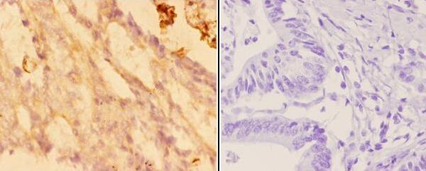
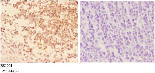
Immunohistochemistry protocol sheet for ACTG2 (IHC for ACTG2): expected localisation Cytoplasmic myoepithelial staining (HPA tissue IHC), antibody A05066-1, validated IHC image, and IHC protocol steps
Printable ACTG2 IHC protocol sheet — expected localisation Cytoplasmic myoepithelial staining (HPA tissue IHC), antibody A05066-1, controls and protocol steps. Open the full ACTG2 IHC guide →

ACTG2 Immunohistochemistry Experimental Design Guide

Expected localisation, validated protocols, controls and antibodies — the at-a-glance facts below, then the full design guide.

Must know before staining
Expected localisation Cytoplasmic myoepithelial staining (HPA tissue IHC)
Staining pattern Myoepithelial cells show cytoplasmic staining (HPA tissue IHC)
Antigen retrieval Citrate pH 6.0 HIER, 95–98 °C, 20 min (rule: cytoplasmic / membrane antigen)
Positive control ⓘ Appendix+4 more · see all
Negative control ⓘ Adipose tissue+4 more · see all
Important caveats
Reasons your staining may differ from the expected pattern.
Fixation Keep fixation consistent across samples. (standard IHC practice; not target-specific)
Caveat Staining may reflect proteins from more than one gene (HPA tissue IHC)
Regulation Abundant in intestinal smooth muscle (UniProt)
Isoform / epitope 2 isoforms; aa 2/3 processing may affect N-terminal epitopes (UniProt)
Section 1

Recommended ACTG2 IHC & IF Protocols

The catalog antibody’s IHC-P protocol is followed by published ACTG2 methods for intestinal tumors, soft-tissue sarcoma, and gastric tissue (PMC4841950; PMC11378013; PMC13289488).

Recommended immunohistochemistry (IHC-P) protocol parameters
SampleParaffin-embedded human colon carcinoma tissue; fixative not specified (datasheet A05066-1)
FixationImage fixative and duration unreported (datasheet A05066-1); verify before use.
Sectioning4–5 µm sections on charged slides (standard)
DeparaffinisationXylene, graded ethanol series to water (standard)
Antigen retrievalHeat-induced epitope retrieval in citrate buffer, pH 6.0, 20 min at 95–98 °C (standard rule: cytoplasmic / membrane antigen)
Peroxidase block3% H2O2, 10 min, room temperature (standard)
Blocking10% normal serum of the secondary host, 30 min, room temperature (standard)
Primary antibodyRabbit anti-ACTG2, 1:50-1:200 (datasheet A05066-1)
Primary incubationOvernight at 4 °C (standard)
DetectionHRP-polymer secondary, DAB chromogen 5–10 min (standard)
CounterstainHematoxylin, blue, dehydrate and mount (standard)
Expected resultACTG2-positive staining in enterocytes - Microvilli of appendix (HPA tissue IHC: High). HPA tissue profile: Cytoplasmic expression of myoepithelial cells. No signal in the no-primary control.
💡Decision noteStart with citrate pH 6.0 HIER at 95–98 °C for 20 min (page antigen-retrieval rule); the gastric study used 100 °C for 10 min (PMC13289488).
Section 2

What Is the Expected ACTG2 Staining Pattern?

ACTG2 is a cytoplasmic cytoskeletal protein with no transmembrane segment (UniProt P63267). In intestinal sections, expect staining in smooth muscle cells of the muscularis mucosa, muscularis propria and vessels (UniProt P63267). HPA reports high smooth muscle cell staining, but rates tissue IHC reliability as Supported with medium consistency and cautions that staining may reflect proteins from more than one gene (HPA tissue IHC).

What am I looking at on my slide?
Strong cytoplasmic staining follows smooth muscle cell bundles in an intestinal section.This fits ACTG2's cytoskeletal location and its abundant expression in intestinal smooth muscle (UniProt P63267). Compare the stained cells with tissue morphology; staining alone cannot prove molecular specificity because HPA cautions that its tissue signal may include proteins from more than one gene (HPA tissue IHC).
The dominant signal appears in nuclei rather than cytoplasm.A nuclear dominant pattern conflicts with the reported cytoplasmic cytoskeletal location (UniProt P63267). Treat it as suspect and review counterstain, detection and controls before scoring it as ACTG2 (general IHC practice).
Strong staining appears in a cell population outside the expected smooth muscle pattern.Check the cell identity before calling cross-reactivity: HPA also reports high enterocyte microvillus staining in several intestinal tissues and a cytoplasmic myoepithelial profile (HPA tissue IHC). An otherwise unexplained pattern may reflect antibody cross-reactivity or endogenous detection activity (general IHC practice).
Color spreads broadly across tissue and spaces without a discernible cell pattern.Diffuse background prevents a reliable compartment or cell type call (general IHC practice). Compare a matched negative detection control and review blocking, washes and detection conditions; do not interpret background intensity as an HPA staining level (general IHC practice).
A smooth muscle positive control shows no discernible signal.This conflicts with HPA's high smooth muscle cell staining and UniProt's intestinal smooth muscle expression (HPA tissue IHC; UniProt P63267). First check section identity and assay controls, then review the antibody's documented IHC conditions (general IHC practice).
💡Expected ACTG2 appearanceCall a positive result when smooth muscle cells show clear, strong cytoplasmic staining consistent with HPA's high level and UniProt's location; isolated nuclear color or shapeless background is suspect (HPA tissue IHC; UniProt P63267; general IHC practice).
How each factor affects the staining
Cellular locationACTG2 is cytoplasmic and cytoskeletal, with no transmembrane segment (UniProt P63267). Score staining within cells and against smooth muscle morphology; a surface only or nuclear dominant pattern needs review (general IHC practice).
Choice of positive comparatorHPA rates smooth muscle cells High, while UniProt reports abundant ACTG2 in intestinal muscularis mucosa, muscularis propria and vascular smooth muscle (HPA tissue IHC; UniProt P63267). Those compartments provide a morphology based reference.
Interpretation of intestinal epitheliumHPA rates enterocyte microvilli High in appendix, colon, duodenum, rectum and small intestine (HPA tissue IHC). Do not automatically attribute that epithelial signal to ACTG2: HPA warns that its antibodies may detect protein from more than one gene (HPA tissue IHC).
Antibody evidenceHPA lists two rabbit polyclonal antibodies, HPA041264 and HPA041271, as Supported for IHC; neither has an ICC status listed (HPA antibodies). Supported tissue staining and medium RNA agreement warrant a pattern based interpretation, not an assumption of absolute specificity (HPA tissue IHC).
Isoforms and processingUniProt lists two isoforms and processed actin chains beginning at residues 2 or 3 (UniProt P63267). Without an antibody epitope in the supplied evidence, these annotations cannot predict which forms the IHC signal detects.
Can IF/ICC confirm this pattern?UniProt places ACTG2 in the cytoplasmic cytoskeleton, but HPA supplies no main ICC-IF location, cell images or ICC status for the listed antibodies (UniProt P63267; HPA subcellular; HPA antibodies). An IF result needs its own controls and interpretation (general IF practice).
Why is my staining missing, weak or wrong?
SituationLikely causeNext action
No color in the expected smooth muscle compartment.A failed detection run, incorrect section or unsuitable assay setting is possible (general IHC practice).Confirm tissue morphology and run controls; check retrieval and dilution against the antibody's documented IHC conditions. No ACTG2 specific retrieval or dilution is supplied here (general IHC practice).
All tissue compartments show similar color.Broad background can obscure the cell pattern; endogenous detection activity is one possible contributor (general IHC practice).Inspect a control lacking primary antibody, then review blocking, washing and detection chemistry as appropriate to the method (general IHC practice).
Nuclei are the strongest positive structures.The distribution conflicts with ACTG2's cytoplasmic cytoskeletal annotation (UniProt P63267).Compare the signal with the counterstain and negative detection control; score only a convincing cell pattern after resolving the discrepancy (general IHC practice).
Enterocyte microvilli stain as strongly as muscle.HPA records High microvillus staining in multiple intestinal tissues and cautions about detection of proteins from more than one gene (HPA tissue IHC).Report epithelial and muscle compartments separately. Avoid using epithelial intensity alone to establish ACTG2 specificity (HPA tissue IHC; general IHC practice).
A presumed negative cell population has focal color.The presumed negative designation may be wrong, or focal signal may reflect cross-reactivity or endogenous activity (general IHC practice).Verify the cell type against morphology and HPA's cell specific observations; compare a negative detection control before assigning target expression (HPA tissue IHC; general IHC practice).
IHC and IF appear to disagree.HPA provides tissue IHC observations but no ICC-IF images or main subcellular location for this target (HPA tissue IHC; HPA subcellular).Assess each assay with its own controls and avoid treating the absent HPA ICC-IF annotation as an IF validation result (general IHC/IF practice; HPA subcellular).

Sample controls for ACTG2 IHC & IF

🧪Run colon first and check for staining in enterocyte microvilli (HPA: High in colon enterocyte microvilli). Run adipose tissue as a negative control (HPA: Not detected in adipocytes); within the colon slide, assess cells without microvillar staining for background, while keeping smooth muscle out of the internal negative reference (UniProt P63267 tissue specificity).
Positive control tissue: Appendix (Enterocytes - Microvilli, HPA High)
Negative control tissue: Adipose tissue (HPA Not detected)
ICC-IF cell lines (HPA subcellular resource): HPA carries no ICC-IF cell line for ACTG2; derive a cell-line control from the positive tissue's cell type (Enterocytes - Microvilli) and confirm it by RNA or western blot first.
Technical controls: Include a no-primary, secondary-only control, matched nonimmune rabbit IgG, and ACTG2 knockout tissue or a peptide-block control if the immunizing peptide is available (caption: Goat Anti-Rabbit IgG secondary; standard IHC practice). Check endogenous peroxidase and biotin background in colon with the stated avidin–peroxidase detection, and assess autofluorescence if using IF (caption: avidin–peroxidase; standard IHC/IF practice).
⚠️Feasibility: A target-specific fixation window and fixation effect are unreported, and the selected A05066-1 paraffin-section caption does not state a fixative (caption: fixative not stated). Antigen retrieval dependency is unreported; the caption establishes staining in a paraffin section but gives no basis to judge whether frozen sections or IF are easier (caption: paraffin-embedded human colon carcinoma). In intestinal sections, distinguish the reported microvillar signal from smooth muscle staining and background associated with avidin–peroxidase detection (HPA: High in colon enterocyte microvilli; UniProt P63267 tissue specificity; caption: avidin–peroxidase).

HPA tissue IHC evidence for ACTG2

Comprehensive Human Protein Atlas IHC scoring per tissue (reliability: Supported — Medium consistency between antibody staining and RNA expression data. Caution, targets protein from more than one gene.). Rows are taken directly from the HPA tissue chart — click any row's HPA link to view the source.

Positive expression · recommended positive controls

TissueCell typeLevelEvidenceSource
Appendix Enterocytes - Microvilli High Protein (IHC) HPA →
Colon Enterocytes - Microvilli High Protein (IHC) HPA →
Duodenum Enterocytes - Microvilli High Protein (IHC) HPA →
Rectum Enterocytes - Microvilli High Protein (IHC) HPA →
Small intestine Enterocytes - Microvilli High Protein (IHC) HPA →

Undetected expression · recommended negative controls

TissueCell typeLevelEvidenceSource
Adipose tissue Adipocytes Not detected Protein (IHC) HPA →
Adrenal gland Glandular cells Not detected Protein (IHC) HPA →
Bone marrow Hematopoietic cells Not detected Protein (IHC) HPA →
Bronchus Respiratory epithelial cells Not detected Protein (IHC) HPA →
Caudate Glial cells Not detected Protein (IHC) HPA →
Section 3

Advanced ACTG2 IHC Tips

Troubleshoot ACTG2 staining in paraffin section chromogenic IHC by checking retrieval, tissue context, cytoskeletal localisation, and antibody specificity (UniProt P63267; HPA tissue IHC).

How should I optimise retrieval when ACTG2 staining is weak in paraffin sections?
Start with citrate buffer at pH 6.0, heated to 95–98 °C for 20 min (page retrieval specification). Compare adjacent sections processed with and without retrieval, keeping antibody incubation and chromogen development constant so the retrieval effect can be judged (standard IHC practice). Assess smooth muscle cells in intestinal muscularis mucosa or muscularis propria as expected positive structures (UniProt P63267: tissue specificity). If staining remains weak, test a different retrieval condition on adjacent sections as an optimisation experiment, without treating it as a validated ACTG2 method (standard IHC practice). Reject conditions that increase diffuse background or damage tissue morphology (standard IHC practice).
Can weak ACTG2 staining be attributed to fixation?
Target-specific ACTG2 sensitivity to fixation is unknown from the supplied evidence; the selected A05066-1 caption identifies paraffin-embedded tissue but does not state its fixative (A05066-1 tissue-IHC caption). Record the actual fixative and fixation duration for each specimen, then compare sections with similar processing histories (standard IHC practice). Apply the page’s citrate pH 6.0 retrieval at 95–98 °C for 20 min consistently before judging between-specimen differences (page retrieval specification). Use intestinal smooth muscle as a within-section reference where present, because that compartment is an expected ACTG2-rich site (UniProt P63267: tissue specificity). Do not infer fixation sensitivity from staining patterns or protein modifications alone (supplied evidence).
Where should convincing ACTG2 staining appear, and how should nuclear signal be handled?
Evaluate ACTG2 primarily as cytoplasmic cytoskeletal staining, with intestinal smooth muscle cells providing an anatomically grounded reference (UniProt P63267: subcellular location and tissue specificity). Its polymerisation into actin filaments makes an organised cytoplasmic pattern biologically plausible, although staining appearance depends on sectioning and detection (UniProt P63267: subunit; standard IHC practice). The selected 1:50 colon carcinoma image describes both cytoplasmic and nuclear staining, but its caption alone does not establish nuclear ACTG2 localisation (A05066-1 tissue-IHC caption). Compare nuclear signal with a no-primary control and with adjacent smooth muscle before interpreting it (standard IHC practice). Report nuclear and cytoplasmic staining separately if both persist (standard IHC practice).
Can this antibody distinguish ACTG2 isoforms or closely related actins in tissue?
ACTG2 has 2 annotated isoforms, but no epitope map or isoform-specific validation is supplied for the catalog antibody (UniProt P63267: isoforms; supplied antibody evidence). The protein also has processed N-terminal chains beginning at residues 2 and 3, with annotated N-terminal acetylation, so epitope location would matter if N-terminal recognition were claimed (UniProt P63267: processing and modified residues). Do not assign staining to one isoform from chromogenic appearance alone (standard IHC interpretation). HPA rates its tissue staining Supported while cautioning that its antibody targets protein from more than one gene (HPA tissue IHC: reliability description). Seek an epitope disclosure or orthogonal specificity evidence before making isoform-level claims (standard IHC practice).
How can IF help assess ACTG2 staining alongside chromogenic IHC?
Use IF as a separate validation experiment and multiplex ACTG2 with a validated smooth muscle cell marker to check whether signal follows the expected cell population (UniProt P63267: intestinal smooth muscle expression; standard IF practice). Choose spectrally separated fluorophores after examining unstained tissue for autofluorescence, and include single-stain controls to assess channel bleed-through (standard IF practice). ACTG2 is cytoplasmic and has no transmembrane segment, so permeabilise fixed cells or sections as needed to expose intracellular epitopes while preserving filament organisation (UniProt P63267: location and topology; standard IF practice). Optimise IF fixation and permeabilisation independently; the A05066-1 paraffin tissue caption supplies no IF fixation condition (A05066-1 tissue-IHC caption). Compare compartment patterns rather than assuming equal chromogenic and fluorescent sensitivity (standard staining practice).
What controls help identify nonspecific ACTG2 staining or excess chromogen?
Run a no-primary section alongside the sample; the selected A05066-1 image used PBS in place of primary antibody as its negative control (A05066-1 tissue-IHC caption). For peroxidase detection, include an endogenous peroxidase block and keep DAB development consistent across sections (standard chromogenic IHC practice). Compare background in expected low-signal compartments with smooth muscle, which HPA reports as High in smooth muscle cells (HPA tissue IHC: smooth muscle). Diffuse staining across unrelated compartments warrants checking antibody concentration, wash stringency, and secondary reagent background before changing retrieval (standard IHC practice). Treat staining at folds, tissue edges, or damaged areas cautiously and confirm it in intact regions (standard IHC practice).
How should ACTG2 IHC be scored across specimens with different muscle content? ⚠ ANSWER MARKED FOR VERIFICATION
Define the anatomical compartment before scoring, because ACTG2 is abundant in intestinal muscularis mucosa, muscularis propria, and vascular smooth muscle cells (UniProt P63267: tissue specificity). Within that compartment, record the percentage of positive cells and an intensity-based H-score using the same thresholds and imaging conditions across specimens (standard IHC quantification practice). Normalise positive counts to the number of eligible smooth muscle cells, or report positive area per mm² of the annotated compartment (standard IHC quantification practice). Exclude folds, necrosis, and poorly preserved regions using prespecified criteria (standard IHC practice). Report compartment size separately so a change in muscle abundance is not mistaken for a change in staining per cell (standard IHC interpretation).
When does ACTG2 staining support a biological conclusion rather than an artefact?
Give greatest weight to reproducible cytoplasmic staining in anatomically identified smooth muscle cells, consistent with ACTG2’s cytoskeletal location and intestinal distribution (UniProt P63267: location and tissue specificity). Investigate nuclear-only or diffuse staining separately: the selected A05066-1 caption reports nuclear staining, while the UniProt location is cytoplasm and cytoskeleton (A05066-1 tissue-IHC caption; UniProt P63267: subcellular location). Check whether signal tracks tissue edges, folds, or necrotic regions, and whether it persists in a no-primary control after peroxidase blocking (standard IHC practice). Interpret epithelial microvillar staining cautiously because HPA lists it as High yet flags staining that targets protein from more than one gene (HPA tissue IHC: positive tissues and reliability description). Base an ACTG2-specific conclusion on anatomical agreement plus independent specificity evidence (standard IHC interpretation).
Boster reagents

Best ACTG2 / Actin, gamma-enteric smooth muscle IHC Antibodies

Two anti-ACTG2 antibodies have IHC images from human paraffin sections and list IF use; both list Human, Mouse and Rat reactivity (catalog: A05066, A05066-1).

Real IHC data Immunohistochemistry (IHC) analyzes of Actin-α3/γ2 (E2) pAb in paraffin-embedded human colon carcinoma tissue at 1:50,showing cytoplasmic and nucleus staining.Negative control (the right)Using PBS instead of primary antibody, secondary antibody is Goat Anti-Rabbit IgG-biotin followed by avidin-peroxidase.
Anti-Actin-Alpha 3/ Gamma 2 (E2) ACTG2 Antibody
Cat # A05066-1
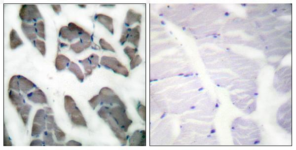
Real IHC data Immunohistochemistry analysis of paraffin-embedded human skeletal muscle tissue, using Actin-gamma2 Antibody. The picture on the right is blocked with the synthesized peptide.
Anti-Actin alpha3 ACTG2 Antibody
Cat # A05066

A05066-1 shows IHC staining in paraffin-embedded human colon carcinoma at 1:50 and lists IF use (A05066-1 image caption; catalog: applications). A05066 shows IHC staining in paraffin-embedded human skeletal muscle, with a peptide-blocked comparison image, and lists IF use (A05066 image caption; catalog: applications).

Which to pick: For tissue IHC, choose A05066-1 when the paraffin-section example in colon carcinoma is relevant; its listed IHC range is 1:50–1:200 (A05066-1 image caption; catalog: IHC dilution). For IF, consider A05066, which lists an IF range of 1:200–1:1000; ICC validation and IF images are unreported (A05066 catalog: applications, IF dilution, image captions). Both list Human, Mouse and Rat reactivity for cross-species planning, but their IHC images show human samples only; neither IHC caption reports the fixative (catalog: reactivity; A05066 and A05066-1 image captions).

Each figure is that product's own IHC / IF validation image from its datasheet.

References

  1. UniProt Consortium. UniProt entry P63267 (ACTH_HUMAN, Actin, gamma-enteric smooth muscle).
  2. Human Protein Atlas. ACTG2 tissue IHC expression (reliability: Supported).
  3. Human Protein Atlas. ACTG2 subcellular location (ICC-IF): Highest expression in U-251MG: 111.8 nTPM.
  4. Human Protein Atlas. ACTG2 antibody validation summary (2 antibodies).
  5. A plausible role for actin gamma smooth muscle 2 (ACTG2) in small intestinal neuroendocrine tumorigenesis. BMC endocrine disorders 2016 — PMC4841950.
  6. Analysis of metastasis‑related risk factors and clinical relevance in adult soft‑tissue sarcoma. Oncology letters 2024 — PMC11378013.
  7. Actin gamma smooth muscle 2 drives proliferation, metastasis, and 5-Fluorouracil resistance in gastric cancer: insights from a Recurrence-Related Gene Signature. Cancer cell international 2026 — PMC13289488.
  8. Clinically Relevant Molecular Subtypes in Leiomyosarcoma. Clinical cancer research : an official journal of the American Association for Cancer Research 2015 — PMC4526352.
  9. PubMed PMID:2377475 — UniProt-cited evidence.
  10. PubMed PMID:1710027 — UniProt-cited evidence.
  11. PubMed PMID:14702039 — UniProt-cited evidence.