This website uses cookies to ensure you get the best experience on our website.


- Table of Contents
Plan ACTN4 chromogenic IHC on paraffin sections using the catalog antibody’s documented conditions. Compare cytoplasmic staining with HPA tissue patterns and include appropriate controls when interpreting cell-specific signal.

Expected localisation, validated protocols, controls and antibodies — the at-a-glance facts below, then the full design guide.
| Expected localisation | Cytoplasmic tissue staining (HPA tissue IHC) | |
| Staining pattern | Cytoplasmic signal across essentially all tissue types (HPA tissue IHC) | |
| Antigen retrieval | EDTA pH 8.0 HIER, heat-mediated (datasheet PB9974) | |
| Positive control | Colon+4 more · see all | |
| Negative control | Adipose tissue+4 more · see all |
| Fixation | Keep fixation consistent across sections (standard IHC practice; not target-specific). Selected-image fixative and duration unreported (datasheet PB9974); verify before use. | |
| Caveat | Staining may reflect more than one gene product (HPA tissue IHC) | |
| Regulation | Widely expressed; no inducer specified (UniProt) | |
| Isoform / epitope | 3 isoforms; verify the antibody epitope across them (UniProt) |
The catalog antibody protocol uses EDTA retrieval (datasheet: PB9974); the published ACTN4 IHC protocols below use citrate retrieval (PMC8798106; PMC10394141).
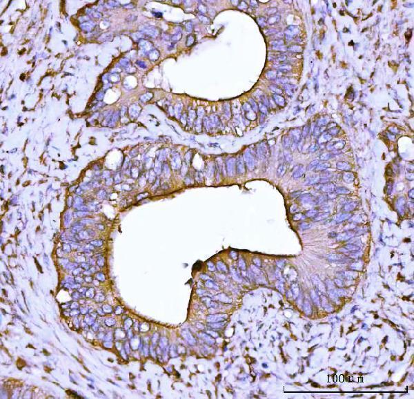
| Sample | Paraffin-embedded human colonic adenocarcinoma tissue; fixative not specified (datasheet PB9974) |
| Fixation | Image fixative and duration unreported (datasheet PB9974); verify before use. |
| Sectioning | 4–5 µm sections on charged slides (standard) |
| Deparaffinisation | Xylene, graded ethanol series to water (standard) |
| Antigen retrieval | Heat retrieval: EDTA pH 8.0 (datasheet PB9974); 20 min, 95–100 °C (standard) |
| Peroxidase block | 3% H2O2, 10 min, room temperature (standard) |
| Blocking | 10% goat serum (datasheet PB9974) |
| Primary antibody | Rabbit anti-ACTN4, 2-5μg/ml (datasheet PB9974) |
| Primary incubation | Overnight at 4 °C (datasheet PB9974) |
| Detection | HRP-conjugated secondary, DAB chromogen (datasheet PB9974) |
| Counterstain | Hematoxylin, blue, dehydrate and mount (standard) |
| Expected result | ACTN4-positive staining in endothelial cells of colon (HPA tissue IHC: High). HPA tissue profile: Ubiquitous cytoplasmic expression in essentially all tissue types. No signal in the no-primary control. |
ACTN4 is a widely expressed, non-transmembrane protein that localises mainly to the cytoplasm, cytoskeleton and cell junctions; nuclear localisation is also reported (UniProt O43707). For paraffin-section IHC, expect cytoplasmic staining across many tissues, with high staining reported in colon endothelial cells, liver cholangiocytes and lung type II alveolar cells (HPA: tissue IHC). HPA rates the tissue pattern Supported, with medium consistency between staining and RNA data, and cautions that the antibodies target proteins from more than one gene (HPA: tissue IHC).
| Clear cytoplasmic staining in colon endothelial cells or liver cholangiocytes, with cell outlines remaining interpretable. | This fits reported high staining in those cell types and the expected cytoplasmic distribution (HPA: tissue IHC; UniProt O43707). Judge the intended cells separately from adjacent cells: a positive tissue section does not mean every cell in it must have the same intensity (HPA: tissue IHC). |
| Signal appears only at an extracellular surface or as a continuous outline around every cell, without discernible cytoplasm. | That is inconsistent with ACTN4's non-transmembrane topology and reported intracellular locations (UniProt O43707). Treat the compartment mismatch as a possible artefact; review the chromogenic detection and staining controls before scoring the section as positive (standard IHC practice). Nuclear signal alone needs separate assessment because nuclear localisation is reported (UniProt O43707). |
| Strong staining is assigned to adipocytes, cardiomyocytes or skeletal myocytes. | HPA reports ACTN4 as not detected in those specific cell types, despite its broad tissue distribution (HPA: tissue IHC). Check cell identity, nearby positive cells and control sections; cross-reactivity or endogenous detection activity can produce misleading signal (HPA: multi-gene antibody caution; standard IHC practice). |
| Brown colour is diffuse across tissue spaces, edges or the whole section, obscuring individual cells. | A diffuse deposit cannot establish the intracellular pattern expected for ACTN4 (UniProt O43707). Assess a no-primary control and the detection reagents, then optimise blocking, washes and chromogen development as needed (standard IHC practice). Avoid scoring intensity where cellular boundaries cannot be resolved (standard IHC practice). |
| No stain appears in a section containing an identified high-staining cell population. | Absent signal in colon endothelial cells, liver cholangiocytes or lung type II alveolar cells conflicts with the reported high IHC levels (HPA: tissue IHC). First verify that the relevant cells are present, then check the antibody's IHC-P instructions, detection run and positive control (standard IHC practice). The HPA level alone does not identify a failure mechanism. |
| Cell and tissue selection | ACTN4 is widely expressed (UniProt O43707), while HPA reports high staining in specified cells and no detection in others within named tissues (HPA: tissue IHC). Select and score controls by cell type, not by tissue name alone (standard IHC practice). |
| Compartment and topology | ACTN4 has no transmembrane segment; UniProt lists cytoplasmic, cytoskeletal, junctional, perinuclear and nuclear locations (UniProt O43707). A nuclear component can be plausible, but an exclusively nuclear IHC pattern should be interpreted in its cell context rather than assumed to be the routine tissue pattern (UniProt O43707; HPA: tissue IHC). |
| Antibody evidence and scope | HPA lists IHC as Supported for HPA001873 and HPA006035, and cautions that its staining evidence involves antibodies targeting proteins from more than one gene (HPA: antibody validation; HPA: tissue IHC). Agreement with an expected pattern supports interpretation but does not, by itself, prove ACTN4-specific binding. |
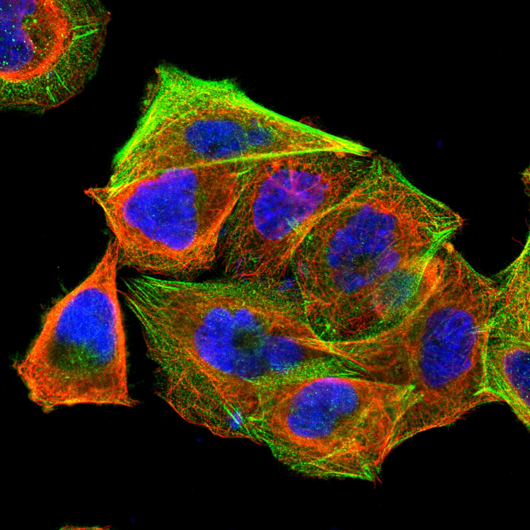
| Can IF/ICC findings guide IHC interpretation? | Yes, for compartment context: HPA reports actin filaments and focal adhesion sites as approved main ICC-IF locations, with cytosol among approved additional locations (HPA: subcellular ICC-IF). Its multi-gene antibody caution applies; fluorescence morphology does not set a chromogenic IHC-P protocol or tissue staining threshold (HPA: subcellular ICC-IF; standard IHC practice). |
| Situation | Likely cause | Next action |
|---|---|---|
| The expected high-staining cells are absent from the field. | A control chosen by organ name may miss the cell population on which the reported level is based (HPA: tissue IHC). | Locate colon endothelial cells, liver cholangiocytes or lung type II alveolar cells before judging the run; choose another supplied high-staining cell population if needed (HPA: tissue IHC). |
| Identified high-staining cells are present but show no signal. | The result conflicts with their reported HPA level; that observation alone does not establish whether antibody, retrieval or detection failed (HPA: tissue IHC). | Review the catalog antibody's IHC-P instructions and the run's positive control, retrieval, primary-antibody step and detection steps (standard IHC practice). Do not infer ACTN4-specific fixation sensitivity from the supplied evidence. |
| The whole slide has weak, diffuse brown background. | Diffuse colour may reflect nonspecific detection, insufficient washing or overdevelopment (standard IHC practice); it is not a resolved ACTN4 compartment pattern (UniProt O43707). | Compare a no-primary control, then check blocking, washes and chromogen development (standard IHC practice). Score only identifiable cells after background is controlled. |
| Reported negative cell types appear strongly positive. | HPA reports no detection in adipocytes, cardiomyocytes and skeletal myocytes, and warns that its antibodies can target proteins from more than one gene (HPA: tissue IHC). Endogenous detection activity is another general IHC possibility (standard IHC practice). | Confirm the cell type and examine no-primary and detection controls before assigning ACTN4 positivity (standard IHC practice). |
| Staining forms a uniform surface rim with little cytoplasmic detail. | A purely surface pattern does not fit ACTN4's non-transmembrane topology or reported intracellular distribution (UniProt O43707). | Inspect tissue morphology and controls, and review chromogen development and detection conditions before interpreting the rim (standard IHC practice). |
| Nuclear staining dominates while the expected cytoplasmic pattern is unclear. | UniProt permits nuclear localisation, including exclusive nuclear localisation in a limited number of cell lines; HPA describes tissue IHC as ubiquitously cytoplasmic (UniProt O43707; HPA: tissue IHC). | Record nuclear and cytoplasmic signal separately, verify the cell population and compare staining controls. Do not score nuclear signal as an automatic failure or as sufficient proof of ACTN4 specificity (standard IHC practice; HPA: multi-gene antibody caution). |



Comprehensive Human Protein Atlas IHC scoring per tissue (reliability: Supported — Medium consistency between antibody staining and RNA expression data. Caution, targets protein from more than one gene.). Rows are taken directly from the HPA tissue chart — click any row's HPA link to view the source.
| Tissue | Cell type | Level | Evidence | Source |
|---|---|---|---|---|
| Colon | Endothelial cells | High | Protein (IHC) | HPA → |
| Liver | Cholangiocytes | High | Protein (IHC) | HPA → |
| Lung | Alveolar cells type II | High | Protein (IHC) | HPA → |
| Placenta | Trophoblastic cells | High | Protein (IHC) | HPA → |
| Urinary bladder | Urothelial cells | High | Protein (IHC) | HPA → |
| Tissue | Cell type | Level | Evidence | Source |
|---|---|---|---|---|
| Adipose tissue | Adipocytes | Not detected | Protein (IHC) | HPA → |
| Caudate | Glial cells | Not detected | Protein (IHC) | HPA → |
| Cerebellum | Cells in granular layer | Not detected | Protein (IHC) | HPA → |
| Heart muscle | Cardiomyocytes | Not detected | Protein (IHC) | HPA → |
| Hippocampus | Glial cells | Not detected | Protein (IHC) | HPA → |
Troubleshoot ACTN4 staining in paraffin sections by checking retrieval, compartment, controls and scoring before interpreting chromogenic signal.
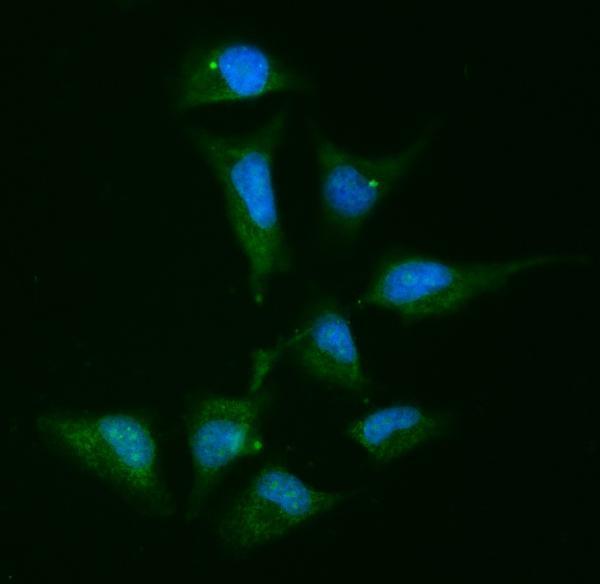
PB9974 has IHC data from human paraffin sections and IF/ICC data from Hela cells; its listed reactivity includes human, mouse and rat (PB9974 image captions and catalog).
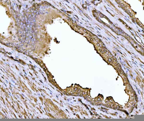
PB9974 is the card that will render, with IHC images from human colonic adenocarcinoma and prostate cancer paraffin sections (PB9974 IHC captions). Its IF image shows Hela cells (PB9974 IF caption).
Which to pick: Choose PB9974 for tissue IHC: its image captions document human paraffin sections, 2 μg/ml antibody and EDTA retrieval at pH 8.0; the fixative is unreported (PB9974 IHC captions). Choose PB9974 for IF/ICC when an imaged example is useful: its caption documents Hela cells at 5 μg/ml (PB9974 IF caption). For cross-species planning, PB9974 and the monoclonal M01975 both list human, mouse and rat reactivity, but the supplied tissue IHC images document human samples only (catalog reactivity; PB9974 IHC captions).