ADAMTS5 / A disintegrin and metalloproteinase with thrombospondin motifs 5 · IHC design guide

Design Immunohistochemistry for ADAMTS5

Use human placenta as a demonstrated paraffin-section specimen (datasheet A02802-1) and bronchial ciliated cells as a high-staining comparator (HPA tissue IHC). This guide covers chromogenic IHC setup and scoring of variable cytoplasmic staining (HPA tissue IHC), with interpretation informed by ADAMTS5 secretion into extracellular space and matrix (UniProt).

Evidence assembled Oct 2026 · For research use; verify linked source records and product datasheet before use
Immunohistochemistry protocol sheet for ADAMTS5 (IHC for ADAMTS5): expected localisation Cytoplasmic tissue staining (HPA tissue IHC); secreted into extracellular space and matrix (UniProt), antibody A02802-1, validated IHC image, and IHC protocol steps
Printable ADAMTS5 IHC protocol sheet — expected localisation Cytoplasmic tissue staining (HPA tissue IHC); secreted into extracellular space and matrix (UniProt), antibody A02802-1, controls and protocol steps. Open the full ADAMTS5 IHC guide →

ADAMTS5 Immunohistochemistry Experimental Design Guide

Expected localisation, validated protocols, controls and antibodies — the at-a-glance facts below, then the full design guide.

Must know before staining
Expected localisation Cytoplasmic tissue staining (HPA tissue IHC); secreted into extracellular space and matrix (UniProt)
Staining pattern Variable cytoplasmic staining in several cell types (HPA tissue IHC)
Antigen retrieval EDTA pH 8.0 HIER, heat-mediated (datasheet A02802-1)
Positive control ⓘ Bronchus+4 more · see all
Negative control ⓘ Adipose tissue+4 more · see all
Important caveats
Reasons your staining may differ from the expected pattern.
Fixation Matched tissue-IHC evidence does not establish the fixation claim. Validate the specimen-specific method before use. (selected-SKU IHC image A02802-1)
Caveat Secreted protein: tissue staining may differ from RNA location (HPA tissue IHC)
Regulation Tissue-enhanced RNA: ovary, placenta (HPA tissue RNA)
Isoform / epitope No isoforms listed; mature chain starts at residue 262 (UniProt)
Section 1

Recommended ADAMTS5 IHC & IF Protocols

The catalog antibody’s IHC-P protocol (datasheet: A02802-1) is accompanied by one published mouse knee cartilage IHC example (PMC12536589: Histological analyses).

Recommended immunohistochemistry (IHC-P) protocol parameters
SampleParaffin-embedded human placenta tissue; fixative not specified (datasheet A02802-1)
FixationImage fixative and duration unreported (datasheet A02802-1); verify before use.
Sectioning4–5 µm sections on charged slides (standard)
DeparaffinisationXylene, graded ethanol series to water (standard)
Antigen retrievalHeat retrieval: EDTA pH 8.0 (datasheet A02802-1); 20 min, 95–100 °C (standard)
Peroxidase block3% H2O2, 10 min, room temperature (standard)
Blocking10% goat serum (datasheet A02802-1)
Primary antibodyRabbit anti-ADAMTS5, 2-5μg/ml (datasheet A02802-1)
Primary incubationOvernight at 4 °C (datasheet A02802-1)
DetectionStreptavidin-biotin complex (SABC), DAB chromogen (datasheet A02802-1)
CounterstainHematoxylin, blue, dehydrate and mount (standard)
Expected resultADAMTS5-positive staining in ciliated cells (cilia axoneme) of bronchus (HPA tissue IHC: High). HPA tissue profile: Cytoplasmic expression at variable levels in several different cell types. No signal in the no-primary control.
💡Decision noteStart with heat-mediated EDTA pH 8.0 retrieval (datasheet: A02802-1), then optimize heating time on matched paraffin sections (standard IHC practice).
Section 2

What Is the Expected ADAMTS5 Staining Pattern?

ADAMTS5 is a secreted extracellular matrix protein with no transmembrane segment (UniProt Q9UNA0 topology). In paraffin section IHC, expect variable cytoplasmic staining in several cell types, with strong staining reported in bronchial and fallopian tube ciliated cells (HPA: tissue IHC). Extracellular signal can fit its biology, but HPA rates tissue staining Approved and reports low consistency with RNA expression; interpret any compartment assignment cautiously (HPA: reliability; UniProt Q9UNA0 subcellular location).

What am I looking at on my slide?
Strong signal in bronchial or fallopian tube ciliated cells, with adjacent cells less prominent (HPA: High in ciliated cells).This matches an observed positive IHC pattern. Check cell identity and tissue structure before scoring: HPA specifies the cilia axoneme in these tissues, while its overall profile describes variable cytoplasmic staining. A positive field need not stain every cell equally (HPA: tissue IHC).
Signal is confined to nuclei, with no plausible cytoplasmic, ciliary or extracellular signal.Treat an isolated nuclear pattern as suspect: HPA reports variable cytoplasmic staining, and UniProt places ADAMTS5 in the extracellular space and matrix (HPA: tissue IHC; UniProt Q9UNA0 subcellular location). Review morphology, counterstain and detection controls before assigning it to ADAMTS5 (general IHC practice).
Strong staining appears in cells listed as undetected, such as skin keratinocytes or adipocytes (HPA: Not detected in these cells).Consider cross-reactivity or endogenous detection activity, then compare with a known positive section and reagent controls (general IHC practice). HPA's result applies to the specified cell type, not every cell in that tissue; unexpected staining alone cannot establish either mechanism (HPA: tissue IHC).
Color covers broad tissue areas, including spaces and cells, without a discernible pattern.Diffuse signal may obscure a true extracellular target or reflect background (UniProt Q9UNA0 subcellular location; general IHC practice). Compare a section processed without primary antibody, inspect tissue edges and assess whether staining follows recognizable matrix or cell structures before calling it positive (general IHC practice).
No signal appears in bronchial or fallopian tube ciliated cells (HPA: High in these cells).First check that the expected cells are present and intact. A blank known positive section points to an assay problem, but HPA's Approved rating and low RNA–protein consistency limit how strongly one section can define biological absence (HPA: reliability; general IHC practice).
💡Expected ADAMTS5 appearanceCall a positive result when staining follows identifiable ciliated cells with strong signal in bronchus or fallopian tube, or shows plausible variable cytoplasmic or extracellular distribution; isolated nuclear staining or uniform, structureless color is suspect (HPA: tissue IHC; UniProt Q9UNA0 subcellular location; general IHC practice).
How each factor affects the staining
Secreted protein and tissue interpretation (UniProt Q9UNA0 subcellular location)Protein can occupy extracellular space or matrix, so its staining need not coincide with the cell that made it; HPA explicitly warns that tissue locations of RNA and protein may differ (HPA: reliability description).
Processing and antibody epitope (UniProt Q9UNA0 processing)ADAMTS5 has a signal peptide at residues 1–16, a propeptide at 17–261 and a mature chain at 262–930. Which form a stain detects depends on the antibody epitope; its position is not supplied here (UniProt Q9UNA0 processing).
Strength of tissue evidence (HPA: antibody validation)HPA lists HPA005661 and CAB025996 as IHC Approved, while reporting low consistency between staining and RNA. Approved should not be read as Enhanced or as proof that every observed cell signal is specific (HPA: antibodies; HPA: reliability).
What should IF/ICC show? (HPA: subcellular summary)A secreted distribution is biologically plausible (HPA: Secreted; UniProt Q9UNA0 subcellular location). HPA provides no main ICC-IF location or cell-line images, so this record cannot define a verified punctate, membrane or nuclear IF pattern (HPA: ICC-IF record).
Negative reference cells (HPA: tissue IHC)Adipocytes, skin keratinocytes and ovarian stromal cells are listed as Not detected. Use the named cells when comparing fields; the entries do not declare whole adipose tissue, skin or ovary ADAMTS5-free (HPA: tissue IHC).
Why is my staining missing, weak or wrong?
SituationLikely causeNext action
The known positive section is blank.The expected ciliated cells may be absent from the section, or a staining step may have failed (HPA: High in bronchus and fallopian tube ciliated cells; general IHC practice).Confirm the cell population by morphology, then check primary antibody, detection reagents and counterstain on a suitable control section (general IHC practice).
The entire slide develops diffuse color.Nonspecific reagent binding or incomplete control of chromogenic detection can obscure a spatial pattern (general IHC practice).Inspect a no-primary control; review blocking, washes and detection exposure. Preserve the distinction between patterned matrix signal and structureless background (UniProt Q9UNA0 subcellular location; general IHC practice).
Only nuclei stain strongly.A nuclear-only result conflicts with the reported cytoplasmic tissue profile and secreted location (HPA: tissue IHC; UniProt Q9UNA0 subcellular location).Check counterstain and no-primary controls, then reassess whether a cytoplasmic or extracellular signal remains (general IHC practice).
Unexpected cells stain more strongly than the expected positive cells.Cross-reactivity, endogenous detection activity or misidentified cells are possible (general IHC practice). HPA cell-level negatives are comparators, not absolute tissue exclusions (HPA: tissue IHC).Verify cell identity, compare the known positive section and run the appropriate detection control before interpreting the unexpected population (general IHC practice).
Protein staining and tissue RNA levels seem discordant.A secreted protein can be found away from its producer, and HPA reports low consistency between antibody staining and RNA expression (UniProt Q9UNA0 subcellular location; HPA: reliability).Score the observed protein location and cell type separately from RNA abundance; describe the discrepancy without using RNA alone to reject staining (HPA: reliability description; general IHC practice).
An IF/ICC image shows a precise intracellular pattern that IHC cannot confirm.HPA gives no main ICC-IF location or cell-line images for ADAMTS5 (HPA: ICC-IF record).Treat the IF compartment assignment as unverified by these sources and assess it with appropriate IF controls in its separate guide (HPA: ICC-IF record; general IF practice).

Sample controls for ADAMTS5 IHC & IF

🧪Run bronchus first and look for staining in ciliated-cell axonemes (HPA: High in bronchus ciliated cells). Use adipose tissue adipocytes as the negative tissue (HPA: Not detected in adipocytes); on the bronchus slide, cells without specific staining should show background only, but HPA does not identify a confirmed negative bronchial cell type.
Positive control tissue: Bronchus (Ciliated cells (cilia axoneme), HPA High)
Negative control tissue: Adipose tissue (HPA Not detected)
ICC-IF cell lines (HPA subcellular resource): HPA carries no ICC-IF cell line for ADAMTS5; derive a cell-line control from the positive tissue's cell type (Ciliated cells (cilia axoneme)) and confirm it by RNA or western blot first.
Technical controls: Include no-primary (secondary-only) and concentration-matched rabbit isotype controls (caption: rabbit primary antibody), plus ADAMTS5 knockout tissue or an immunogen-peptide block if available. Quench endogenous peroxidase and check for endogenous biotin background when using the caption’s biotin-based, DAB detection (caption: biotinylated secondary, SABC and DAB).
⚠️Feasibility: No target-specific fixation window or fixation effect is reported, and the exact A02802-1 tissue-IHC caption does not state a fixative (caption: fixative unreported). Heat retrieval in EDTA at pH 8.0 was used for the paraffin-section example, but a requirement for retrieval is unestablished (caption: placenta IHC). The evidence does not establish whether frozen sections or IF are easier; assess possible staining along the narrow ciliary edge against controls when scoring bronchus (HPA: High in ciliated-cell axonemes).

HPA tissue IHC evidence for ADAMTS5

Comprehensive Human Protein Atlas IHC scoring per tissue (reliability: Approved — Low consistency between antibody staining and RNA expression data. Secreted protein, tissue location of RNA and protein is expected to differ.). Rows are taken directly from the HPA tissue chart — click any row's HPA link to view the source.

Positive expression · recommended positive controls

TissueCell typeLevelEvidenceSource
Bronchus Ciliated cells (cilia axoneme) High Protein (IHC) HPA →
Fallopian tube Ciliated cells (cilia axoneme) High Protein (IHC) HPA →
Nasopharynx Ciliated cells (cell body) High Protein (IHC) HPA →
Cervix Glandular cells Medium Protein (IHC) HPA →
Colon Glandular cells Medium Protein (IHC) HPA →

Undetected expression · recommended negative controls

TissueCell typeLevelEvidenceSource
Adipose tissue Adipocytes Not detected Protein (IHC) HPA →
Bone marrow Hematopoietic cells Not detected Protein (IHC) HPA →
Liver Cholangiocytes Not detected Protein (IHC) HPA →
Oral mucosa Squamous epithelial cells Not detected Protein (IHC) HPA →
Ovary Ovarian stroma cells Not detected Protein (IHC) HPA →
Section 3

Advanced ADAMTS5 IHC Tips

Troubleshooting ADAMTS5 staining in paraffin-section chromogenic IHC, with one IF/ICC application note.

Which retrieval conditions should I start with for weak ADAMTS5 staining?
Start with heat-mediated antigen retrieval in EDTA at pH 8.0 for paraffin sections (datasheet A02802-1). The selected tissue-IHC image used this retrieval before 2 μg/ml primary antibody overnight at 4°C; keep those conditions consistent when comparing runs (datasheet A02802-1). If staining remains weak, optimise heating and cooling against a known positive section before testing another buffer as a fallback (standard IHC practice; datasheet A02802-1). Compare tissue morphology and no-primary controls alongside signal, because excessive retrieval can damage sections or increase background (standard IHC practice). Record retrieval conditions separately from antibody concentration so that a changed signal has an identifiable cause (standard IHC practice).
Could fixation explain variable ADAMTS5 staining between paraffin blocks?
The selected paraffin-section caption does not report its fixative, so ADAMTS5-specific fixation sensitivity is unknown (datasheet A02802-1). Record each block’s fixation method and processing history, then compare sections using the same EDTA pH 8.0 retrieval and antibody conditions (datasheet A02802-1; standard IHC practice). Include similarly processed control tissue in each run, since differences in fixation and processing can alter antigen accessibility in paraffin sections (standard IHC practice). Check morphology and background before attributing a weak result to fixation; retrieval, antibody incubation, and detection can also affect staining (standard IHC practice). Do not infer a fixation effect from reported tissue staining patterns or ADAMTS5 protein features alone (HPA tissue IHC; UniProt Q9UNA0).
Should ADAMTS5 staining appear inside cells or in the surrounding matrix?
Assess both cellular and extracellular staining: ADAMTS5 is annotated as secreted and extracellular, with no transmembrane segment (UniProt Q9UNA0 topology and subcellular location). Variable cytoplasmic staining across several cell types is also reported in tissue IHC, so cytoplasmic signal is plausible but does not establish where the secreted protein acts (HPA tissue IHC; UniProt Q9UNA0). Map staining separately in cell bodies, cilia, and nearby matrix rather than combining these compartments into one score (HPA tissue IHC; standard IHC practice). In bronchus, strong cilia-axoneme staining is reported, providing a specific pattern to examine alongside morphology (HPA: High in bronchus ciliated cells). Treat exclusively nuclear staining cautiously because it conflicts with the reported localisation (UniProt Q9UNA0 subcellular location; standard IHC practice).
How could ADAMTS5 processing affect what this antibody detects?
ADAMTS5 has a signal peptide at residues 1–16, a propeptide at 17–261, and an annotated mature chain at 262–930 (UniProt Q9UNA0 processing). An antibody recognising the propeptide could give a different spatial pattern from one recognising the mature chain, so establish the immunogen or mapped epitope before comparing stains (UniProt Q9UNA0 processing; standard IHC practice). The supplied record lists 7 glycosylation sites and 0 isoforms, but provides no epitope location for the selected antibody (UniProt Q9UNA0; datasheet A02802-1). If patterns disagree across antibodies, compare epitope information and controls before assigning the difference to a distinct ADAMTS5 form (standard IHC practice; UniProt Q9UNA0 processing).
How should I assess ADAMTS5 by IF alongside cell identity markers?
Treat IF/ICC as a separate application, since the selected antibody evidence here describes paraffin-section chromogenic IHC and supplies no IF/ICC validation (datasheet A02802-1). For bronchus sections, multiplex ADAMTS5 with a ciliated-cell marker to test whether signal follows the reported ciliated-cell pattern (HPA: High in bronchus ciliated cells; standard IF practice). Select a fluorophore channel with low tissue autofluorescence, and inspect single-channel controls before interpreting overlap (standard IF practice). Because ADAMTS5 is secreted and has no transmembrane segment, choose permeabilisation according to the epitope being tested: intracellular access may require it, while extracellular staining may be assessed without it (UniProt Q9UNA0 topology and subcellular location; standard IF practice).
How can I troubleshoot diffuse brown staining in ADAMTS5 IHC?
The selected detection workflow uses biotinylated goat anti-rabbit secondary antibody, streptavidin-biotin complex, and DAB, so inspect background at each detection stage (datasheet A02802-1; standard IHC practice). Run no-primary and secondary-only controls to distinguish primary-antibody staining from detection-related signal (standard IHC practice). Include a peroxidase block before DAB development and assess endogenous biotin where the biotin-based system produces unexplained staining; these are general workflow checks (standard IHC practice). Review blocking and washing if broad nonspecific colour persists; the selected image used 10% goat serum as its block (datasheet A02802-1; standard IHC practice). Compare suspicious deposits with tissue morphology because ADAMTS5 can occur extracellularly (UniProt Q9UNA0 subcellular location; standard IHC practice).
What should I score when ADAMTS5 staining spans cells and matrix? ⚠ ANSWER MARKED FOR VERIFICATION
Define cellular and extracellular compartments before scoring, because ADAMTS5 is secreted and extracellular while tissue IHC also reports variable cytoplasmic staining (UniProt Q9UNA0 subcellular location; HPA tissue IHC). For cellular staining, record the percentage of positive cells and intensity, or calculate an H-score on a consistent 0–300 scale (standard IHC practice). For matrix staining, quantify DAB-positive area or integrated optical density per mm² of annotated matrix, using the same threshold across sections (standard IHC practice). Normalise cellular results to the relevant cell count and matrix results to assessed matrix area, rather than total section area (standard IHC practice). Report compartment, region selection, and control-based background correction with each comparison (standard IHC practice).
How do I distinguish credible ADAMTS5 signal from staining artefacts?
Prioritise reproducible cellular or extracellular patterns consistent with secreted ADAMTS5, and examine bronchial cilia when that tissue is used (UniProt Q9UNA0 subcellular location; HPA: High in bronchus ciliated cells). Treat isolated nuclear staining, section-edge colour, and staining confined to necrotic areas as suspicious until controls and morphology support them (UniProt Q9UNA0 subcellular location; standard IHC practice). Compare no-primary and detection controls when DAB deposits could reflect endogenous enzyme activity or the biotin-based detection workflow (datasheet A02802-1; standard IHC practice). HPA reports low consistency between staining and RNA expression and notes that secretion can separate protein location from RNA location, so RNA mismatch alone does not resolve specificity (HPA tissue IHC).
Boster reagents

Best ADAMTS5 / A disintegrin and metalloproteinase with thrombospondin motifs 5 IHC Antibodies

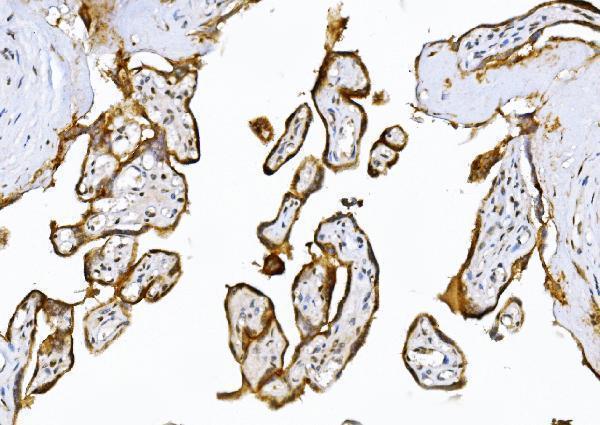
A02802-1 has a human paraffin-section IHC image (A02802-1 image caption); M02802 lists IF/ICC applications (catalog: M02802 applications). Both list Human, Mouse and Rat reactivity (catalog: reactivity).

Real IHC data IHC analysis of ADAMTS5 using anti-ADAMTS5 antibody (A02802-1). ADAMTS5 was detected in a paraffin-embedded section of human placenta tissue. Heat mediated antigen retrieval was performed in EDTA buffer (pH 8.0, epitope retrieval solution). The tissue section was blocked with 10% goat serum. The tissue section was then incubated with 2 μg/ml rabbit anti-ADAMTS5 Antibody (A02802-1) overnight at 4°C. Biotinylated goat anti-rabbit IgG was used as secondary antibody and incubated for 30 minutes at 37°C. The tissue section was developed using Strepavidin-Biotin-Complex (SABC) (Catalog # SA1022) with DAB as the chromogen.
Anti-ADAMTS5 Antibody ®
Cat # A02802-1

A02802-1 lists IHC and Human, Mouse and Rat reactivity, with an IHC image from human paraffin-embedded placenta (catalog: A02802-1 applications/reactivity; A02802-1 image caption). M02802 lists IHC and IF/ICC applications and Human, Mouse and Rat reactivity; no IHC or IF image is supplied (catalog: M02802 applications/reactivity/image alts).

Which to pick: For tissue IHC, choose A02802-1 for its documented human paraffin-section workflow, including EDTA retrieval at pH 8.0 and DAB detection; the fixative is unreported (A02802-1 image caption). For IF/ICC, M02802 is the listed option and is monoclonal clone 33A47, although no IF image is supplied (catalog: M02802 applications/clone/image alts). Both list Human, Mouse and Rat reactivity, but only A02802-1 has a tissue IHC image, and that image shows human tissue (catalog: reactivity/image alts; A02802-1 image caption).

Each figure is that product's own IHC / IF validation image from its datasheet.

References

  1. UniProt Consortium. UniProt entry Q9UNA0 (ATS5_HUMAN, A disintegrin and metalloproteinase with thrombospondin motifs 5).
  2. Human Protein Atlas. ADAMTS5 tissue IHC expression (reliability: Approved).
  3. Human Protein Atlas. ADAMTS5 subcellular location (ICC-IF): Secreted.
  4. Human Protein Atlas. ADAMTS5 antibody validation summary (2 antibodies).
  5. Mulberroside A alleviates osteoarthritis via restoring impaired autophagy and suppressing MAPK/NF-κB/PI3K-AKT-mTOR signaling pathways. iScience 2023 — PMC9868682.
  6. TRPM2-mediated feed-forward loop promotes chondrocyte damage in osteoarthritis via calcium-cGAS-STING-NF-κB pathway. Journal of advanced research 2025 — PMC12536589.
  7. TBK1 pharmacological inhibition mitigates osteoarthritis through attenuating inflammation and cellular senescence in chondrocytes. Journal of orthopaedic translation 2024 — PMC11260960.
  8. Adamts5, the gene encoding a proteoglycan-degrading metalloprotease, is expressed by specific cell lineages during mouse embryonic development and in adult tissues. Gene expression patterns : GEP 2009 — PMC2725439.
  9. PubMed PMID:10438522 — UniProt-cited evidence.
  10. PubMed PMID:10830953 — UniProt-cited evidence.
  11. PubMed PMID:15489334 — UniProt-cited evidence.