Click image to see more details
-

-
-
-
-
+3
Product Info Summary
| SKU: | A02802-1 |
|---|---|
| Size: | 100 μg/vial |
| Reactive Species: | Human, Mouse, Rat |
| Host: | Rabbit |
| Application: | ELISA, IHC, WB |
Customers Who Bought This Also Bought

Product info
Product Name
Anti-ADAMTS5 Antibody Picoband®
SKU/Catalog Number
A02802-1
Size
100 μg/vial
Form
Lyophilized
Description
Boster Bio Anti-ADAMTS5 Antibody Picoband® catalog # A02802-1. Tested in ELISA, IHC, WB applications. This antibody reacts with Human, Mouse, Rat. The brand Picoband indicates this is a premium antibody that guarantees superior quality, high affinity, and strong signals with minimal background in Western blot applications. Only our best-performing antibodies are designated as Picoband, ensuring unmatched performance.
Storage & Handling
Store at -20˚C for one year from date of receipt. After reconstitution, at 4˚C for one month. It can also be aliquotted and stored frozen at -20˚C for six months. Avoid repeated freeze-thaw cycles.
Cite This Product
Anti-ADAMTS5 Antibody Picoband® (Boster Biological Technology, Pleasanton CA, USA, Catalog # A02802-1)
Host
Rabbit
Contents
Each vial contains 4mg Trehalose, 0.9mg NaCl and 0.2mg Na2HPO4.
Clonality
Polyclonal
Isotype
Rabbit IgG
Immunogen
E.coli-derived human ADAMTS5 recombinant protein (Position: D747-K780).
Cross-reactivity
No cross-reactivity with other proteins
Reactive Species
A02802-1 is reactive to ADAMTS5 in Human, Mouse, Rat
Observed Molecular Weight
75 kDa
Calculated molecular weight
101.7 kDa
Background of ADAMTS5
ADAMTS5 (A Disintegrin-Like and Metalloproteinase with Thrombospondin Type 1 Motif, 5), is an enzyme that in humans is encoded by the ADAMTS5 gene. ADAMTS5 is a member of the large ADAMTS family of zinc-dependent proteases. The enzyme encoded by this gene contains two C-terminal TS motifs and functions as aggrecanase to cleave aggrecan, a major proteoglycan of cartilage. By somatic cell hybrid analysis, the human ADAMTS5 gene is mapped to chromosome 21. Used mouse models, it is showed that Sdc4 controls a pathway that activates Adamts5 at the chondrocyte cell surface through Erk1/Erk2 activation of Mmp3.
Antibody Validation
Boster validates all antibodies on WB, IHC, ICC, Immunofluorescence, and ELISA with known positive control and negative samples to ensure specificity and high affinity, including thorough antibody incubations.
Application & Images
Applications
A02802-1 is guaranteed for ELISA, IHC, WB Boster Guarantee
Recommend Dilution
| Application | Dilution | Species |
|---|---|---|
| Western blot | 0.25-0.5μg/ml | Human, Mouse, Rat |
| Immunohistochemistry (Paraffin-embedded Section) | 2-5μg/ml | Human |
| ELISA | 0.1-0.5μg/ml | - |
Tested application
Suggested blocking solution with 5% non-fat milk or BSA; (*)Recommended protein loading: 20-40 µg per lane
Use TE buffer pH 9.0 for antigen retrieval; (*) citrate buffer pH 6.0 is an alternative.
Validation Images & Assay Conditions
Click image to see more details
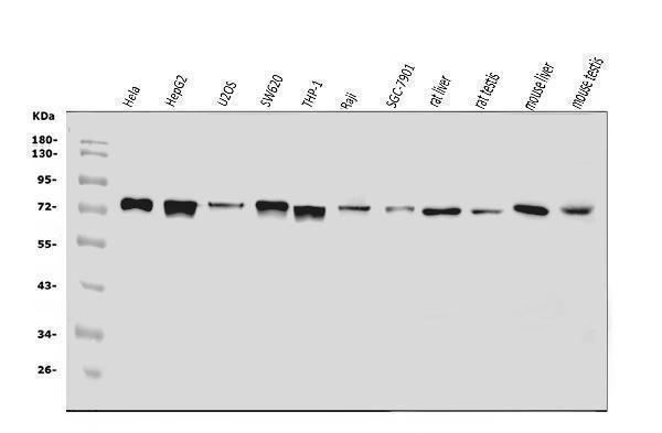
Western blot analysis of ADAMTS5 using anti-ADAMTS5 antibody (A02802-1).
Electrophoresis was performed on a 5-20% SDS-PAGE gel at 70V (Stacking gel) / 90V (Resolving gel) for 2-3 hours. The sample well of each lane was loaded with 30 ug of sample under reducing conditions.
Lane 1: human Hela whole cell lysates,
Lane 2: human HepG2 whole cell lysates,
Lane 3: human U20S whole cell lysates,
Lane 4: human SW620 whole cell lysates,
Lane 5: human THP-1 whole cell lysates,
Lane 6: human Raji whole cell lysates,
Lane 7: human SGC-7901 whole cell lysates,
Lane 8: rat liver tissue lysates,
Lane 9: rat testis tissue lysates,
Lane 10: mouse liver tissue lysates,
Lane 11: mouse testis tissue lysates,
After electrophoresis, proteins were transferred to a nitrocellulose membrane at 150 mA for 50-90 minutes. Blocked the membrane with 5% non-fat milk/TBS for 1.5 hour at RT. The membrane was incubated with rabbit anti-ADAMTS5 antigen affinity purified polyclonal antibody (Catalog # A02802-1) at 0.5 μg/mL overnight at 4°C, then washed with TBS-0.1%Tween 3 times with 5 minutes each and probed with a goat anti-rabbit IgG-HRP secondary antibody at a dilution of 1:5000 for 1.5 hour at RT. The signal is developed using an Enhanced Chemiluminescent detection (ECL) kit (Catalog # EK1002) with Tanon 5200 system. A specific band was detected for ADAMTS5 at approximately 75 kDa. The expected band size for ADAMTS5 is at 102 kDa.

Click image to see more details
PA suppressed excess expression of the catabolic indicators of chondrocytes induced by IL-1β, including ADAMTS5, MMP1, MMP3, and MMP13. Mice chondrocytes were treated with 5 ng/ml of IL-1β, alone or with PA (2.5, 5, and 10 μM) for 24 h (A) Western blotting results and (B–E) quantitative analysis of ADAMTS5, MMP1, MMP3, and MMP13. (F) MMP13 expression was observed by immunofluorescence staining when chondrocytes were treated with 5 ng/ml of IL-1β, alone or with 10 μM of PA (scale bar 200 μm). (G–I) Relative mRNA levels of ADAMTS5, MMP3, and MMP13 in chondrocytes stimulated with 5 ng/ml of IL-1β, alone or with PA (2.5, 5, and 10 μM) for 24 h. GAPDH was used as an internal reference. Data are presented as means ± SD ( n = 3). The exact p value was marked in the corresponding figure and p < 0.05 was considered statistically significant.
Index in PubMed under a CC BY license. PMID: 34925020

Click image to see more details
Knockdown of integrin αVβ3 weakened the anti-inflammatory, anabolism enhancing, and catabolism inhibiting effect of PA on IL-1β-induced chondrocytes. (A,B) Relative mRNA levels of integrin αV (Itg αV) and integrin β3 (Itg β3) in chondrocytes stimulated with 5 ng/ml of IL-1β, alone or with PA (2.5, 5, and 10 μM) for 24 h (C,D) Itg αV and Itg β3 were knocked down by siRNA transfection, and the knockdown efficiency was verified by RT-PCR. (E,F) Inflammatory markers (COX2, iNOS) were detected by western blotting and the band density of protein levels were quantified after mice chondrocytes were added with or without 5 ng/ml of IL-1β, 10 μM of PA, and Itg αVβ3 siRNA. (G–I) Western blotting was applied to measure the anabolic (aggrecan, collagen II) and catabolic markers (MMP1, MMP3, MMP13, and ADAMTS5) in the Itg αVβ3-deficiency mice chondrocytes along with or without the administration of 5 ng/ml of IL-1β and 10 μM of PA, and the band density of these protein levels were quantified in the histogram. GAPDH was used as an internal reference. Data are presented as means ± SD ( n = 3). The exact p value was marked in the corresponding figure and p < 0.05 was considered statistically significant.
Index in PubMed under a CC BY license. PMID: 34925020

Click image to see more details
ALT alleviated excess MMPs and ADAMTS5 expression in IL-1β-stimulated mouse chondrocytes. Chondrocytes were exposed to ALT (2.5, 5, and 10 μM) with or without IL-1β (10 ng/ml) for 24 h. (A) Western blot was employed to determine the expression of MMP1, MMP3, MMP13, and ADAMTS5. (B) Relative protein expression was qualified by ImageJ software, GAPDH was used as the internal control ( n = 3). # p < 0.05 vs. control group; * p < 0.05 vs. IL-1β group; ** p < 0.01 vs. IL-1β group.
Index in PubMed under a CC BY license. PMID: 34650433

Click image to see more details
Schematic diagram of the effect of ALT on cartilage degeneration. IL-1β induces the expression of pro-inflammatory factors, including iNOS, COX2, MMPs and ADAMTS5. IL-1β stimulates the degradation of Collagen II and the impairment of autophagy. Furthermore, IL-1β functions by activating the PI3K/AKT/mTOR, NF-κB signaling pathways, phosphorylating and translocating STAT3. As shown in the figure, the levels of P-PI3K, P-AKT, P-mTOR, P-IKB, P-P65, P-STAT3 and the translocation of P65 and STAT3 are increasing. However, ALT can attenuate these effects.
Index in PubMed under a CC BY license. PMID: 34650433

Click image to see more details
Effects of Schisandrin A on IL-1β-induced MMPs and ADAMTS5 protein expression. Chondrocytes were exposed to Schisandrin A (25, 50 μM) with or without IL-1β (10 ng/ml) for 24 h. (A) Western blot was employed to determine the expression of MMP1, MMP3, MMP13, and ADAMTS5. (B) Relative protein expression was qualified by Image-J software, GAPDH was used as the internal control ( n = 3). # P < 0.05 vs. control group; ∗ P < 0.05 vs. IL-1β group; ∗∗ P < 0.01 vs. IL-1β group.
Index in PubMed under a CC BY license. PMID: 30761007

Click image to see more details
IHC analysis of ADAMTS5 using anti-ADAMTS5 antibody (A02802-1).
ADAMTS5 was detected in a paraffin-embedded section of human placenta tissue. Heat mediated antigen retrieval was performed in EDTA buffer (pH 8.0, epitope retrieval solution). The tissue section was blocked with 10% goat serum. The tissue section was then incubated with 2 μg/ml rabbit anti-ADAMTS5 Antibody (A02802-1) overnight at 4°C. Biotinylated goat anti-rabbit IgG was used as secondary antibody and incubated for 30 minutes at 37°C. The tissue section was developed using Strepavidin-Biotin-Complex (SABC) (Catalog # SA1022) with DAB as the chromogen.
Specific Publications For Anti-ADAMTS5 Antibody Picoband® (A02802-1)
Loading publications
Recommended Resources
Here are featured tools and databases that you might find useful.
- Boster's Pathways Library
- Protein Databases
- Bioscience Research Protocol Resources
- Data Processing & Analysis Software
- Photo Editing Software
- Scientific Literature Resources
- Research Paper Management Tools
- Molecular Biology Software
- Primer Design Tools
- Bioinformatics Tools
- Phylogenetic Tree Analysis
Customer Reviews
Have you used Anti-ADAMTS5 Antibody Picoband®?
Share your experimental results or join a short interview to earn up to $1,000 in product credits or other rewards.
0 Reviews For Anti-ADAMTS5 Antibody Picoband®
Customer Q&As
Have a question?
Find answers in Q&As, reviews.
Can't find your answer?
Submit your question
