This website uses cookies to ensure you get the best experience on our website.
- Table of Contents
Facts about Adhesion G-protein coupled receptor V1.
Human | |
---|---|
Gene Name: | ADGRV1 |
Uniprot: | Q8WXG9 |
Entrez: | 84059 |
Belongs to: |
---|
G-protein coupled receptor 2 family |
Adhesion G-protein coupled receptor V1
Mass (kDA):
693.069 kDA
Human | |
---|---|
Location: | 5q14.3 |
Sequence: | 5; NC_000005.10 (90558796..91164437) |
Expressed at low levels in adult tissues.
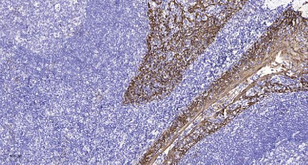
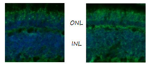
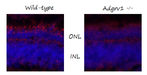
Cell membrane; Multi-pass membrane protein. Cell projection, stereocilium membrane. Photoreceptor inner segment. Localizes at the ankle region of the stereocilia. In photoreceptors, localizes at a plasma membrane microdomain in the apical inner segment that surrounds the connecting cilia called periciliary membrane complex.
This article explains the many uses of the ADGRV1 markers in different types and types of scientific research. This article also describes various methods of interpreting the results. Learn about Western blots, Protein transfer efficacy, and Autoradiography. These methods can be applied to all types scientists around the globe. Let's see some examples of the ADGRV1 mark's different uses.
Medical research is becoming increasingly important in molecular diagnostics. These tests analyze DNA and sequences of RNA to identify abnormalities that could indicate a disease. Molecular diagnostics can detect diseases that are related to particular gene mutations. Molecular diagnostics are crucial in early diagnosis for a variety of diseases, such as cancer.
Molecular diagnostics were developed as a way to improve the accuracy, reproducibility, and reproducibility of pathological diagnoses. Many tumors can be diagnosed today using a single marker that is isolated from small amounts of tissue. This test has become a vital tool in lymphoma diagnosis and has been used to detect small quantities of malignant cells in biopsies and cytological preparations. Other molecular diagnostics methods are used in a variety of clinical settings, including detecting minimal residual disease, classifying subtypes, and identifying specific chromosomal translocations.
Molecular tests can be used in routine diagnosis for hereditary diseases. Patients with known cancer-causing genes benefit from close medical monitoring and preventative intervention. The personalized drug selection process has become an important part in cancer therapy. For instance, patients with BRAF mutations receive EGFR inhibitors or PARP inhibitors. Anti-tumor medications are used to treat specific mutations in DNA and proteins.
Recent advances in molecular cancer diagnosis are making RNA and DNA markers more useful in determining the primary site of a cancer. The ADGRV1 marker is a highly sensitive and specific marker of CUPs. DNA tests are less likely that protein-based markers will give false-positive results than DNA tests. These markers can often detect tissue-specific mutations. These markers may be more suited for detecting tumor-specific CUPs.
A Western blot of the ADGRV1, also known as PDZD7, is an immunochemical test which detects the presence of the ADGRV1 protein. The ADGRV1 marker is an antibody to the Rat Adgrv1 protein, which is composed of seven transmembrane receptor domains and binds calcium. The central nervous sistem expresses the ADGRV1 marker. It is implicated with Usher syndrome type 2C (family febrile seizures) and Usher syndrome 2C (Usher syndrome). It is a 254-amino-acid long protein.
It serves as a marker of ADGRV1 protein and helps in identification of FAs. FAs are signaling hubs that transform signals from the ECM to the cell's interior. The VLGR1 marker can be found at the FA protein cluster. This marker is critical for the signaling process. It is also necessary for cell spreading and migration. Failure to have FAs or FA signaling mediators can cause cell spreading and migration problems.
There are several methods for western blots. The first step is to prepare the sample using the primary antigen. The second step is to use a secondary antibody that recognizes primary antibody. After the secondary antibody has bound the target protein, it will combine with an enzyme or another material to detect the target proteins. The target protein appears as an area in the blot after it has been processed. The luminescent, fluorescent, and colorimetric methods are all options to detect the target proteins.
It is possible to identify the expression of other USH Proteins by performing Western blots of the ADGRV-1 protein marker. Similar expression patterns are observed for VLGR1 and ADGRV1 proteins. In addition to phosphorylation, ADGRV1 also interacts with a large number of other proteins in the USH-related interactome.
Double-label immunofluorescence shows that VLGR1 co-localizes with F-actin. It also appears as a component of different FA forms. The Pearson correlation coefficient, R is used for determining the co-localization ADGRV1/FA. It is important to note that both Factin and vinculin share similar peaks on the immunofluorescence intensities plots. These results indicate that ADGRs are involved with FA mechanosensation.
Both mice as well as humans are carriers of the ADGRV1 mutation. A mutation in this gene could cause febrile seizures in both humans and animals. Boster's gene Infographics provide basic information on each gene. The gene infographics can be accessed for all genes, from human to mouse. You can quickly search for the gene you are interested in using the gene search bar. The price of a blocking peptide depends on its length and the immunogen.
Adhesion to a gel surface is critical to effective protein transfer. Gels with high molecular weights need higher electric fields, and thinner ones require smaller ones. The rate at which elution occurs can be affected by adjusting the distance between voltage and electrodes. Boster Bio's ADGRV1 marker enhances the efficiency and effectiveness of protein transfer using tank-transfer systems. Tank systems are used for routine work while semi-dry and rapid systems can be used to transfer larger amounts of protein. They can be easily used in combination with sample plates.
Transfer steps are required for Western blotting to move proteins from the gel matrix into the membrane support. A transfer sandwich is a gel placed between two filter papers. Transfer buffers contain a conductive strong buffering agent to maintain the pH and conductivity of the gel during transfer. Transfer buffers are required to prevent protein loss in membranes. While traditional tank transfer requires buffers, advanced rapid semidry systems don't.
PMID: 10976914 by Nikkila H., et al. Sequence similarities between a novel putative G protein-coupled receptor and Na+/Ca2+ exchangers define a cation binding domain.
PMID: 11606593 by McMillan D.R., et al. Very large G protein-coupled receptor-1, the largest known cell surface protein, is highly expressed in the developing central nervous system.