This website uses cookies to ensure you get the best experience on our website.
- Table of Contents
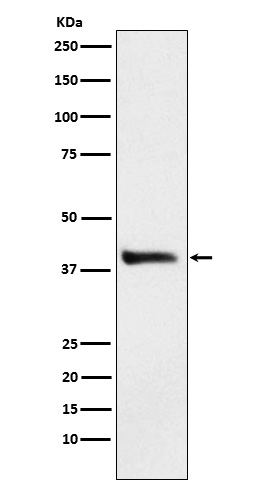
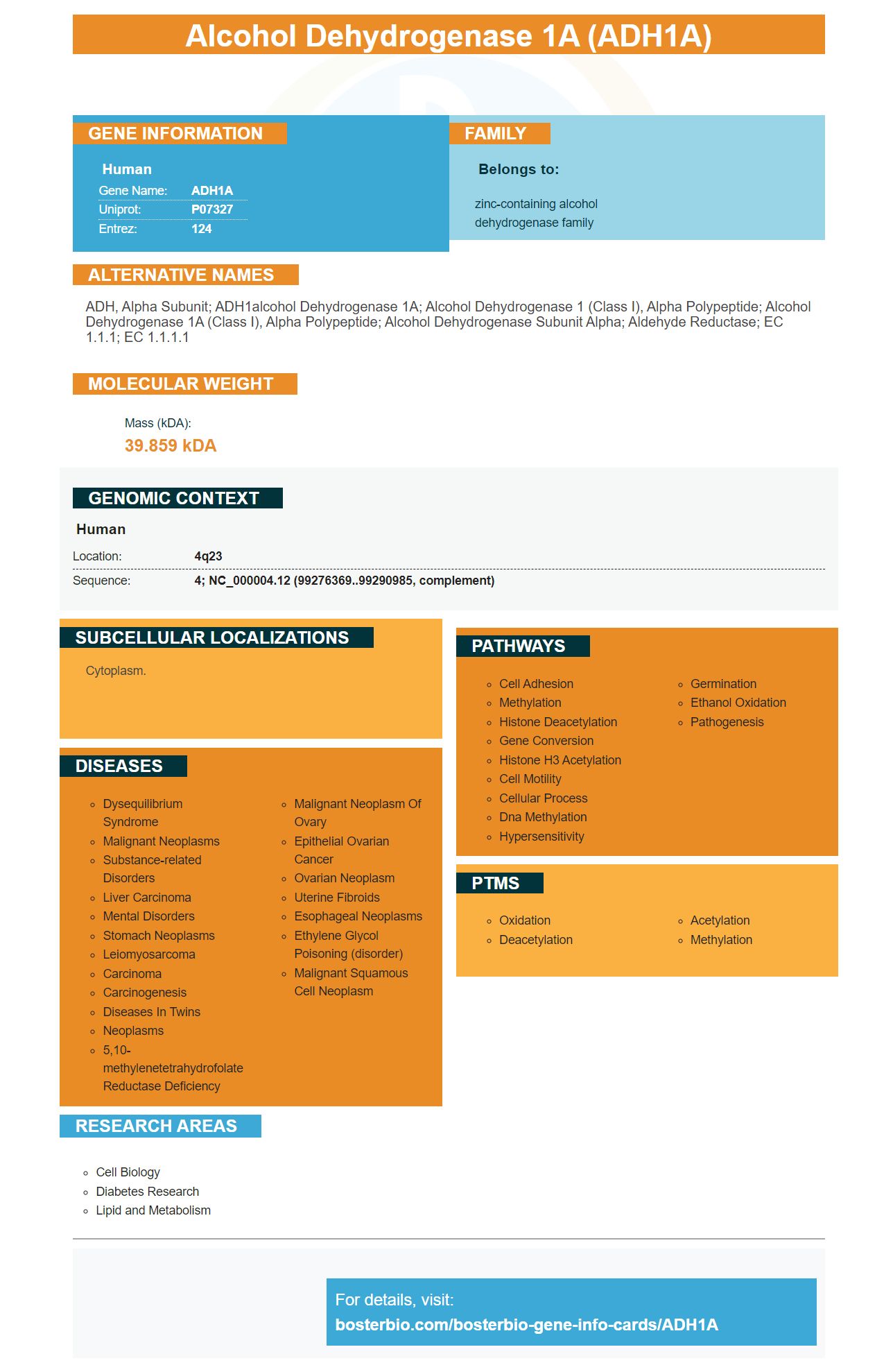
Facts about Alcohol dehydrogenase 1A.
Human | |
---|---|
Gene Name: | ADH1A |
Uniprot: | P07327 |
Entrez: | 124 |
Belongs to: |
---|
zinc-containing alcohol dehydrogenase family |
ADH, alpha subunit; ADH1alcohol dehydrogenase 1A; alcohol dehydrogenase 1 (class I), alpha polypeptide; alcohol dehydrogenase 1A (class I), alpha polypeptide; Alcohol dehydrogenase subunit alpha; aldehyde reductase; EC 1.1.1; EC 1.1.1.1
Mass (kDA):
39.859 kDA
Human | |
---|---|
Location: | 4q23 |
Sequence: | 4; NC_000004.12 (99276369..99290985, complement) |
Cytoplasm.
Boster Bio antibodies are tested on multiple platforms and have been proven to be valid. Boster ensures that their antibodies have high specificity as well as affinity. The company also offers a credit towards purchases for the first reviewers who review each product. Likewise, Boster awards scientists around the world for their contributions to the scientific community.
Boster Bio's Anti-Alcohol Dehydrogenase/ADh2A Antibody is a reagent-grade product that reacts with ADh2A proteins in Rat, Mouse, Human. The antibody was developed by Steven Boster, who was known as "the lavatory scientist." His company eventually became one the largest Chinese catalog antibody companies. Boster developed PicoKine(tm) an ELISA platform that is high-affinity and uses trade secrets.
ADh2A is also known as ADh2, and is a protein with seven different isozymes. Three of these are class-II while two are class-II. ADh2A cannot be expressed in all cells, which can cause problems with the assay. In order to determine its specificity, it should be expressed at endogenous levels. This method has many advantages over other immunosorbent assays.
The datasheet should include detailed information about the antibody. This includes its source, purification method, and type of immunogen. It should also contain the specific conditions under which the antibody's specificity was evaluated. Validation data should also be available from multiple cell line types. Scientists must carefully verify the product's legitimacy once the datasheets have been completed.
The context inwhich an antibody is being used will greatly affect its specificity and selectivity. Different types of immunoassays need different validation methods. Assay-specific validation is especially important to confirm an antibody's specificity and selectivity. This validation aims to produce reproducible results. Primary antibodies' performance is affected by assay context. Therefore, recommendations can differ depending on this.
Boster Bio ADh2A Marker, a comprehensive assay, demonstrates the validity and reliability of all antibodies IHC. The ad-hoc group responsible for this project examined seven antibody manufacturers to determine how closely their antibodies replicate the Boster Bio ADh2A Marker. They found that all IHC antibodies were valid and validated. The other factors were not well defined.
The ADh2A marker is a monoclonal antibody that recognizes the protein Cytokeratin 19. This antibody displays the expected band, along with numerous additional bands at higher molecular weights. It also recognizes nuclear staining in FFPE lung cancer tissue. The BosterBio IHC Marker ADh2A is fully validated, even in vitro.
Boster Bio ADh2A marker is more specific than commercially available ADh2A markers. Its immunogen binds to ADh2A in both human and murine tissues. It is highly sensitive and specific, so it can be used for many purposes. Boster Bio ADh2A Marker is validated on IHC, ELISA, and FC.
Boster Bio ADh2A markers offer IHC and biochemical assays that can be used to validate the antibody against specific biochemical markers. Boster Bio ADh2A Marker validates IHC using biochemical or cell culture methods. It can be used in many areas of cancer research. It is highly versatile and reliable in detecting cancer biomarkers via IHC.
Boster Bio ADh2A markers include a protocol for antibody validation. The datasheet includes information on the animal host as well as the source of the synthetic peptide. To verify phospho-specificity, the antibody is tested in paraffin-embedded cells with siRNA and phosphatase. The Company also tests its antibody on xenografts of the target protein.
WB is an appropriate initial validation stage. Monoclonal antibodies perform better on native proteins, than denatured. WB results do not provide absolute proof of the antibody’s specificity. Monoclonal monoclonal antibodies can also be validated by IHC and flowcytometry. Both WB and IHC are complemented by the negative controls. Negative controls are particularly important in immunohistochemical studies, because they help to determine if secondary antibodies are binding non-specifically or detecting false negatives.
Despite a growing number of antibody vendors, not every antibody manufacturer validates all its products. There are many reasons this could happen, including nonspecific antibody binding, differences between batches and experimental conditions. Research can be lost if the antibody is used in an inappropriate manner. Fortunately, some vendors are making strides to address this problem by developing more stringent antibody validation guidelines. There are many bodies and organizations that can validate antibodies in addition to the manufacturer's guidelines.
Orthogonal validation involves manually evaluating the correlation between the intensity of staining by a single target antibody and the mRNA expression of a cell line or tissue. Independent antibodies must not have overlapping epitopes in order to be considered independent. The orthogonal validation of antibodies must show similar patterns across multiple types of cells and tissues. These antibodies must show similar mRNA levels across all specimens.
Human Protein Atlas is an organization of distinguished researchers that provides a framework for evaluating the effectiveness and safety of antibodies. To validate antibodies, the Human Protein Atlas employs five pillars in addition to the IWGAV guidelines. The company bases its Enhanced Validation on these pillars. Atlas Antibodies employs the most appropriate validation strategy for your experiment to offer enhanced validation. This provides greater security in antibody specificity.
Orthogonal Validation, which is a comparison in protein expression levels from two tissues, was used to assess whether an antibody correlates or not with mRNA levels. Many proteins showed similar patterns in the expressions of different antibodies. However, a larger number of antibodies may need independent validation in order to qualify for orthogonal validation. Orthogonal validation can require more experiments, however these are the best guidelines.
PMID: 3013304 by von Bahr-Lindstroem H., et al. cDNA and protein structure for the alpha subunit of human liver alcohol dehydrogenase.
PMID: 2935875 by Ikuta T., et al. Three human alcohol dehydrogenase subunits: cDNA structure and molecular and evolutionary divergence.