ADH1A / Alcohol dehydrogenase 1A · IHC design guide

Design Immunohistochemistry for ADH1A

Plan paraffin-section chromogenic IHC for ADH1A using hepatocyte cytoplasmic staining as a reference (HPA tissue IHC). This guide covers fixation, controls and scoring, including the possibility that HPA staining reflects proteins from more than one gene (HPA tissue IHC).

Evidence assembled Oct 2026 · For research use; verify linked source records and product datasheet before use
Immunohistochemistry protocol sheet for ADH1A (IHC for ADH1A): expected localisation Hepatocyte cytoplasm (HPA tissue IHC), antibody PB10029, validated IHC image, and IHC protocol steps
Printable ADH1A IHC protocol sheet — expected localisation Hepatocyte cytoplasm (HPA tissue IHC), antibody PB10029, controls and protocol steps. Open the full ADH1A IHC guide →

ADH1A Immunohistochemistry Experimental Design Guide

Expected localisation, validated protocols, controls and antibodies — the at-a-glance facts below, then the full design guide.

Must know before staining
Expected localisation Hepatocyte cytoplasm (HPA tissue IHC)
Staining pattern Selective cytoplasmic staining in hepatocytes (HPA tissue IHC)
Antigen retrieval EDTA pH 8.0 HIER, heat-mediated (datasheet PB10029)
Positive control ⓘ Appendix+3 more · see all
Negative control ⓘ Adipose tissue+4 more · see all
Important caveats
Reasons your staining may differ from the expected pattern.
Fixation Keep fixation consistent across sections (standard IHC practice; not target-specific)
Caveat HPA staining may reflect proteins from more than one gene (HPA tissue IHC)
Regulation Expression regulation is not annotated (UniProt)
Isoform / epitope No annotated isoforms; chain spans residues 2–375 (UniProt)
Section 1

Recommended ADH1A IHC & IF Protocols

The catalog antibody protocol uses EDTA pH 8.0 retrieval (datasheet: PB10029). The published options below report ADH1A IHC staining in paraffin sections (PMC13357096; PMC13326605).

Recommended immunohistochemistry (IHC-P) protocol parameters
SampleParaffin-embedded human liver cancer tissue; fixative not specified (datasheet PB10029)
FixationImage fixative and duration unreported (datasheet PB10029); verify before use.
Sectioning4–5 µm sections on charged slides (standard)
DeparaffinisationXylene, graded ethanol series to water (standard)
Antigen retrievalHeat retrieval: EDTA pH 8.0 (datasheet PB10029); 20 min, 95–100 °C (standard)
Peroxidase block3% H2O2, 10 min, room temperature (standard)
Blocking10% goat serum (datasheet PB10029)
Primary antibodyRabbit anti-ADH1A, 2-5μg/ml (datasheet PB10029)
Primary incubationOvernight at 4 °C (datasheet PB10029)
DetectionHRP-conjugated secondary, DAB chromogen (datasheet PB10029)
CounterstainHematoxylin, blue, dehydrate and mount (standard)
Expected resultADH1A-positive staining in endocrine cells of appendix (HPA tissue IHC: Medium). HPA tissue profile: Selective cytoplasmic expression in hepatocytes. No signal in the no-primary control.
💡Decision noteStart with EDTA pH 8.0 heat retrieval for the catalog antibody (datasheet: PB10029); treat the published citrate conditions as antibody-specific alternatives (PMC13357096; PMC13326605).
Section 2

What Is the Expected ADH1A Staining Pattern?

ADH1A is a cytoplasmic protein with no transmembrane segment (UniProt P07327). In paraffin-section IHC, expect selective cytoplasmic staining of hepatocytes at a medium level (HPA: liver tissue IHC). HPA rates the tissue pattern Enhanced because staining agrees with RNA data, but cautions that the assessed antibodies can target proteins from more than one gene (HPA: tissue IHC reliability).

What am I looking at on my slide?
Cytoplasmic staining in hepatocytes, with little staining in surrounding liver tissue.This matches the selective hepatocyte pattern; HPA reports a medium staining level, so a very dark signal is unnecessary for a positive call (HPA: liver tissue IHC).
Predominantly nuclear or membrane-only staining in liver hepatocytes.This differs from the expected cytoplasmic IHC pattern (HPA: liver tissue IHC; UniProt P07327: cytoplasm). Check controls and staining distribution before treating it as ADH1A. ICC-IF reports additional uncertain locations, which do not establish a nuclear liver IHC pattern (HPA: subcellular).
Strong staining in adipocytes or another cell type scored not detected by HPA.Consider antibody cross-reactivity or endogenous detection activity; adipocytes were scored not detected in adipose tissue (HPA: tissue IHC). Staining outside hepatocytes is not automatically wrong: HPA also reports medium signal in selected intestinal and endocrine cells (HPA: tissue IHC).
Diffuse brown color across cells and empty areas of the section.A signal that ignores cell boundaries is difficult to score as cytoplasmic ADH1A. Check background from the detection system and nonspecific binding with appropriate controls (general IHC practice); compare against the selective hepatocyte pattern (HPA: liver tissue IHC).
No staining in hepatocytes of a liver section expected to serve as a positive control.This conflicts with HPA's medium hepatocyte staining (HPA: liver tissue IHC). Review the positive-control section, antibody and detection reagents, and retrieval conditions before interpreting a test section as negative (general IHC practice).
💡Expected ADH1A appearanceCall a section positive when hepatocytes show selective, predominantly cytoplasmic chromogenic staining around the medium level reported by HPA; widespread extracellular color or isolated nuclear staining does not match that liver pattern (HPA: liver tissue IHC; UniProt P07327: cytoplasm).
How each factor affects the staining
Cellular compartmentUniProt places ADH1A in the cytoplasm and lists no transmembrane segment (UniProt P07327). Use cytoplasmic hepatocyte staining as the main IHC readout (HPA: liver tissue IHC); a membrane outline alone is insufficient.
Tissue and cell selectionLiver hepatocytes are a documented medium-level positive control (HPA: tissue IHC). Appendix and duodenal endocrine cells and small-intestinal enterocytes also have reported medium staining, while adipose-tissue adipocytes were not detected (HPA: tissue IHC).
Antibody interpretationThe overall tissue profile is rated Enhanced, yet HPA cautions that the staining assessment involves antibodies targeting proteins from more than one gene (HPA: tissue IHC reliability). Concordant staining supports the expected pattern but cannot, by itself, prove ADH1A-specific signal.
Subcellular evidence from ICC-IFQ: Should an IF image define the paraffin-section IHC pattern? A: Use it as context only: cytosol is supported, whereas plasma membrane, nucleoplasm and ciliary sites are uncertain; the ICC-IF summary carries a multiple-gene antibody caution (HPA: subcellular).
Fixation sensitivityTarget-specific effects of fixation on ADH1A staining are unreported in the supplied UniProt and HPA records. Retrieval choices therefore require empirical evaluation under the local IHC workflow (general IHC practice), without assuming a particular ADH1A epitope is masked.
Why is my staining missing, weak or wrong?
SituationLikely causeNext action
Liver positive control is blank.A failed staining run or inadequate antibody access is possible; HPA reports medium hepatocyte staining (HPA: liver tissue IHC).Confirm that the positive-control section and detection reagents worked, then review the antibody conditions and retrieval used in that run (general IHC practice). Do not infer target-specific fixation sensitivity from this result.
Hepatocytes stain only in nuclei.A nuclear-only liver result conflicts with the cytoplasmic tissue profile (HPA: liver tissue IHC; UniProt P07327: cytoplasm).Inspect the counterstain and antibody-omission control, then compare cellular boundaries and the expected hepatocyte pattern (general IHC practice). Treat uncertain ICC-IF nucleoplasm staining as context, not IHC confirmation (HPA: subcellular).
Adipocytes show strong chromogen.HPA scored adipose-tissue adipocytes not detected; cross-reactivity or endogenous detection activity may explain unexpected staining (HPA: tissue IHC; general IHC practice).Compare the signal with an antibody-omission control and assess whether it follows cell boundaries (general IHC practice). Recheck any antibody-based conclusion against HPA's multiple-gene-target caution (HPA: tissue IHC reliability).
Brown haze covers the section.Nonspecific binding or detection background can obscure the selective cytoplasmic pattern (general IHC practice; HPA: liver tissue IHC).Inspect a control without primary antibody, the blocking and wash steps, and chromogen development (general IHC practice). Score hepatocytes only where intracellular signal is distinguishable from the haze.
A liver section shows crisp membrane outlines but little cytoplasmic signal.That distribution differs from HPA's cytoplasmic hepatocyte IHC profile; UniProt lists no transmembrane segment (HPA: liver tissue IHC; UniProt P07327).Check the positive-control pattern and detection background before calling the outlines positive (general IHC practice). HPA's uncertain plasma-membrane ICC-IF location does not establish a membrane-only liver IHC result (HPA: subcellular).
A non-liver section stains in a limited cell population.This can be compatible with the record: HPA reports medium staining in appendix and duodenal endocrine cells and small-intestinal enterocytes (HPA: tissue IHC).Identify the stained cell type before calling the result nonspecific. Compare it with the matching HPA tissue entry and use appropriate staining controls; avoid extending those observations to unreported tissues (HPA: tissue IHC; general IHC practice).

Sample controls for ADH1A IHC & IF

🧪Run liver first: hepatocytes should stain (HPA: Liver hepatocytes, Medium). Run adipose tissue as the negative comparator; adipocytes should be unstained (HPA: Adipose tissue adipocytes, Not detected). On the liver slide, use unstained non-hepatocyte areas as an internal background reference without assuming that every non-hepatocyte is ADH1A-negative.
Positive control tissue: Appendix (Endocrine cells, HPA Medium)
Negative control tissue: Adipose tissue (HPA Not detected)
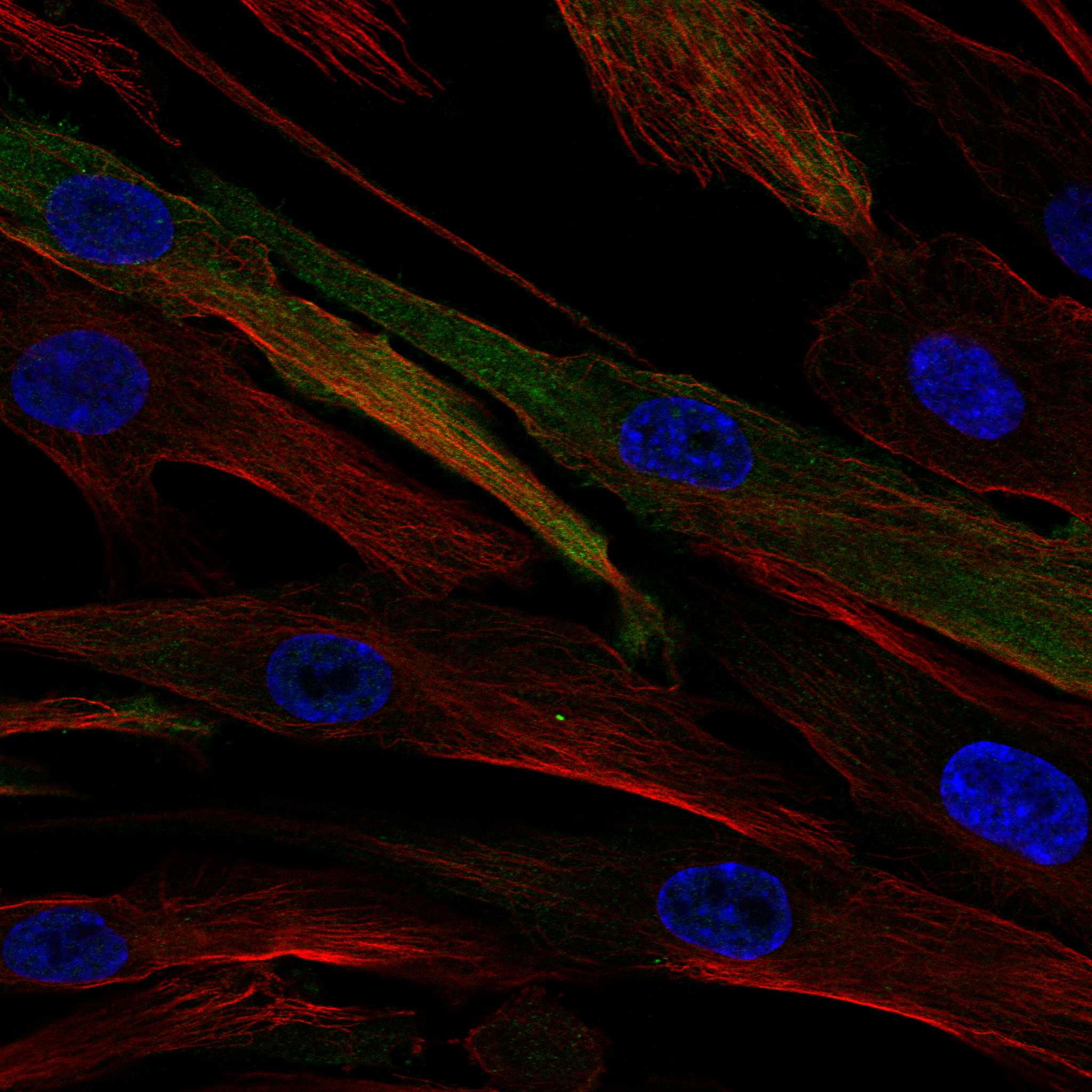
ICC-IF cell lines (HPA subcellular resource): HPA ICC-IF images show ADH1A in ASC52telo, SiHa, U2OS, RPTEC/TERT1, hTERT-RPE1 (serum starved), with annotated localisation: Plasma membrane (uncertain), Cytosol (supported) (HPA subcellular).
Technical controls: Include no-primary (secondary-only), a rabbit IgG isotype control matched to the primary antibody, and ADH1A knockout material as a biological negative (caption: rabbit primary antibody; standard IHC practice). Quench endogenous peroxidase in liver before HRP/DAB detection and distinguish pigment from specific chromogen (caption: liver tissue and HRP/DAB detection; standard IHC practice).
⚠️Feasibility: No target-specific fixation window or fixation effect is reported, and the selected PB10029 paraffin-section caption does not state a fixative (caption: fixative not stated). The example uses heat-mediated EDTA retrieval at pH 8.0, but does not establish that retrieval is required; the supplied evidence does not show frozen sections or IF to be easier (caption: EDTA retrieval in a paraffin section). For IF/ICC, assess cytosolic staining cautiously: cytosol is supported, while the reported plasma membrane and additional locations are uncertain and the HPA localization evidence carries a multiple-gene antibody caution (HPA: subcellular localization).

HPA tissue IHC evidence for ADH1A

Comprehensive Human Protein Atlas IHC scoring per tissue (reliability: Enhanced — High consistency between antibody staining and RNA expression data. Caution, targets protein from more than one gene.). Rows are taken directly from the HPA tissue chart — click any row's HPA link to view the source.

Positive expression · recommended positive controls

TissueCell typeLevelEvidenceSource
Appendix Endocrine cells Medium Protein (IHC) HPA →
Duodenum Endocrine cells Medium Protein (IHC) HPA →
Liver Hepatocytes Medium Protein (IHC) HPA →
Small intestine Enterocytes - Microvilli Medium Protein (IHC) HPA →

Undetected expression · recommended negative controls

TissueCell typeLevelEvidenceSource
Adipose tissue Adipocytes Not detected Protein (IHC) HPA →
Adrenal gland Glandular cells Not detected Protein (IHC) HPA →
Bone marrow Hematopoietic cells Not detected Protein (IHC) HPA →
Breast Adipocytes Not detected Protein (IHC) HPA →
Bronchus Respiratory epithelial cells Not detected Protein (IHC) HPA →
Section 3

Advanced ADH1A IHC Tips

Troubleshoot ADH1A staining in paraffin sections using the catalog antibody’s IHC conditions and compartment aware controls.

How should I adjust retrieval when ADH1A staining is weak or uneven?
Start with heat mediated retrieval in EDTA at pH 8.0 for paraffin sections (datasheet PB10029). The selected tissue image used this retrieval before overnight primary antibody incubation at 4°C, but its fixative was not reported (datasheet PB10029). If staining is weak, compare a modestly longer heating interval with the original interval on adjacent sections, keeping cooling and subsequent staining steps identical (standard IHC practice). If tissue lifts or morphology deteriorates, reduce heating and inspect section adhesion before changing antibody concentration (standard IHC practice). Judge improvement by selective cytoplasmic staining in hepatocytes, while remembering that the reference tissue image is liver cancer (HPA tissue IHC; datasheet PB10029).
Could fixation explain weak ADH1A staining in my paraffin sections?
Target specific sensitivity to fixation is unknown: the selected paraffin tissue caption does not state its fixative (datasheet PB10029). Record the fixative, fixation duration and processing conditions for each specimen, then compare matched sections processed under the same conditions (standard IHC practice). Keep retrieval at EDTA pH 8.0 initially and use the catalog antibody’s reported 2 μg/ml concentration as a reference while assessing the fixation variable (datasheet PB10029). If signal differs between processing batches, check morphology and a same run positive control before attributing the difference to ADH1A abundance (standard IHC practice). Do not use hepatocyte staining patterns alone to assign a fixation effect (HPA tissue IHC).
What staining pattern should count as plausible ADH1A in liver?
Prioritise cytoplasmic staining in hepatocytes when evaluating liver sections (HPA tissue IHC; UniProt P07327 localisation). ADH1A has no annotated transmembrane segment or signal peptide, so a sharply membrane restricted IHC pattern needs independent confirmation before assignment to this protein (UniProt P07327 topology and processing). Subcellular data support cytosol but mark plasma membrane localisation uncertain, with a caution that the antibodies may recognise proteins from multiple genes (HPA subcellular). Compare tumour cells with adjacent tissue where available and score the compartments separately rather than combining all brown signal into one result (standard IHC practice). Treat isolated nuclear or ciliary staining cautiously because those locations are also marked uncertain (HPA subcellular).
How can I assess whether the stain identifies ADH1A specifically?
ADH1A has 0 annotated isoforms in the supplied record, but an antibody epitope is not specified here (UniProt P07327 record; datasheet PB10029). Closely related ADH1A, ADH1B and ADH1C subunits can form dimers, so dimer biology does not establish antibody specificity (UniProt P07327 subunit). HPA rates its tissue staining Enhanced while cautioning that its antibody targets proteins from more than one gene (HPA tissue IHC). Before assigning a disputed IHC signal to ADH1A alone, check the catalog antibody’s epitope and cross reactivity documentation, and compare with an independently validated epitope where available (standard IHC validation practice). Record which antibody produced each result rather than transferring HPA specificity claims to PB10029 (HPA tissue IHC; datasheet PB10029).
How would I use IF to investigate an ambiguous IHC pattern?
Use IF as a secondary localisation check and multiplex ADH1A with a validated hepatocyte marker when examining liver, because hepatocytes show selective cytoplasmic expression in tissue IHC (HPA tissue IHC). Select spectrally separated fluorophores and place the weaker expected signal in a channel with low tissue autofluorescence; include single label and no primary controls (standard IF practice). For a cytosolic epitope, use controlled permeabilisation and compare it with an otherwise matched condition, since the supplied record places ADH1A in the cytoplasm and lists no transmembrane segment (UniProt P07327 localisation and topology). Confirm the marker and ADH1A signals within individual cells, then compare compartment patterns with chromogenic IHC without assuming identical assay performance (standard IF/IHC practice).
What should I check when DAB appears throughout the section?
First separate diffuse DAB background from cell restricted cytoplasmic signal, using a no primary control and a known positive section in the same run (standard IHC practice; HPA tissue IHC). The selected protocol used 10% goat serum blocking, a rabbit primary at 2 μg/ml overnight at 4°C, and peroxidase based DAB detection (datasheet PB10029). Check peroxidase blocking, washes, secondary antibody concentration and DAB development time if the no primary control stains (standard chromogenic IHC practice). If background is confined to damaged or necrotic areas, exclude those areas from scoring and inspect adjacent intact tissue (standard IHC practice). Retain cytoplasmic hepatocyte staining as the main biological reference for liver (HPA tissue IHC).
How should I quantify ADH1A staining across paraffin sections? ⚠ ANSWER MARKED FOR VERIFICATION
Define the cell population and cytoplasmic compartment before scoring, because liver tissue IHC reports selective cytoplasmic staining in hepatocytes (HPA tissue IHC). For chromogenic sections, record the percentage of positive eligible cells and intensity categories, then calculate an H-score on a 0–300 scale if ordinal intensity scoring is reproducible (standard IHC practice). Where cells are sparse or architecture differs, report positive cell density per mm² of viable tissue as a complementary measure (standard IHC practice). Normalise counts to eligible cells or viable tissue area, use consistent thresholds and imaging settings across batches, and exclude necrosis and section edges (standard IHC practice). Keep tumour and non tumour regions separate when both are present (standard IHC practice).
When is an apparent ADH1A positive result likely to be artefact?
A convincing liver result shows reproducible cytoplasmic staining in the intended cell population, consistent with hepatocyte tissue staining and ADH1A cytoplasmic localisation (HPA tissue IHC; UniProt P07327 localisation). Be cautious with isolated nuclear, ciliary or sharply membrane restricted signal: the subcellular source marks those locations uncertain and warns about antibodies recognising multiple genes (HPA subcellular). Compare section edges, necrotic areas and the no primary control; staining restricted to damaged regions or persisting without primary antibody suggests an assay artefact (standard IHC practice). Check endogenous peroxidase blocking when DAB appears independent of primary antibody, then repeat with matched positive and negative controls (standard chromogenic IHC practice). Interpret an unexpected cell type only after confirming morphology and antibody specificity (standard IHC validation practice).
Boster reagents

Best ADH1A / Alcohol dehydrogenase 1A IHC Antibodies

PB10029 has IHC images from human liver cancer and mouse and rat liver paraffin sections, plus an IF image from U20S cells (catalog image captions).

Real IHC data IHC analysis of ADH1A using anti-ADH1A antibody (PB10029). ADH1A was detected in a paraffin-embedded section of human liver cancer tissue. Heat mediated antigen retrieval was performed in EDTA buffer (pH 8.0, epitope retrieval solution). The tissue section was blocked with 10% goat serum. The tissue section was then incubated with 2 μg/ml rabbit anti-ADH1A Antibody (PB10029) overnight at 4°C. Peroxidase Conjugated Goat Anti-rabbit IgG was used as secondary antibody and incubated for 30 minutes at 37°C. The tissue section was developed using HRP Conjugated Rabbit IgG Super Vision Assay Kit (Catalog # SV0002) with DAB as the chromogen.
Anti-Alcohol Dehydrogenase/ADH1A Antibody ®
Cat # PB10029

PB10029 is the SKU that will render; its IHC images show human liver cancer and mouse and rat liver paraffin sections (PB10029 IHC captions). Its IF image shows U20S cells (PB10029 IF caption).

Which to pick: Choose PB10029 for tissue IHC: its images document paraffin sections from all three listed species, while the fixative is unreported (PB10029 IHC captions). Choose PB10029 for IF/ICC because it lists both applications and has a U20S cell IF image (PB10029 catalog applications; PB10029 IF caption). For cross-species IHC, PB10029 has human, mouse and rat images; M06473-1 lists those species and IHC/IF applications and is monoclonal clone 19A72, but has no IHC or IF image in the payload (catalog reactivity, applications, clone and image fields).

Each figure is that product's own IHC / IF validation image from its datasheet.

References

  1. UniProt Consortium. UniProt entry P07327 (ADH1A_HUMAN, Alcohol dehydrogenase 1A).
  2. Human Protein Atlas. ADH1A tissue IHC expression (reliability: Enhanced).
  3. Human Protein Atlas. ADH1A subcellular location (ICC-IF): Mainly localized to the plasma membrane and cytosol. In addition localized to the nucleoplasm, primary cilium, primary cilium transition zone and basal body. Caution: Based on antibodies targeting proteins from multiple genes..
  4. Human Protein Atlas. ADH1A antibody validation summary (3 antibodies).
  5. Overexpression of alcohol dehydrogenase 1 A inhibits the progress of triple negative breast cancer via Wnt/β-catenin signaling. Scientific reports 2025 — PMC12475006.
  6. A pan-cancer functional atlas of ADH1A reveals its conserved role in the tumor microenvironment and immunity. Translational cancer research 2026 — PMC13357096.
  7. mTOR/HDAC1 Crosstalk Mediated Suppression of ADH1A and ALDH2 Links Alcohol Metabolism to Hepatocellular Carcinoma Onset and Progression in silico. Frontiers in oncology 2019 — PMC6787164.
  8. HBx-mediated GPT2 suppression promotes liver cancer development by downregulating ADH1A. Cancer biology & medicine 2026 — PMC13326605.
  9. PubMed PMID:3013304 — UniProt-cited evidence.
  10. PubMed PMID:2935875 — UniProt-cited evidence.
  11. PubMed PMID:2920825 — UniProt-cited evidence.